|
|
 |
| Metridinidae Sars, 1902 ( Arietelloidea ) | | Syn.: | Metridiidae Sars, 1902 (1903) (p.110); Sewell, 1932 (p.247); Gurney, 1931 a (p.84); Rose,1933 a (p.175); Sewell, 1947 (p.165), Rem.; Brodsky, 1950 (1967) (p.81, 289); Mazza, 1967 (p.185); Andronov, 1974 a (p.1005); Brodsky & al., 1983 (p.141, 145); Zheng Zhong & al., 1984 (1989) (p.242) | | Ref.: | Dunn & Hulsemann, 1979 (p.53: emend.); Razouls, 1982 (p.405); 1993 (p.307); Bowman & Abele, 1982 (p.9); Bull. Zool. Nomencl, 1984 ( N°41,1, p.19); Sazhina, 1985 (p.114, Nauplius); Mauchline, 1988 (p.711: cuticular pores); Huys & Boxshall, 1991 (p.464); Madhupratap & al., 1996 (p.863, Table 5: %/copepods); Chihara & Murano, 1997 (p.837); Cuoc & al., 1997 (p.651, Fig.6: female genital area); Bradford-Grieve & al., 1999 (p.883, 901, 903, 947, Genera Key); Bradford-Grieve, 1999 b (p.110, Déf., Rem.); Soh & al., 1999 (p.122, phylogeny); Ohtsuka & Huys, 2001 (p.461); Boxshall & Halsey, 2004 (p.13; 49; 145: Def.; p.146: Genera Key); Vives & Shmeleva, 2007 (p.356, clé des Genera Key); Blanco-Bercial & al., 2011 (p.103, Table 1, Fig.2, 3, 4, Biol. mol, phylogeny); Laakmann & al., 2019 (p.330, fig. 2, 3, phylogenetic relationships); Hirai & al., 2020 (p.1, Fig.4: metabarcoding, Fig.8: OTUs distribution patterns, Fig.9: phylogenetic analysis )
Bradford-Grieve J.M., (2002 onwards). Key to calanoid copepod families. Version 1 : 2 oct 2002. http://www.crustacea.net/crustace/calanoida/index.htm  | | Rem.: | 3 G.: Gaussia, Metridia, Pleuromamma.
For numerous authors, after mouthpart morphology and gut contents, and dietary preferences of laboratory reared specimens, this family appears to be omnivorous.
Key to genera after Boxshall & Halsey (2004, p.146) :
1 - Prosome with conspicuous black or dark brown spot on one side of 1st pedigerous somite ........ Pleuromamma.
1' - Prosome lacking such spot ......... 2.
2 - Posterolateral angles of prosome produced into wing-like expansions, tapering to point; anal somite with long posterior processes located dorsolateral to caudal rami ...... Gaussia.
2' - Posterolateral angles of prosome usually rounded, if pointed then not with wing-like expansions; anal somite typically with short posterior processes .......... Metridia.
Definition after Bradford-Grieve (1999b, p.110) Female :
- Body elongate.
- Cephalosome and pediger 1 separate, pedigers 4 and 5 fused.
- Posterolateral corners of the last thoracic segment usually rounded, expanded in Gaussia.
- Pediger segment 1 Pleuromamma.
- Urosome 3-segmented.
- Caudal rami often asymmetrical with up to 6 setae.
- Genital segment with copulatory pores and seminal receptacles paired in Gaussia and Metridia, unpaired in Pleuromamma ; gonopores and egg laying ducts paired.
- Rostrum with paired filaments.
- A1 23- or 34-segmented (segments 1 and 2 fused, 5 and 7 separate, 8 and 9 fused, 10-22 separate, apical segment double).
- A2 with basipods 1 and 2 separate with 1 and 2 setae respectively ; endopod 2-segmented, bilobed distal segment with 1-2, 8-9 + 6-7 setae ; exopod 7-segmented with 1, 2
, 1, 1, 1, 1, 4 setae respectively.
- Md with well-developed blade, palp basipod 2 with 4 setae ; endopod 2-segmented with 4 and 10 setae respectively ; exopod 5-segmented with 1, 1, 1, 1, 2 setae respectively.
- Mx1 inner lobe 1 with about 15 spines and setae, inner lobes 2 and 3 with 4-5 and 4 setae ; basipod 2 with 5-7 setae ; endopod 2-segmented with 6, 9-12 setae ; exopod with 11 setae ; outer lobes 1 and 2 with 9 and 1 setae.
- Mx2 with lobes 1-5 with 5 (9 in P. xiphias), 3, 3, 3, 4 setae respectively ; endopod 4-segmented with 3-4, 3, 2, 2 setae or reduced.
- Mxp 7-segmented, basipod 1 with 1, 2, 4, 4 setae ; basipod 2 with 3 setae plus 2 setae on the incorporated 1st endopodal segment ; endopod 5-segmented with 4, 4, 3, 3, 4 setae respectively.
- Swimming legs with both rami 3-segmented.
- Exopods of P2-P4 large, and flat, outer edge spines small.
- P1 basipod 2 inner seta situated on its anterior surface and passing across the face of endopodal segment 1
- Endopod segment 1 of P2 typically incised and ornamented with 1 or more hook-like spinous processes ; endopods typically small, cylindrical.
Male :
- Urosome 5-segmented, sometimes strongly asymmetrical.
- A1 usually prehensile on the left.
- Mouthparts similar to those of female.
|  issued from :C. Cuoc, D. Defaye, M. Brunet, R. Notonier & J. Mazza in Mar. Biol., 1997, 129. [p.13, Fig.6]. Diagrammatic representation of main evolutionary trends in organization of female genital area in calanoïds for Metridinidae (modified after Huys & Boxshall, 1991 and Ohtsuka & al., 1994). *: seminal duct of Pleuromamma may be situated on left or right, according to individual. |
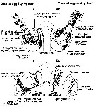 issued from :C. Cuoc, D. Defaye, M. Brunet, R. Notonier & J. Mazza in Mar. Biol., 1997, 129. [p.661, Fig.5]. Metridinidae. Schematic three-dimensional interpretation of structure and function of egg-laying ducts. A1, A2 Dorsal representations of egg-laying duct when closed (A1) and open (A2). B1, B2 details of part duct when closed (B1) and open (B2), showing how contraction and relaxation of muscles of egg-laying duct resulut in coordinated opening and closing of the gonopores and seminal ducts (black arrows), allowing release of gametes (open arrows) (the shell ducts, which open into the egg-laying ducts, are not shown on diagram). |
 Issued from : G.A. Boxshall & S.H. Halsey in An Introduction to Copepod Diversity. The Ray Society, 2004, No 166, Part. I. [p.145]. Armature formula of swimming legs P1 to P4. Nota: Female P5 with transverse plate formed by fusion of coxae and intercoxal sclerite; each leg uniramous, comprising basis plus 1, 2 or 3-segmenhted exopod; basis and 1st exopodal segment typically with outer seta and spine respectively, 2nd exopodal segment with outer spine and inner seta, 3rd segment with 2 to 4 distal elements (I-0; II,I,3 in Metridia); 2nd and 3rd exopodal segments often fused, which setation reduced. - Male P5 asymmetrical, carried on plate formed by fused coxae and intercoxal sclerite. Right leg comprising basis with outer seta and 3-segmented exopod; 2nd exopodal segment with inner spinous process in some genera; 3rd segment with 1 or 2 minute distal setae. left leg comprising basis with outer seta and 2 or 3-segmented exopod; 1st exopodal segment bearing curved inner process, distal segment swollen, often curved or claw-like. - Eggs released into water. |
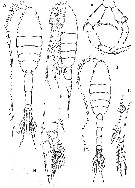 Issued from : G.A. Boxshall & S.H. Halsey in An Introduction to Copepod Diversity. The Ray Society, 2004, No 166, Part. I. [p.147, Fig.31]. Metridinidae: A, Metridia princeps habitus female; B, P2; C, female P5; D, habitus male; E, male P5; F, Pleuromamma xiphias habitus female. [Dars, 1924]. |
 Issued from : J.M. Bradford-Grieve in NIWA Biodiversity memoir 111, 1999. [p.110]. Spine and seta formula typically (but sometimes reduced) of the swimming legs P1 to P4. - Female P5 uniramous , small, symmetrical 2-4-segmented, basipod 1 and coupler (= intercoxal sclerite) fused; basipod 2 and exopod segment 1 usually with an outer setae and spine respectively; exopod segment 2 with 2-4 setae, terminal exopod segments may be fused. - Male P5 assymmetrical, attached to a plate formed from fusion of basipod 1 and coupler; right leg (or left) comprising basipod 2 with an outer seta and a 2?-3-segmented exopod; exopod segment 2 with an inner spinous process in some genera; exopod segment 3 with 1 or 2 minute distal setae; left leg (or right) comprising basipod 2 with an outer seta and a 2-or 3-segment exopod; exopod segment 1 bearing a curved inner process, distal segment swollen, often curved or claw-like. | | | | | (1) Gaussia Wolfenden, 1905 | |
| | Ref.: | Wolfenden, 1905 a (p.5); 1911 (p.289); Sewell, 1932 (p.176); Davis, 1949 (p.50); Brodsky, 1950 (1967) (p.312); Tanaka, 1963 (p.26); Barnes & Case, 1972 (p.66: Rem.); Saraswathy, 1973 (p.190, Redef.); Razouls, 1982 (p.418); Gardner & Szabo, 1982 (p.347); Mauchline, 1988 (p.709); Björnberg & Campaner, 1988 (p.351); Hulsemann, 1988 (p.188); Razouls, 1993 (p.307); Mauchline, 1998 (p.67); Bradford-Grieve & al., 1999 (p.948: clé spp.); Bradford-Grieve, 1999 b (p.110, Déf., Rem.); Soh & al., 1999 (p.122, phylogeny); Boxshall & Halsey, 2004 (p.146); Vives & Shmeleva, 2007 (p.357) | | Rem.: | Type: Pleuromma princeps T. Scott, 1894. Total: 4 spp.
Diagnosis after Bradford-Grieve (1999b, p.110) :
-As for family definition.
- Head with a small conical knob anteriorly.
- Metasome without a differentiated luminous organ.
- Posterolateral corners of metasome produced into spines which are more prominent in female than male.
- Genital segment of female symmetrically or asymmetrically inflated, sometimes with a black mass ventrally
- Urosome female segment 2 with distolateral borders produced into small flaps.
- Anal segment in both sexes produced into large pterygoid processes terminated with a pore and with fine hairs on both sides of segment which arise from ventral surface.
- Caudal rami with a blunt process on distal border.
- Right A1 of male geniculate, segment 12 with a small rounded glandular structure in addition to an aesthetasc and a seta.
- Mx1 with a small but distinct outer lobe 2 bearing a single plumose seta.
- Endopod segment 1 of P2 with 2 inner hooks.
- Female P5 3-4-segmented, segment 3 with a long plumose seta, segment 4 with 2 shorter plumose setae.
- Male P5 3-segmented; left segment 3 with 2 strong processes, 1 directed distally and the other proximally; right segment 3 with 4 setae, distal half of segment with an undulating inner margin. | | Remarks on dimensions and sex ratio: | | The mean female size is 10,09 mm (n= 4; S= 0,477; Cv= 0,047) and the mean male size is 9,60 mm (n= 3; S= 0,854; Cv= 0,089). The size ratio (M/F) is 0,948 or 94,8 % (n= 3; S= 0,046; Cv= 0,049) | | | | | | Ref.: | Giesbrecht, 1892 (p.61, 339); Giesbrecht & Schmeil, 1898 (p.105, spp. Key); Sars, 1900 (p.99); Wheeler, 1901 (p.175); Sars, 1902 (1903) (p.110); Esterly, 1905 (p.177); van Breemen, 1908 a (p.107, spp. Key); A. Scott, 1909 (p.120); Esterly, 1924 (p.96); Wilson, 1932 a (p.117, spp. Key); Sewell, 1932 (p.247); Rose, 1933 a (p.175, spp. Key); Mori, 1937 (1964) (p.67); Sewell, 1947 (p.165, 166); Farran, 1948 c (n°14, p.3); Davis, 1949 (p.47); Brodsky, 1950 (1967) (p.289, spp. Key); Tanaka, 1963 (p.15); Björnberg, 1972 (p.30, 31); Björnberg & al., 1981 (p.640); Razouls, 1982 (p.405); Gardner & Szabo, 1982 (p.321); Mauchline, 1988 (p.708); Razouls, 1993 (p.307); Bucklin & al., 1995 (p.655); Chihara & Murano, 1997 (p.837); Mauchline, 1998 (p.67); Bradford-Grieve & al., 1999 (p.948, spp. Key); Bradford-Grieve, 1999 b (p.112, Def.); Soh & al., 1999 (p.122, phylogeny); Boxshall & Halsey, 2004 (p.146); Vives & Shmeleva, 2007 (p.359, spp. Key) | | Rem.: | Type: Metridia armata Boeck, 1865 (= Calanus longus (Lubbock, 1854). Total: 25 spp. (of which 3 doubtful) + 3 unidentified.
Diagnosis after Bradford-Grieve (1999 b, p.112) :
- As in family definition.
- Urosome narrow.
- Body without a differentiated luminous organ.
- A2 with exopod a little longer than endopod, 6-segmented.
- P2 endopod segment 1 with a strong pair of internal hooks one larger than the other.
- Female P5 3- or 4-segmented with 2 or 3 long plumose terminal setae.
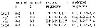
- Male P5 5-segmented, more or less curved ; on right (or left) the last segment is slightly widened, the antepenultimate segment with a long curved inner appendage. | | Remarks on dimensions and sex ratio: | | The mean female size is 4,858 mm (n= 23; S= 2,580; Cv= 0,53) and the mean male size is 4,050 mm (n= 22; S= 2,61; Cv= 0,64). The coefficients of variance indicate a strong heterogeneity of the sizes (7 species have sizes > 7 mm and 6 species 3 mm or less) The size ratio (M/F) is 0,848 ou 84,8 % (n= 20; S= 0,101; Cv= 0,119). The sex-ratio (F/M) is 1,05.
In the first group (body size < 6 mm) from all measurements: the mean female size is 3.167 mm (n = 31; SD = 1.0338), and for male 2.426 mm (n = 31; SD = 0.7570). The size ratio male: female is 0.798 (n = 15; 0.0996).
In the second group (body size > 6.5 mm): the mean female size is 8.584 (n = 13; SD = 1.1045), and for male 8.106 (n = 12; SD = 1.0588). The size ratio male: female is 0.938 (n = 6; SD = 0.0522). The ex ratio is nearest of 1. | | | | (3) Pleuromamma Giesbrecht, 1898 | |
| | Syn.: | Diaptomus (part.) Lubbock,1856; Pleuromma Claus,1863 (p.195); Giesbrecht, 1892 (p.61, 347); Dahl, 1893 (p.105, clé spp.); Wheeler, 1901 (p.76) | | Ref.: | in Giesbrecht & Schmeil, 1898 (p.108, spp. Key); Sars,1902 (1903) (p.114); Esterly, 1905 (p.174); van Breemen, 1908 a (p.103, clé spp.); A. Scott, 1909 (p.122); Sewell, 1932 (p.263); Wilson, 1932 a (p.123, spp. Key); Steuer, 1932 a (p.42, 62, 83, spp. Key); Rose, 1933 a (p.179, spp. Key); Mori, 1937 (1964) (p.68, spp. Key); Dakin & Colefax, 1940 (p.84, spp. Key); Farran, 1948 f (n°17, p.3); Davis, 1949 (p.51); Brodsky, 1950 (1967) (p.305, key spp.); Tanaka, 1963 (p.22); Owre & Foyo, 1967 (p.70, clé spp.); Björnberg, 1972 (p.30,31); Björnberg & al., 1981 (p.640); Razouls, 1982 (p.412); Gardner & Szabo, 1982 (p.333); Ferrari, 1984 a (p.166: asymmetry); Mauchline, 1988 (p.709); Huys & Boxshall,1991 (p.63, 68); Razouls, 1993 (p.307); Park (J.S) & Mauchline, 1994 (p.107, Rem.: cuticular pores); Beckmann, 1995 (in Monoculus, n°29, p.6: asymmetry); Chihara & Murano, 1997 (p.838); Mauchline, 1998 (p.67); Bradford-Grieve & al., 1999 (p.947, 948: spp. Key); Bradford-Grieve, 1999 b (p.118, Def.); Soh & al., 1999 (p.122, phylogeny); Boxshall & Halsey, 2004 (p.146); Mulyadi, 2004 (p.10, spp. Key for Indonesian Seas); Vives & Shmeleva, 2007 (p.372, spp. Key) | | Rem.: | Type: Diaptomus abdominalis Lubbock, 1856. Total: 11 spp. + 1 (juv.?).
Diagnosis after Bradford-Grieve (1999 b, p.118) :
- As in family definition.
- Urosome shorter than in Metridia, often asymmetrical, with curved segments and setal bundles.
- Head with a short acute apical process.
- Rostrum massive with 2 hairy filaments.
- Proximal part of A1 with large outer denticles.
- Asymmetrically situated circular, convex black spot on right or left side of cephalothorax (probably a luminous organ).
- Endopod segment 1 of P2 with an uncinateinner border as in Metridia, but right and left legs ofthen differ in size ; only one leg uncinate in some cases.
- Outer spine on exopod segment 1 of P3 with an elongate base, marked off by a deep notch.
- P5 of two types in female with 3 free segments and 3 long setae on distal segment ; or with 1 free segment and 3 short spines on distal segment
- Right P5 of male distal segment strongly curved, round ; preceding segment of same leg with a long curved inner spine. | | Remarks on dimensions and sex ratio: | | The mean female size is 3.219 mm (n = 22; SD = 1.2217), and the mean male size is 2.973 mm (n = 22; SD = 1.2018). The size ratio (Male: Female) is 0.918 (n = 11; SD = 0.0893). The sex-ratio (Female: Male) is 1. | | | | |
|
|
 Any use of this site for a publication will be mentioned with the following reference : Any use of this site for a publication will be mentioned with the following reference :
Razouls C., Desreumaux N., Kouwenberg J. and de Bovée F., 2005-2024. - Biodiversity of Marine Planktonic Copepods (morphology, geographical distribution and biological data). Sorbonne University, CNRS. Available at http://copepodes.obs-banyuls.fr/en [Accessed April 19, 2024] © copyright 2005-2024 Sorbonne University, CNRS
|
|
|
|


;)
;)
;)
