|
|
 |
|
Calanoida ( Order ) |
|
|
|
Diaptomoidea ( Superfamily ) |
|
|
|
Pontellidae ( Family ) |
|
|
|
Pontellina ( Genus ) |
|
|
| |
Pontellina sobrina Fleminger & Hulsemann, 1974 (F,M) | |
| | | | | | | Ref.: | | | Fleminger & Hulsemann, 1974 (p.84, figs.F,M); Hulsemann & Fleminger, 1975 (p.174, fig.F,M, juv.); Silas & Pillai, 1973 (1976) (p.778); Hulsemann & Fleminger, 1990 (p.99, figs.F); Mulyadi, 2002 (p.160, figs.F,M, Rem.) | 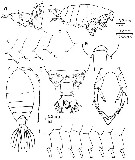 issued from : A. Fleminger & K. Hulsemann in Fishery bulletin, 1974, 72 (1). [p.85, Fig.13]. Female (from different area): a, Th 4-5 and urosome (lateral right side); b, habitus (lateral view; same as a); c, variation Th 4-5 spine (different area); d, variation in left Th 4-5 (lateral view: specimen with 2 spines); e, variation in left Th 4-5 (dorsal view; f, habitus (dorsal; same as a); g, Th 4-5 and urosome (dorsal; same as a); h, variation in lateral margin of right caudal ramus (dorsal view); i, P5 (anterior view; same as a). Nota: Most similar in appearance to P. morii. Right caudal ramus somewhat shorter relative to its width as well as to length of prosome, median ratio length to width 1.12:1, range 1.02-1.31:1, lateral edge usually with broad point immediatlely anterior to base of outermost seta, glandular tissue within ramus as in P. plumata. P5 similar to that in P. morii except that ratio of lengths of exopod to endopod tends to be smaller, median1.29:1, range 1.07-1.50:1; endopod typically with 2 relatively equal apical spines.
|
 issued from : A. Fleminger & K. Hulsemann in Fishery bulletin, 1974, 72 (1). [p.86, Fig.14]. Male: a, Th 4-5, part of urosome and P5 (lateral right side); b, variation in Th 4-5 spine (lateral); c, P5 (posterior view); d, chela P5 (lateral view; another specimen). Nota: Right caudal ramus similar to that in P. morii in both relative length and in proportion of length to width, median 1.88:1, range 1.71-2.07:1. Endopod 1 of left P5, compared to length of right caudal ramus, relatively longer than that in P. morii, this ratio in P. sobrina ranging from 0.96-1.17:1
|
 issued from : A. Fleminger & K. Hulsemann in Fishery bulletin, 1974, 72 (1). [p.103, Fig.34, a-c]. Female: Th 4-5 and urosome with attached spermatophore (dorsal, lateral and ventral, respectively).
|
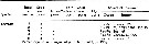 issued from : A. Fleminger & K. Hulsemann in Fishery bulletin, 1974, 72 (1). [p.111, Table 19]. List of identified particles from microscopic analysis of stomach contents in adulte female.
|
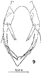 issued from : K. Hulsemann & A. Fleminger in Bull. Mar. Sci., 1975, 25 (2). [p.178, Fig.9]. Female: P5.
|
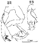 issued from : K. Hulsemann & A. Fleminger in Bull. Mar. Sci., 1975, 25 (2). [p.181, Figs.22-23]. Male: 22, P5 (posterior view); 23, chela in lateral view.
|
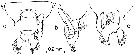
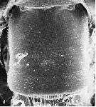 issued from : K. Hulsemann & A. Fleminger in Mar. Biol., 1990, 105. [p.101, Fig.1, d]. Female: d, genital segment (dorsal view). Nota: Scanning electron microscope examination revealed on the genital segment many patches of small spinules ornamenting the integument dorsally and laterally. The spinules are flattened, triangular, non-articulated, rigid structures rising from the integument, usually pointing obliquely posteriad.
|
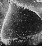 issued from : K. Hulsemann & A. Fleminger in Mar. Biol., 1990, 105. [p.102, Fig.2, d]. Female: d, genital segment (right laterall view). Nota: The spinules are arranged in polygonal patches. Within a patch they are of similar size and tend to be arranged in rows.
|
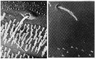 issued from : K. Hulsemann & A. Fleminger in Mar. Biol., 1990, 105. [p.103, Fig.3, b-c]. b, Peg sensillum and pore of integumental gland on genital segment of female (dorsal view; 2600 x); c, hair sensillum originating in clear space between patches of spinules. Shallow depressions indicate missing spinules. From right dorsal sector of third prosome segment of female (1000 x).
|
 issued from : K. Hulsemann & A. Fleminger in Mar. Biol., 1990, 105. [p.104, Fig.5-7]. Female: genital segment (left to right: dorsal view, right latera viewl and ventral view).Generalized distribution of peg sensilla (o) and pores of integumental glands (filled circle); symbols are larger than organs. Organs shown occurred in at least 40 % of specimens examined (n = 25).
|
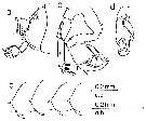
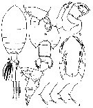 issued from : Mulyadi in Treubia, 2002, 32; [p.161, Fig.60]. Female (from Celebes Sea): a, habitus (dorsal); b-c, metasomal somite 5 and urosome (dorsal and lateral, respectively); d, posterolateral end of right metasomal somite 5 (lateral); e, P5. Nota: Cephalon and metasomal segment 1 separated, 4th and 5th metasomal segment fused. Posterolateral ends of metasome almost symmetrical, produced into short spine-like processes. Urosome 2-segmented; urosomal segment 1 with posterolateral cluster coarse hairs both margins, posterior margin fringed by hairs. Right caudal ramus fused with urosomal segment 2, somewhat shorter than wide, posterolateral end with broad point immediatelly anterior to base of outer seta; Endopod of P5 bifurcated, more than a half length of exopod, exopod ending in 3 long setae and 1 serrated flagella medially and 1 seta on posterior surface at 1/3 distal end. Male: f, metasomal somite 5 and urosome (lateral); g, P5. Nota: Cephalon as in female. Right caudal ramus fused with anal somite, less than 2 times as long as wide; left caudal ramus separated from anal somite. P5 asymmetrical; right leg, thumb of exopodal segment 1 (chela) stout with 1 seta at base, concave surface with 1 rounded lamella and 1 distal seta; exopodal segment 2 (claw) curved inwards, rounded at apex with 2 inner and 1 outer setae. Left leg, exopodal segment 1 with distolateral outer spine; exopodal segment 2 short, with 1 outer spinule, 1 leaf-like inner spine proximally, and terminating in 3 spine-like processes.
| | | | | Compl. Ref.: | | | Brinton & al., 1986 (p.228, Table 1); Chen Y.-Q., 1986 (p.205, Table 1: abundance %); Suarez-Morales & Gasca, 1998 a (p.111); Uysal & al., 2002 (p.17, tab.1) | | | | NZ: | 4 | | |
|
Distribution map of Pontellina sobrina by geographical zones
|
| | | | | | 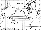 issued from : A. Fleminger & K. Hulsemann in Fishery bulletin, 1974, 72 (1). [p.87, Fig.15]. issued from : A. Fleminger & K. Hulsemann in Fishery bulletin, 1974, 72 (1). [p.87, Fig.15].
Geographical distribution of captures recorded diuring the present study. |
| | | | Loc: | | | Indonesia (Ambon Bay, Celebes Sea: Manado Bay, Pacif. (E equatorial), Gulf of California, Galapagos, E Medit. (NW Lebanon Basin) (in Uysal & al., 2002) | | | | N: | 8 | | | | Lg.: | | | (275) F: 1,78-1,42; M: 1,64-1,18; (1087) F: 1,5-1,55; M: 1,4-1,45; {F: 1,42-1,78; M: 1,18-1,64} | | | | Rem.: |
According to Fleminger & Hulsemann (1974, p.86) the species is indigenous to the eastern tropical Pacific Ocean (found only at Pacific stations east of 130° W). Occurrences at latitudes higher than 20° were restricted to a few samples taken near the mouth of the Gulf of California. The apparent boundaries coincide in general with the North and South Equatorial Currents, and its westernmost limits lie in the path of the Equatorial Countercurrent.
The occurence of this species in the Eastern Méditerranean Sea is surprising. | | | Last update : 16/11/2020 | |
|
|
 Any use of this site for a publication will be mentioned with the following reference : Any use of this site for a publication will be mentioned with the following reference :
Razouls C., Desreumaux N., Kouwenberg J. and de Bovée F., 2005-2024. - Biodiversity of Marine Planktonic Copepods (morphology, geographical distribution and biological data). Sorbonne University, CNRS. Available at http://copepodes.obs-banyuls.fr/en [Accessed April 19, 2024] © copyright 2005-2024 Sorbonne University, CNRS
|
|
|
|


;)
;)
;)
;)
;)
;)
;)
;)

;)
