|
|
 |
|
Calanoida ( Order ) |
|
|
|
Clausocalanoidea ( Superfamily ) |
|
|
|
Scolecitrichidae ( Family ) |
|
|
|
Pseudoamallothrix ( Genus ) |
|
|
| |
Pseudoamallothrix ovata (Farran, 1905) (F,M) | |
| | | | | | | Syn.: | Scolecithrix ovata Farran, 1905 (p.37, Descr.F, figs.F); Sars, 1925 (p., figs.F); Farran, 1926 (p.264); 1929 (p.209, 246, Rem.); Hardy & Gunther, 1935 (1936) (p.164, Rem.); Cabal & al., 2008 (289, Table 1);
? Scolecithricella ovata (M) : Minoda, 1971 (p.31, figs.M: juv.);
Scolecithrix subdentata Esterly, 1905 (p.167, figs.F);
Scolecithricella subdentata : Sewell, 1948 (p.557, 563); Davis, 1949 (p.46, figs.F); Brodsky, 1950 (1967) (p.272, figs.F); Ahlstrom & Thrailkill, 1963 (p.57, Table 5, abundance); Shih & al., 1971 (p.152, Rem.); Björnberg & al., 1981 (p.639, figs.F); Gardner & Szabo, 1982 (p.304, figs.F); Shih & Marhue, 1991 (tab.3); in CalCOFI regional list (MDO, Nov. 2013; M. Ohman, comm. pers.);
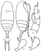
Scolecithricella ovata : Pearson, 1906 (p.18); Farran, 1908 b (p.51); With, 1915 (p.208, figs.F, juv.); Lysholm & Nordgaard, 1921 (p.20); Sars, 1925 (p.188, figs.F); Wilson, 1932 a (p.84, figs.F); Rose, 1933 a (p.157, figs.F); Jespersen, 1934 (p.91); Farran, 1936 a (p.97, Rem.); Jespersen, 1939 (p.59, Rem.); 1940 (p.40); Wilson, 1942 a (p.208, fig.F); Rose, 1942 ( p.147, Rem.F); Lysholm & al., 1945 (p.29 ); Sewell, 1948 (p.502, 514, 546, 549); C.B. Wilson, 1950 (p.334, fig.F); Brodsky, 1950 (1967) (p.270, figs.F, Rem.); Vervoort, 1951 (p.99, figs.F, Rem.); Østvedt, 1955 (p.15: Table 3, p.70); Vervoort, 1957 (p.102, Rem.); Minoda, 1958 (p.253, Table 2, abundance); Tanaka, 1962 (p.55, figs.F,M); Shmeleva, 1963 (p.141); Grice, 1963 a (p.495); Unterüberbacher, 1964 (p.25); Grice & Hulsemann, 1965 (p.224); Furuhashi, 1966 a (p.295, vertical distribution in Kuroshio region, Table 10) ; Mazza, 1966 (p.71); Grice & Hulsemann, 1967 (p.17); Fleminger, 1967 a (tabl.1); De Decker, 1968 (p.45); Vinogradov, 1968 (1970) (p.257-258); Park, 1968 (p.555, figs.F); Park, 1970 (p.476); Vaupel-Klein, 1970 (p.20); Bradford, 1970 a (p.356, fig.F); Minoda, 1971 (p.31, figs. juv.M); Binet & al., 1972 (p.55); Roe, 1972 (p.277, tabl.1, tabl.2); 1972 b (p.537, Rem.); Björnberg, 1973 (p.332, 389); Bradford, 1973 (p.142); Vives & al., 1975 (tab.II); Carter, 1977 (1978) (p.35); Park, 1980 (p.58, figs.F, Rem.); Dawson & Knatz, 1980 (p.6, figs.F); Björnberg & al, 1981 (p.639, figs.F); Gardner & Szabo, 1982 (p.306, figs.F); Kovalev & Shmeleva, 1982 (p.84); Vives, 1982 (p.292); Huntley & al., 1983 (p.143, Table 2); Bradford & al., 1983 (p.109, Rem., figs.F,M); Tremblay & Anderson, 1984 (p.6, Rem.); Guangshan & Honglin, 1984 (p.118, tab.); Roe, 1984 (p.357); Brenning, 1984 (p.5, Rem.); Campaner, 1984 a (p.178, Rem., figs.F); Brenning, 1985 a (p.28, Table 2); Rudyakov, 1986 (tab.1); Lozano Soldevilla & al., 1988 (p.59); Shih & Marhue, 1991 (tab.3); Hattori, 1991 (tab.1, Appendix); Gopalakrishnan & Balachandran, 1992 (p.167, figs.1, 6, Table 1, 2); Shih & Young, 1995 (p.73); Errhif & al., 1997 (p.423); Chihara & Murano, 1997 (p.901, Pl.174,175: F,M); Hure & Krsinic, 1998 (p.49, 101); Bradford-Grieve & al., 1999 (p.881, 932, figs.F,M); Voronina & Kolosova, 1999 (p.71); Razouls & al., 2000 (p.343, Appendix); Pakhomov & al., 2000 (p.1663, Table 2, transect Cape Town-SANAE antarctic base); Holmes, 2001 (p.60); Yamaguchi & al., 2002 (p.1007, tab.1); Labat & al., 2002 (p.741, tab.3); Sameoto & al., 2002 (p.13); Conway & al., 2003 (p.194, figs.F,M, Rem.); Hsiao & al., 2004 (p.326, tab.1); Sedova & Grigoriev, 2005 (p.112); Hopcroft & al., 2005 (p.198, table 2); Lavaniegos & Jiménez-Pérez, 2006 (p.155, tab.2, 3, Rem.); Vives & Shmeleva, 2007 (p.784, figs.F,M, Rem.); Valdés & al., 2007 (p.104: tab.1); Gaard & al., 2008 (p.59, Table1, N Atlantic Mid-Ridge); Park & Ferrari, 2009 (p.143, Table 4, fig.1, Appendix 1, biogeography); Galbraith, 2009 (pers. comm.); Hidalgo & al., 2010 (p.2089, Table 2); Mazzocchi & Di Capua, 2010 (p.427); Homma & al., 2011 (p.29, Table 2, 3, 4, 5, abundance, feeding pattern: detritivores); Ohashi & al., 2013 (p.44, Table 1, Rem.); Bonecker & a., 2014 (p.445, Table II: frequency, horizontal & vertical distributions); Mazzocchi & al., 2014 (p.64, Table 5, abundance) | | | | Ref.: | | | Vyshkvartzeva, 1999 (p.228, Rem.) | 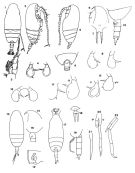 Female: 1, habitus (dorsal view); 2, idem (lateral view); 3, P1; 4, P5; issued from : Campaner, 1984 a); 5, habitus (lateral view); 6, head (lateral view, left side); ; 7, rostrum (anterior aspect); 8, 8', P5 (Dte = right); 9, Th5 and urosome segments 1 and 2 (dorsal view); 10, Th5 and urosome (lateral view); issued from Park, 1968, 1980); 11, 11', P5 (variant forms); 12, genital segment (lateral view, right side); ; issued fom : Bradford, Haakonssen & Jillet, 1983. 13, habitus (dorsal view); 14, forehead (lateral view); 15, P5; 16, Th5 and urosme segment 1; issued from : Tanaka, 1962. Male: 17, habitus (dorsal view); 18, rostrum (anterior aspect); 19, Th5 and urosome (lateral view, left side); 20, P5 (Dt: right, G: left); 21, right P5 (variant form); issued from : Tanaka, 1962. 22, P5; issued from Bradford & al., 1983.
|
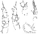
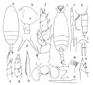 issued from : O. Tanaka in Publ. Seto Mar. Biol. Lab., 1962, X (1). [p.56, Fig.137]; As Scolecithricella ovata. Female: a, habitus (dorsal); b, forehead (left lateral side); c, last thoracic segment and genital somite (left lateral side); d, rostrum (dorsal view); e, exopod of P1; f, P2; g, endopod of P3; h, terminal spine of exopod of P4; i, P5. Male: j, habitus (dorsal); k, last thoracic segment and urosome (left lateral side); l, rostrum (dorsal view); m, Mx1; n, P4 (base); o, 5th pair of legs; p, right P5 (other specimen). Right P5 is 4-jointed and short, but another specimen has an elongated distal segment; the left leg 5-jointed, and very long. Nota Female: - Abdomen contained 4.6 times in the length of cephalothorax. - Head and 1st thoracic segment fused, 4th and 5th thoracic sefgments fused. - Posterolateral corners of the last thoracic segment slightly emarginate. - Rostrum with a divergent base to which rather strong spines attached. - Propotional lengths of the abdominal segments and caudal rami 38 : 20 : 20 : 5 : 17 = 100. - Genital segment as long as wide; slightly produced ventrally. - Caudal rami 1.4 times as long as wide. - A1 23-segmented; extends to the distal end of anal segment. Segments 8-9 and 24-25 fused. The spinulation on the segments described by With. - A2 exopod 1.6 times as long as endopod. - Mx1 with 9 setae on the outer lobe; 5 setae on exopod; 5 setae on endopod; 4 setae on the 2nd basal; 3 setae on the 3rd inner lobe; 2 setae on the 2nd inner lobe; 11 setae on the 1st inner lobe (Praecoxal arthrite). - Mx2 with a large 5th lobe; endopod furnished with 3 long vermiform filaments and several short amalliform setae. - Mxp with 1 short amalliform seta, 2 long sensory filaments, and 1 ordinary seta on the proximal half ofg the 1st basal segment. -P1 with a well developed outer edge spine on the 1st exopodal segment. - P2 with the coxa indented on the outer margin; posterior surface of segments of both exopod and endopod furnished with groups of small spines; terminal spine of exopod with 43 teeth. - P3 with endopod fusnished with rows of spines. - P4 terminal toothed spine of the exopod makes fenestella (fig. h). Nota Male: - Cephalothorax about 2.53 times the length of abdomen. - Head and 1st pedigerous segment fused, 4th and 5th thoracic segments incompletely fused (line of fusion faintly visible on the mid-dorsal region. - Frontal margin of forehead slightly produced in the mid-dorsal area. - Posterolateral corners of last thoracic segment rounded. - Basal part of rostrum produced ventrally in a spoon-shaped projection to which 2 parallel spines are attached. - Abdominal segments and caudal rami in the proportional lengths 14 : 30 : 20 : 24 : 4 : 8 = 100. - Caudal rami divergent, about as long as wide. - A1 19-segmented on the left side; extends to the distal margin of the 3rd abdominal segment. The 2nd segment has a large and characteristic aesthetasc on the anterior distal margin. - A2 exopod 1.3 times as long as endopod; 1st exopodal segment with fine hairs on the anterior margin. - Md robust. - Mx1 reduced except the outer lobe which has 7 long setae on the anterior margin. - Mx2 with only long vermiform filaments on endopod. - Mxp 1st basal segment conspicuous , spinulation reduced much. - P1 with an outer edge spine on the 1st exopodal segment; 1st basal segment (= coxa) with an indentation on the distal 1/4 of the outer margin. - P2 with an outer edge spipe on the 1st exopodal segment, reaching the middle of the outer margin of the next segment; terminal spine of exopod with 40 teeth. - P3 and P4 with the 2nd basal segment (= basis) each with a sharp ridge in the inner distal corner of the segment.; posterior surface of exopod and endopod provided with rows of small spines. - P4 with the 1st basal segment (= coxa) smooth on the outer margin; terminal spine of the exopod with 40 serrations. - P5 reaches back of the end of the anal segment. Right leg 4-segmented and short, terminal segment rounded at apex (but another specimen has an elongated distal segment. Left leg 5-segmented, very long; 2nd segment is as long as the combined lengths of the next 2 segments; distal segment furnished with 2 spines at apex, and a row of short hairs on the inner margin. Tanaka remarks that the species differs from the other members of Scolecithricella (stricto sensu) in having an outer edge spine on the 1st exopodal segment of P1. The male P5 has, in the main, the similar structure as is found in Scolecithricella ctenopus (= Scolecitrichopsis ctenopus).
|
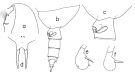
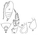 Issued from : T.S. Park in Fishery Bull. Fish Wild. Serv. U.S., 1968, 66 (3). [p.553, Pl.8, Figs.17-21]. As Scolecithricella ovata. Female: 17, habitus (left lateral side); 18, last thoracic segment and urosome (dorsal); 19, P2; 20, left P5; 21, right P5. Nota: One specimen had a small second internal spine close to the distal end of the right P5.
|
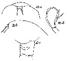
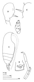 issued from : J.M. Bradford, L. Haakonssen & J.B. Jillett in Mem. N.Z. Oceanogr. Inst., 1983, 90. [p.110, Fig.66]. As Scolecithricella ovata. Female: A, habitus (lateral right side); B, genital segment (lateral view); C, endopod of P2; D, P5; E, P5 (specimen from another station). Male: G, P5. Nota: The left P5 terminal segment appears to be damaged. Appendages from different specimens of different stations.
|
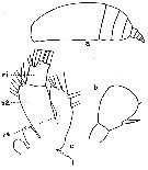
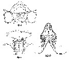
 issued from : T. Park in Antarct. Res. Ser. Washington, 1980, 31 (2). [p.58, Fig.17]. As Scolecithricella ovata. Female: a, forehead (lateral); b, last thoracic segment and urosome (lateral left side); c, genital segment (lateral left side); d, rostrum (anterior); e, , f, P5 (posterior). Nota: The species identified have a short spermathecal vesicle and occured mainly in waters north to the Antarctic Convergence.
|
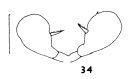 issued from : J.M. Bradford in N.Z. Jl Mar. Freshw. Res., 1970, 4 (4). [p.356, Fig. 34]. Female (off Kaikoura, New Zealand): 34, P5 Scale bar represents 0.1 mm.
|
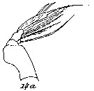
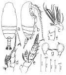 issued from : A.F. Campaner in Bolm. Zool., Univ. S. Paulo, 1984, 6 . [p.179, Fig.7]. Female (off Rio de Janeiro): a, habitus (dorsal); b, idem (lateral right side); c, Mx1 (Ba = basipodite 2, Ri = endopodite; Re = exopodite); d, P1 (posterior); e, P2 (posterior); f, basipodal segment 1 of P4; g, P5.
|
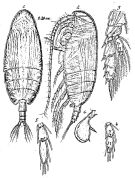 Issued from : G.O. Sars in Résult. Camp. Scient. Prince Albert I, 69, pls.1-127 (1924). [Pl.LII, figs.1-6]. As Scolecithrix ovata. Female from Canary Islands): 1, habitus (dorsal); 2, idem (lateral left side); 3, P2; 4, endopodal segments of P3; 5, endopodal segments of P4; 6, P5. Nota Female: - head and 1st pedigerous segment fused, 4th and 5th pedigers fused. - Rostrum short splitted at apex, with 2 little filaments. - Posterolatgeral corners of the last thoracic segment rounded with distinctly on the posterior edge a sinuous shape. - Urosome, short and narrow, reaching barely 1/4 the prosome lenth. - Genital segment slightly swollen ventrally, about as long as the two following segments together. - Caudal rami short and little divergent, with 4 setae of about equal length. - A1 23-segmented (the distal two segments not separated), extend about back to the 3rd abdominal segment. - A2, mouthparts and swimming legs, almost identical with the structures of the species type of the genus Scolecithricella minor- P5 showing a particular shape, discoid-like almost circular, attached at base by a narrow neck. external margin curved, naked; inner marlin more or less straight provided in middle with 1 short and thick spine, a little inner tooth near the apex. Remarks: For Sars (1925, p.189) this species resemnles Amallothrix obtusifrons (= Pseudoamallothrix obtusifrons).
|
 Issued from : K.A. Brodskii in Calanoida of the Far Eastern Seas and Polar Basin of the USSR. Opred. Fauna SSSR, 1950, 35 (Israel Program for Scientific Translations, Jerusalem, 1967) [p.271, Fig.179]. As Scolecithricella ovata. Female (from NW Pacif.): habitus (dorsal and lateral right side); R, rostrum; S1, P1; S5, P5.
|
 issued from : G.P. Farran in Ann. Rep. Fish. Brch., Ireland, 1902-1903, II, App. 2, 1905. [Plate VII, Figs.1-5]. As Scolecithrix ovataFemale (from W ireland): 1-5, P1 to P5.
|
 issued from : W. Vervoort in Verh. K. ned. Akad. Wet., Afd. Natuurk., 1951, (Sect. 2) 47 (2). [p.100, Fig.53]. As Scolecithricella ovata. Female (from 66°58'S, 16°03'.5W): a, right Mx2 (appendages of endopod omitted); b, apical portion of Mx2, showing the arrangement of the sensory appendages on the endopod; c, left P5; d, right P5. Nota: - Mx2 has a much and lengthened 5th lobe; lobe 1 and 2 have 2 strong, spiniform setae and 1 small curved, haired seta; lobe 3 with 2 strong setae, one of which is spine-like; lobe 4 with 1 strong, curved spine-like seta and 2 additional, smaller setae; lobe 5 has 1 strong, spiniform seta, slightly longer than the 2 additional setae found on that lobe; the spiniform setae on the 3rd to 5th lobes each have a dense row of small spinules along the inner edge; ventral border of Mx2 with a big tubercle but without seta; endopod 3-segmented, there are 9 sensory appendages, of these appendages 3 are very long, curved and swollen over a considerable portion of their length, one is short and club-like, whilst the remaining 5 are more or less capitate and rather short.
|
 issued from : C.O. Esterly in Univ. Calif. Publs Zool., 1905, 2 (4). [p.168, Fig.29]. As Scolecithrix subdentata. Female (from San Diego Region): a, habitus (lateral); b, P5; c, Mx1 (b2 = 2nd basal; ri = endopod; re = exopod). Nota: Last two thoracic segments fused, each side with a small indentation in the lateral margin. A1 23-segmented, not much longer than the cephalothorax. Endopod of A2 3/4 as long as the exopod. 2nd basal of Mx1 with 4 bristles, rami each with 5. Appendages of Mx2 vermiform.
|
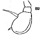 issued from : C.C. Davis in Univ. Wash. Publs Biol., 1949, 14. [Pl.7, Figs.89]. As Scolecithricella subdentata. Female (from NE Pacific): 89, P5. Nota : The last two thoracic segments fused, on each of the lateral border is a small indentation or notch, which is not as deep as in S. dentata. Genital segment nearly as long as the two following segments (9 :11). A1 23-segmented, reach to the posterior edge of the 2nd urosomal segment.
|
 issued from : C. With in The Danish Ingolf-Expedition, Copepoda I, 1915, III, 4. [Pl. VIII, Fig. 12, a-d]. As Scolecithricella ovata. Female (from 60°29'N, 34°14'W): a, forehead (ventral ); b, same (lateral); c, genital segment (lateral); d, right P5. Nota : Rostrum consists of 2 short, slightly divergent teeth, arising from a short lamelliform undivided process, to which are attaced fairly slender and apparently obtuse rostral filaments, several times longer than the rostrum proper and, in direction downwards and backwards, reaching beyond the insertion of A1 (filaments wanting in most specimens). Genital segment only slightly produced ventrally ; receptaculum seminis elongated pear-shaped, and generally very prominent. Comparative length between the abdominal segments and the caudal rami, almost twice as long as wide 22 : 12 : 14 : 3 : 12. A1 23-segmented ( segments 24 and 25 completely fused), reachingabout to the end of the 3rd urosomal segment. A2 with 7 setae on the inner lobe of the endopodite. Mx1 with 7+2 setae in the outer lobe. Inner lobe 1 with 2 setae, inner lobe 2 with 3 setae. Basis with 4 setae. Endopodite 1-3 with 3+3 setae. Exopodite with 5 setae. P5 : 1st basal segment well distinguished, and with the second more or less well separated from the broad lamellar 3rd segment. The legs are generally assymmetrical, partly because the articulation is better developed on the left side, and partly because the number of setae is rather variable.
|
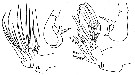
 issued from : C. With in The Danish Ingolf-Expedition, Copepoda I, 1915, III, 4. [Pl. VII, Fig.14, b-d]. As Scolecithricella ovata. Female: b, labrum and lobi labiales (anterior view); c, same (oral view); d, serrula 6-dentata. Nota : The anterior portion of the labrum is distinctly prominent in front ; the anterior surface shows a rather simple arrangement of the bristles (fig. 14b) ; in addition to the marginal rows of bristles an anterior transverse distinctly convex row, composed of several units, and a posterior shorter one are observed. The oral surface of the labrum has the anterior lateral group fairly well separated from the marginal rows (fig.14c) ; the posterior groups of the longitudinal series are more or less fused. The transverse median groups are rather poorly developed. The arrangement of the hairs upon and behind the labial lobes shows (fig. 14b) a very marked difference between a central and two lateral groups. The intestine is not straight, but distinctly twisted, at least vertically.
|
 issued from : C. With in The Danish Ingolf-Expedition, Copepoda I, 1915, III, 4. [Pl. VII, Fig.14 a]. As Scolecithricella ovata. Female: a, left Mx2 (anterior view). Nota : Mx2 possess numerous vermiform, but not amalliform, setae.
|
 Issued from : C. Razouls in Ann. Inst. océanogr., Paris, 1994, 70 (1). [p.197]. As Scolecithricella ovata.
Caractéristiques morphologiques de Pseudoamallothrix ovata femelle et mâle adultes.
Terminologie et abbréviations: voir à Calanus propinquus.
|
 issued from : O. Tanaka in Publ. Seto Mar. Biol. Lab., 1962, X (1). [p.57]. Male: Proportional lengths of segments of A1.
| | | | | Compl. Ref.: | | | Kuriyama & Nishida, 2006 (p.299: Tab.II; fig.7, 10, vertical distribution, Rem.: p.313); Schnack-Schiel & al., 2010 (p.2064, Table 2: E Atlantic subtropical/tropical); in CalCOFI regional list (MDO, Nov. 2013; M. Ohman, pers. comm.); El Arraj & al., 2017 (p.272, table 2); Belmonte, 2018 (p.273, Table I: Italian zones); Dias & al., 2019 (p.1, Table II, IV, occurrrence, vertical distribution). | | | | NZ: | 22 | | |
|
Distribution map of Pseudoamallothrix ovata by geographical zones
|
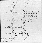
| | | | | | | | | | | | | | |  issued from : T.C. Gopalakrishnan & T. Balachandran in Oceanogr. Indian Ocean, 1992. [p.171, fig.6, D]. As Scolecithricella ovata. issued from : T.C. Gopalakrishnan & T. Balachandran in Oceanogr. Indian Ocean, 1992. [p.171, fig.6, D]. As Scolecithricella ovata.
Distribution of Pseudoamallothrix ovata in the Indian Ocean.
Nota: Main occurnces between 10° N - 20° S. |
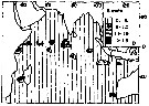
 issued from : U. Brenning in Wiss. Z. Wilhelm-Pieck-Univ. Rostock - 33. Jahrgang 1984. Mat.-nat. wiss. Reihe, 6. [p.6, Fig.2]. issued from : U. Brenning in Wiss. Z. Wilhelm-Pieck-Univ. Rostock - 33. Jahrgang 1984. Mat.-nat. wiss. Reihe, 6. [p.6, Fig.2].
Spatial distribution for Scolecithricella ovata (= Pseudoamallothrix ovata) and other scolecithrids from 8° S - 26° N; 16°- 20° W, for different expeditions (V1: Dec. 1972- Jan. 1973; V2: Feb/Mar. 1973; VI: May 1974; IV: Jun./Jul. 1972). |
| | | | Loc: | | | Cosmopolite: Antarct. (Atlant., Indian, Pacif.), Atlant., Medit. (W-E, Ligurian Sea, Tyrrhenian Sea, S, Adriatic), Indian, China Seas (East China Sea, South China Sea), Pacif., Calfornia, Chile (S), Arct., E Greenland, Flemish Cape, St. Margaret's Bay, Bering Sea , Aleutian Basin, S Aleutian Is. | | | | N: | 101 | | | | Lg.: | | | (1) F: 2,2; (7) F: 2,32; (9) F: 2,3-1,7; M: 1,8; (25) F: 2,32-1,94; (31) F: 2,39-2,08; (34) F: 1,75; (35) F: 2,16-2,04; (38) F: 2,13-2,08; (45) F: 2,5-2,3; (59) F: 1,9-1,5; (72) F: 1,98; 1,88; (117) F: 2,13-1,8; M: 1,59; (142) F: 1,48; (199) F: 2,05-1,82; M: 1,9-1,75; (205) F: 1,85-1,75; (208) F: 2,38-1,98; (313) F: 2,2-1,8; (432) F: 2,3-1,95; (523) F: 2,34-1,76; (532) F: 1,8-1,73; (866) M: 1,38-1,8; (991) F: 1,7-2,39; M: 1,38-1,8; (1000) F: 1,9 ± 0,1; M: 1,8 ±0,1; (1111) F: 2,05-2,21; {F: 1,48-2,50; M: 1,38-1,90} | | | | Rem.: | Epipelagic to bathypelagic. Distributional range (m) from Roe (1972) Day: 350-950, Night: 100-960; from Grice & Hulsemann (1965): 200-1000 (in Kuriyama & Nishida, 2006).
According to Østvedt (1955) this species has not previously been reported from the Norwegian Sea.
Vervoort (1951, 1957) mentions the variability of the female P5.
For Vervoort (1957, p.103) there is practically no variability in the general shape of the body ; all specimens in Banzare collection have a vaulted, rounded back ; the postero-lateral thoracic border has a slight but distinct incurvation. They show, however, the same variability in the structure of P5 as is also shown by the Willem Barendsz specimens described in Vervoort (1951, p.101). Females with 2 spines (a rather long proximal and a much smaller distal spine) on the pallet shaped terminal segment of the P5 dominate. Occasionally this pallet shaped terminal segment is separated by an intermediate segment from the communal basal portion. Females in the Vth copepodid stage are common in the collection ; they have about the same overall length as the adult females
For Park (1980, p.59) This species agrees with P. cenotelis in all anatomical details except for the spermatheca.
W. Zhang confirms the presence of this species in the China's Seas (pers. comm., 2006).
See in DVP Conway & al., 2003 (version 1) | | | Last update : 24/10/2022 | |
|
|
 Any use of this site for a publication will be mentioned with the following reference : Any use of this site for a publication will be mentioned with the following reference :
Razouls C., Desreumaux N., Kouwenberg J. and de Bovée F., 2005-2024. - Biodiversity of Marine Planktonic Copepods (morphology, geographical distribution and biological data). Sorbonne University, CNRS. Available at http://copepodes.obs-banyuls.fr/en [Accessed April 19, 2024] © copyright 2005-2024 Sorbonne University, CNRS
|
|
|
|


;)
;)
;)
;)
;)
;)
;)
;)
;)
;)
;)
;)
;)
;)
;)
;)
;)

;)
;)
