|
|
 |
|
Harpacticoida ( Order ) |
|
|
|
Clytemnestridae ( Family ) |
|
|
|
Clytemnestra ( Genus ) |
|
|
| |
Clytemnestra scutellata Dana, 1848 (F,M) | |
| | | | | | | Syn.: | Clytemnestra Hendorffi Poppe, 1891 (p.132, figs.).
Clytemnestra rostrata : T. Scott, 1894 b (p.106, figs.F,M);
Non Clytemnestra scutellata : Giesbrecht, 1892 (p.568,Pl. 1, fig.9; Pl. 45, figs. 16-18, 21, 23-24, 27-30, 32, 34-38); Farran, 1936; ? Mori, 1929; 1937; ? Legaré, 1964; ? Owre & Foyo, 1967; ? Chen & al., 1974; ? Kazmi & Muniza, 1994; ? Campos Hernadez & Suarez Morales, 1994;
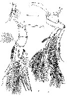
? Clytemnestra scutelata : Obuid Allah & al., 2005 (p.123, occurrence % vs metal contamination); Lavaniegos & al., 2012 (p. 11, Appendix); Zaafa & al., 2014 (p.67, Table I, occurrence); doubtful identifications because no reference from Huys & Conroy-Dalton (2000). | | | | Ref.: | | | Huys & Conroy-Dalton, 2000 (p.5, 45, Redescr., figs.F,M); Boxshall & Halsey, 2004 (p.278, 279: figs.F,M); Vives & Shmeleva, 2010 (p.95, figs.F,M, Rem.) | 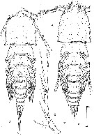 issued from : Huys and Conroy-Dalton in Bull. nat. Mus. Lond., 2000, 66 (1). [p.6, Fig.1]. Neotype from Great barrier Reef Expedition 1928-29 (Farran, 1936), stations 19, 20 or 28. Female: A, habitus (dorsal); C, P5 (anterior). Nota : Rostrum triangular, with rounded anterior margin, completely fused to cephalothorax ; with numerous dorsal pores, none on ventral surface ; with minute lateral sensillae near apex. - Urosomites without dorsal ornamentation. - P5 uniramous, laterally displaced ; 2-segmented ; not extending beyond posterior margin of genital double-somite. Basis with short outer seta and anterior pore. Exopod about twice as long as basis, slightly curved inwards ; outer margin with 4 pinnate setae ; inner margin each with 1 long pinnate seta ; anterior surface with 3 pores and spinules near apex and in proximal third. Male: B, habitus (dorsal). Nota : Body with similar projections as in female, urosome more slender with genital and 1st abdominal somites separate. Rostrum more obtuse than in female. Scale bars in micrometers. A, C based on neotype.
|
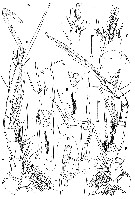 issued from : Huys and Conroy-Dalton in Bull. nat. Mus. Lond., 2000, 66 (1). [p.8, Fig.2]. Female (neotype): A, A1 (dorsal). Nota : A1 slender, 7-segmented; segment 7 longest. Plumose setae present on segments 1-4. Segment 1 with small pore near seta and few short spinules along anterior margin. Apical acrothek consisting of aesthetasc, long transformed seta and short bare seta. Transformed setae on segments 3, 4 and 7 long and aesthetasc-like, with rounded tip; those on segments 4 and 7 basally fused to aesthetascd. Rudimentary element present at base of acrothek. Male: B, A1 (ventral); C, antennulary segment 3 (anterior); D, antennulary segments 4-7 (anterior; distal portion of segment 7 and proximal portion of segment 4 omitted); E, antennulary segments 5-6 (ventral); F, antennulary segment 7, distal portion (dorsal; arrow indicating rudimentary element). Nota : A1 distinctly 7-segmented with ancestral segment XIII completely incorporated into segment 4 ; haplocer, with geniculation located between segment 6 and 7. Plumose setae present on segments 1-4. Segment 1 with small pore near seta and few tiny spinules along anterior margin. Apical acrothek consisting of aesthetasc, long transformed seta and short bare seta. Transformed setae on segments 3, 4 and 7 long and aesthetasc-like, with rounded tip ; those on segments 4 and 7 basally fused to aesthetasc. Rudimentary element present at base of acrothek (arrowed in fig. 2F). Segment 6 with 2 patches of spinules on anterior surface. Segment 7 with 2 fused elements near geniculation (in fig. 2D). Spermatophore with very long, recurved neck. Scale bars in micrometers.
|
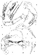 issued from : Huys and Conroy-Dalton in Bull. nat. Mus. Lond., 2000, 66 (1). [p.9, Fig.3]. Female: A, A2 (outer); B, distal endopod segment of A2 (inner); C, oral area showing position of labrum, paragnaths, mandibles, maxillules and right maxilla (ventral; position of Mxp indicated); D, Md (posterior); E, Mx1 (posterior); F, Mx2 (posterior; exit of maxillary gland arrowed). Nota : A2 4-segmented (coxa, basis, endopod 2-segmented). Exopod inserted in membranous area between basis and endopod ; represented by small, well defined segment bearing 2 strong recurved setae apically ; exopodal setae multipinnate with long setules in proximal third. Distal endopod segment with several surface frills and minute spinules on outer surface and patch of long setules on medial surface ; lateral armature consisting of 1 naked seta ; distal armature consisting of 5 apical, non-geniculate, bipinnate or multipinnate elements, 2 of which spiniform, recurved and bearing long spinules proximally. - Labrum large, with 6 secretory pores on anterior surface ; distal margin spinulose medially and with spinular patch on either lateral lobe. - Paragnaths well developed hirsute lobes. - Md reduced. Palp represented by single naked seta. Gnathobase long and narrow, stylet-like ; produced into number of cuspidate processes apically and subapically ; without dosal seta. - Mx1 reduced ; represented by small bolobed segment bearing 2 naked apical spines and raised seta along outer margin ; posterior surface with distinct pore. - Mx2 2-segmented, comprising elongate syncoxa and allobasis. Syncoxa with expanded basal portion and 2 endites ; exit of maxillary gland large (arrowed), partly concealed under lobate extension ; proximal endite represented by small cylindrical process bearing very long plumose seta, distal endite cylindrical, with 1 naked and 2 pinnate spines apically. Allobasis with large articulating claw distally, smaller inner pinnate spine and naked seta along outer margin. Scale bars in micrometers. All based on neotype.
|
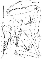 issued from : Huys and Conroy-Dalton in Bull. nat. Mus. Lond., 2000, 66 (1). [p.10, Fig.4]. Female: A, Mxp (posterior); B, distal half of basis and endopod of Mxp (anterior; proximal palmar element arrowed); E, right caudal ramus (dorsal). Nota : Mxp very large, articulating with well developed pedestal ; 3-segmented (syncoxa, basis, endopod) ; Syncoxa extremely elongate, longer than basis ; without ornamentation but with 1 anterior, plumose seta near membranous articulation with basis. Basis elongate ; distal third of palmar margin with double spinule row (anterior spinules coarser than posterior ones) and 2 elements located closely to articulation with endopod ; proximal element spiniform and bare (arrowed in fig. 4B), distal element pad-like and spinulose. Endopod represented by short segment bearing short naked claw ; accessory armature consisting of 3 anterior and 2 posterior elements. - Caudal rami about twice as long as wide, parallel ; slightly tapering towards rear margin, with steppe douter margin marking insertion sites of setae I, II and III ; produced into conical process bearing terminal pore. Male: C, Mxp (anterior); D, distal portion of basis and endopod of Mxp (posterior; proximal palmar element arrowed); F, right caudal ramus (dorsal). Nota : Mxp much larger than in female, articulating with well developed pedestal ; 3-segmented (syncoxa, basis, endopod). Syncoxa extremely elongate but not distinctly longer than basis ; without ornamentation but with 1 short anterior seta near membranous articulation wit basis. Basis elongate, more swollen than in female, middle and distal thirds of palmar margin forming longitudinal furrow bordered by single row of spinules on both anterior and posterior sides ; with 2 elements located closely to articulation with endopod ; proximal element spiniform and bare (arrowed in fig. 4D), distal element pad-like and spinulose. Endopod represented by short segment produced into very long naked claw which in reflexed position typically fits in palmar furrow with the apical part closely adpressed onto the anterior surface of the basis ; accessory armature consisting of 3 anterior and 2 posterior setae ; claw with spatulate apex. - Caudal rami somewhat shorter than in female ; seta II relatively longer ; seta III more slender and with longer pinnules ; setae IV-V long (60 % of urosome length) and plumose ; seta VI much longer than in female and sparsely plumose. Scale bars in micrometers. A, B, E based on neotype.
|
 issued from : Huys and Conroy-Dalton in Bull. nat. Mus. Lond., 2000, 66 (1). [p.13, Fig.6]. Female: A, P1 (anterior); B, P2 (anterior). Male: C, exopodite 3 of right P2 (anterior; aberrant setation). Nota: The right distal exopod segment of male P2 has only 2 outer spines (variability). Scale bars in micrometers. A, B based on neotype.
|
 issued from : Huys and Conroy-Dalton in Bull. nat. Mus. Lond., 2000, 66 (1). [p.14, Fig.7]. Female: A, P3 (anterior); B, P4 (anterior). Male: C, P5 (anterior). Nota : P5 very similar to that of female, with identical proportions, pore pattern and setation. Scale bars in micrometers. A, B based on neotype.
|
 issued from : Huys and Conroy-Dalton in Bull. nat. Mus. Lond., 2000, 66 (1). [p.12]. Female: Spine and setal formula of swimming legs.
|
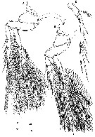
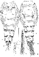 issued from : Huys and Conroy-Dalton in Bull. nat. Mus. Lond., 2000, 66 (1). [p.11, Fig.5]. Female: A, urosome (ventral). Nota : Penultimate and anal somites with multiple rows of spinules around ventral hind margin. Posterior third of the conical process of caudal rami with ventral spinular patch. Male: B, urosome (ventral (inset showing setae IV-V at full length); C, P6 (ventral). Nota : Urosomites 4-5 and anal somite with spinules around ventral hind margin. P6 weakly asymmetrical, forming highly membranous midventral area covering single, large median genital aperture ; each P6 produced into cylindrical process with 1 apical and 2 outer bare setae ; few spinules along inner margin. Scale bars in micrometers. A based on neotype.
|
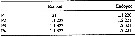
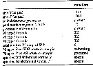 issued from : Huys and Conroy-Dalton in Bull. nat. Mus. Lond., 2000, 66 (1). [p.45, Table 1]. Diagnostic characters. A1 = antennule; GDS = genital double-somite; AS = first abdominal somite. Measurements based on material examined in the publication.
| | | | | Compl. Ref.: | | | ? Baker & al., 2005 (tabl.); Hwang & al., 2006 (p.943, tabl. I); ?T. Zuo & al., 2006 (p.159, tab. 1, abundance, fig.8: stations group); McKinnon & al., 2008 (p.844: Tab.1); Galbraith, 2009 (pers. comm.); C.E. Morales & al., 2009 (Table 1); ? Medellin-Mora & Navas S., 2010 (p.265, Tab. 2); ? Fazeli & al., 2010 (p.153, Table 1); Hsiao S.H. & al., 2011 (p.475, Appendix I); ? Johan & al., 2012 (2013) (p.1, Table 1); ? in CalCOFI regional list (MDO, Nov. 2013; M. Ohman, comm. pers.); Hwang & al., 2014 (p.43, Appendix A: seasonal abundance); Dias & al., 2015 (p.483, Table 2, abundance, biomass, production); Marques-Rojas & Zoppi de Roa, 2017 (p.495, Table 1); Jerez-Guerrero & al., 2017 (p.1046, Table 1: temporal occurrence); El Arraj & al., 2017 (p.272, table 2); | | | | NZ: | 9 + 9 doubtful | | |
|
Distribution map of Clytemnestra scutellata by geographical zones
|
| | | | | | | | | | | | | | | | Loc: | | | Indo-Pacific Ocean, Malaysia (Sarawak: Bintulu coast), China Seas (Yellow Sea, East China Sea, South China Sea) in Zhang & al., 2010), off SW Taiwan, Taiwan (E: Kuroshio Current), ? Oregon-California (rare), ? G. of California, Bahia Cupica (Colombia), Chile (Concepcion), Australia (West Australian Basin, North West Cape, Great Barrier Reef), ? Brazil (Campos Basin, Bracui estuary), Mochima Bay (Venezuela).
Neotype: 16°19-20' S, 146°3-7' E. | | | | N: | 17 ? | | | | Lg.: | | | (931) F: 1,121; M: 1,064; {F: 1,121; M: 1,064}
? (1255): 1.45 (without sex indicated) | | | | Rem.: | The revision of the genus by Huys & Conroy-Dalton (2000) introduces a serious doubt on most of the references given, as for the locality records.
The presence of this species in the Atlantic and in the Mediterranean needs confirmation regarding a confusion with C. gracilis. The authors had been unable to confirm the presence of this species in the Atlantic or the Mediterranean Sea.
According to Huys & Conroy-Dalton (2000, p.12) there are very few published records of C. scutellata that can be verified absolutely. There is little doubt that the species descibed by Poppe (1891) under the name C. hendorffi is synonymous with C. scutellata. Poppes material came from two localities in the Indian Ocean (West Australian Basin, south of Madagascar), three localities in the southwest Atlantic off the coasts of Brazil and Argentina, and the Karimata Strait in the Java Sea. He also re-identified Thompsons material of Goniopsyllus from the Maltese Sea as C. hendorffi, but it appears more conceivable that Thompson had collected the species described by Claus (1891) under the name Goniopelte gracilis, the description of which was unknown to Poppe (the same year 1891). After us the presence of C. scutellata in the Atlantic or the Mediterranean Sea is suspect because Poppes records from the southwest Atlantic might have been based on another species, possibly C. gracilis. The redescription by Giesbrecht (1892) has long been accepted as the basis for identification of C. scutellata even though his material was not from the type locality. It is clear that Giesbrecht has redescribed Goniopelte gracilis Claus,1891 (= Clytemnestra gracilis) ; both species are closely related, sharing the posterolateral projections on the cephalothorax and the presence of 3 outer spines on P2-P4 exopodal segment 3 and 6 elements on the P5 exopod in both sexes. They can be separated by body size, length of caudal ramus setae IV-V, length of the P5 in both sexes and urosome ornamentation in the female.
Fazeli & al., 2010 (p.153, Table 1) note the occurrence of this species in Chabahar By (Gulf of Oman) but without identification reference from Huys and Conroy-Dalton (2000); as in Araujo & al. (2016, Table III) on theBrazil coast.
Identification characters after G.A. Boxshall & S.H. Halsey (2004, p.278):
1- Exopod of P1 with 4 distal setal elements; female A1 7-segmented.
2- Exopod of P5 in both sexes armed with 6 setae; 3rd exopodal segment of P2 with total of 7 spines/setae.
3- Female lacking patches of spinules posteriorly on ventral surface of 1st free abdominal somite. | | | Last update : 28/10/2022 | |
|
|
 Any use of this site for a publication will be mentioned with the following reference : Any use of this site for a publication will be mentioned with the following reference :
Razouls C., Desreumaux N., Kouwenberg J. and de Bovée F., 2005-2024. - Biodiversity of Marine Planktonic Copepods (morphology, geographical distribution and biological data). Sorbonne University, CNRS. Available at http://copepodes.obs-banyuls.fr/en [Accessed April 20, 2024] © copyright 2005-2024 Sorbonne University, CNRS
|
|
|
|


;)
;)
;)
;)
;)
;)
;)
;)

