|
|
 |
|
Calanoida ( Order ) |
|
|
|
Clausocalanoidea ( Superfamily ) |
|
|
|
Aetideidae ( Family ) |
|
|
|
Euchirella ( Genus ) |
|
|
| |
Euchirella truncata Esterly, 1911 (F,M) | |
| | | | | | | Syn.: | Euchirella venusta : Sars, 1905 b (p.4);
E. propria (M) Esterly, 1911 (p.321, Descr.M, figs.M); Sewell, 1948 (p.556, 563); Brodsky, 1950 (1967) (p.179, figs.M;
Euchirella gracilis Wolfenden, 1911 (p.237, figs.F); Sewell, 1948 (p.500);
Euchirella intermedia With, 1915 (p.124, figs.F,M); Sars, 1925 (p.68, figs.F,M); Rose, 1929 (p.20); 1933 a (p.106, figs.F,M); Wilson, 1942 a (p.185); Lysholm & al., 1945 (p.17); Sewell, 1948 (p.348, 500, 566); Vervoort, 1949 (p.28, figs.F); C.B. Wilson, 1950 (p.225); Vervoort, 1952 f (n°47, p.4, figs.F,M); Tanaka, 1957 b (p.183, figs.F); Fagetti,1962 (p.19); Grice & Hart, 1962 (p.287, table 3); Grice, 1963 a (p.495); Grice & Hulsemann, 1965 (p.223); Fleminger, 1967 a (tabl.1); Deevey, 1971 (p.224); Bainbridge, 1972 (p.61, Appendix Table III: occurrence); Björnberg & al., 1981 (p.605, 632, figs.F); Vives, 1982 (p.291); Kovalev & Schmeleva, 1982 (p.83); Gaudy & Boucker, 1983 (p.37, Table 1, Rem.: metabolism); Greze & al., 1985 (p.7); Pancucci-Papadopoulou & al., 1990 (p.199); Hidalgo & al., 2010 (p.2089, Table 2); Andersen N.G. & al., 2011 (p.71, Fig.3: abundance);
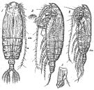
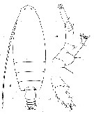
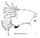
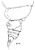
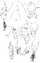
E. acadiana Willey, 1918 (1919) (p.190, figs.F) | | | | Ref.: | | | Esterly, 1911 (p.322, Descr.F, figs.F); Sewell, 1947 (p.69, 82, figs.F, Rem.); Brodsky, 1950 (1967) (p.179, figs.F); Vervoort, 1952 f (n°47, p.4: Rem.); 1963 b (p.135: Rem.); Park, 1968 (p.545, Redescr.M, figs.F,M); Tanaka & Omori, 1969 (p.57, figs.F,M); Vaupel-Klein, 1972 (p.502, 505, fig.F); Park, 1975 (p.294, figs.F, Rem.); Bradford & Jillett, 1980 (p.43, figs.F,M, fig. 73, distribution chart); Vaupel Klein, 1984 a (p.47, Table II: characters, Rem.: synonymies.); Markhaseva, 1996 (p.170, figs.F,M); Chihara & Murano, 1997 (p.686, Pl. 37, 41: F,M); Bradford-Grieve & al., 1999 (p.879, 921, figs.F,M); Vives & Shmeleva, 2007 (p.571, figs.F,M, Rem.) | 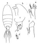 issued from : Tanaka O. in Publ. Seto mar. Biol., Lab., 1957, 6 (2). [Fig.48, p.184]. As Euchirella intermedia. Female (from Suruga): a, habitus (dorsal aspect); b, head (lateral aspect); c, last thoracic segment and urosome (lateral aspect); d, protuberance on left margin genital segment; e, endopodite and lobes of Mx1; f, basal joints of P4. Nota Female: - Cephalothorax about 5.59 times the abdomen length (5.25 : 0.94). - Forehead margin produced triangularly. - Head and 1st pedigerous segment faintly visible, last two thoracic segments incompletely fused. - Lateral corners of last thoracicsegment rounded. - Rostrum slightly curved posteriorly; rostral spine slender. - Abdomen 4-segmented; segments and caudal rami in the proportional lengths 51 : 12 : 8 : 15 : 13 = 100. - Genital segment asymmetrical, left side , in dorsal view, more inflated about the middle of the segment; lateral distal corner of the left side with a process wihch is concave at the apical portion. ^A1 24-segmented, extends to distal end of caudal rami. - Mx1 with 5 setae on endopod; 3 setae on 2nd basal segment, of which 2 are very short. - P4 coxa with a single spine extending to the distal margin of the segment on the inner margin at the base of the inner marginal seta.
|
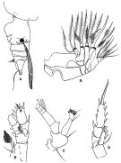
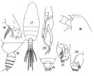 Issued from : T.S. Park in Fishery Bull. Fish Wild. Serv. U.S., 1968, 66 (3). [p.543, Pl.5, Figs.15-22]. Female: 15, last thoracic segment and urosome (dorsal); 16, basipod of P4. Male: 17, habitus (dorsal); 18, forehead (left lateral side); 19, last thoracic segment and urosome (left lateral side); 20, P5 (posterior); 21, terminal part of left P5 (posterior); 22, idem (anterior). Nota: Proportional lengths of the urosome segments and furca as 23:22:18:19:6:12.
|
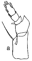
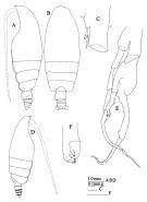 issued from : J.M. Bradford & J.B. Jillett in Mem. N.Z. Oceanogr. Inst., 86, 1980. [p.45, Fig.29]. Female: A, habitus (lateral left side); B, idem (dorsal); C, basipod 1 of P4. Male: D, habitus (lateral riht side); E, P5; F, terminal part of left exopod of P5.
|
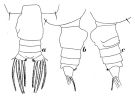 Issued from : W. Vervoort in Zool. Verh., Leiden, 1949, 5. [p.29, Fig.13]. As Euchirella intermedia. Female (from Indo-Malaysian Seas): a, urosome (dorsal); b, idem (lateral right side); c, idem (lateral left side).
|
 Issued from : G.O. Sars in Résult. Camp. Scient. Prince Albert I, 69, pls.1-127 (1924). [Pl.XX, figs.1-4]. As Euchirella intermedia. Female: 1, habitus (dorsal); 2, idem (lateral left side); 3, basipodal segment of P4. Male: habitus (lateral left side).
|
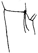
 issued from : R.B.S. Sewell in The John Murray Expedition, 1933-34, Scientific Reports, VIII (1), 1947. [p.84, Fig.17]. Female (from Arabian Sea): A, urosome with spermatophore (lateral left side); B, Mx2; C, A2; D, P2; E, P4 (abnormal). Nota: Proportional lengths of cephalothorax and abdomen as 4.5 to 1. The proportional lengths of the various segments of the body (cephalon to caudal rami) as 356:161:99:91:85:97:28:22:18:38=1000. The line of separation between the head and the 1st pediger segment is in one specimen quite clear both dorsally and laterally; in other examples it is only clear in the lateral region (as stated by With). The left side of the genital segment is more swollen than the right, and behind this swelling there is a depression that is bordered posteriorly by a lamellar ridge (in two specimens the head of a attachment of a spermatophore was lodged in this depression). Anal segment bears a tuft of hairs ventrally. A1 23-segmented (segments 8-9 and 24-25 respectively fused) reaches back to about the posterior end of the caudal rami. In A2 the endopod reaches nearly to the distal end of the 2nd segment of exopod, the two proximal segments are only partly fused. In Mx1 the 2nd inner lobe with 4 setae and the 3rd lobe 2, of which one is quite small; the 2nd basal segment bears 2 small and 1 well developed setae; endopod with 5 setae, of which the inner is very small; exopod with 11 setae and the outer lobe 8 setae, the posterior being smaller than the others.
|
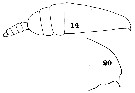
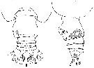 issued from : T. Park in Crustaceana, 1975, 28 (3). [p.295, Fig.3]. Female (drawn from Esterly's type specimens from the San Diego region): A-B, posterior part of body (dorsal and lateral, respectively).
|
 issued from : R.N. Wolfenden in Die Marinen Copepoden der Deutschen Südpolar-Expedition 1901-1903, 1911. [p.237, Fig.22]. As Euchirella gracilis. Female: basipod of P4.
|
 issued from : R.N. Wolfenden in Die Marinen Copepoden der Deutschen Südpolar-Expedition 1901-1903, 1911. [Pl.XXVII, Figs.8-10]. As Euchirella gracilis. Female: 8, habitus (dorsal); 9, A2; 10, last segment of exopod of P5.
|
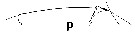
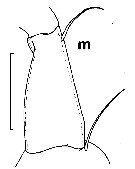 issued from : J.C. von Vaupel Klein in Crustaceana, Supplt 9, Studies on Copepoda, III, 1984. [p.63, Fig.5, m]. Female: m, setal armature of endopodite 1 of A2 (note: a single, long seta). Scale bar 0.2 mm.
|
 issued from : C.O. Esterly in Univ. Calif. Publs Zool., 1911, 6 (14). [Pl.27, Figs;14, 20]. As Euchirella propria. Male (from San Diego Region): 14, habitus (lateral); 20, forehead (lateral).
|
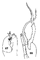 issued from : C.O. Esterly in Univ. Calif. Publs Zool., 1911, 6 (14). [Pl.30,, Figs.67, 83]. As Euchirella propria. Male: 67, tip of left P5; 83, P5 (exclusive of basipod).
|
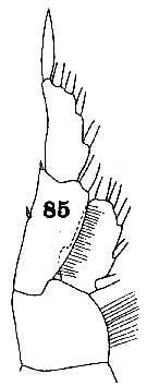 issued from : C.O. Esterly in Univ. Calif. Publs Zool., 1911, 6 (14). [Pl.31, Fig.85]. As Euchirella propria. Male: 85, P1.
|
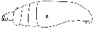 issued from : C.O. Esterly in Univ. Calif. Publs Zool., 1911, 6 (14). [Pl.26, Fig.5]. Female (from San Diego Region): 5, habitus (lateral). Nota: A1 23-segmented, reach beyond the end of the caudal rami. Urosome very short, being a little more than one-sixth the length of the cephalothorax.
|
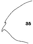 issued from : C.O. Esterly in Univ. Calif. Publs Zool., 1911, 6 (14). [Pl.28, Fig.35]. Female: 35, forehead (lateral).
|
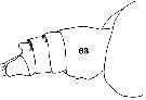 issued from : C.O. Esterly in Univ. Calif. Publs Zool., 1911, 6 (14). [Pl.29, Fig.63]. Female: 63, last part of thorax and urosome (lateral, right side). Nota: Genital segment longer than the last three, and the two middle segments are of about equal lengths. Caudal rami about as broad as long, widely divergent and provided with 4 bristles richly plumose to their ends and of equal lengths.
|
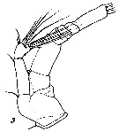
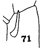 issued from : C.O. Esterly in Univ. Calif. Publs Zool., 1911, 6 (14). [Pl.30, Fig.71]. Female: 71, 1st basal of P4 showing the spine.
|
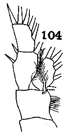 issued from : C.O. Esterly in Univ. Calif. Publs Zool., 1911, 6 (14). [Pl.31, Fig.104]. Female: 104, P1.
|
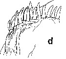
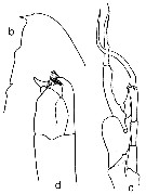
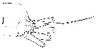
 issued from : O. Tanaka & M. Omori in Publ. Seto mar. Biol. Lab., 1969, XVII, 1. [p.58, Fig.9]. Female (from off Peru and Chile): a, forehead (lateral); b-c, last thoracic segment and urosome (lateral and dorsal, respectively). Male: d, forehead (lateral); e, P1; f, P5; g, left P5 (distal segment of exopod).
|
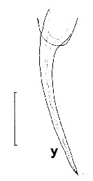 issued from : J.C. von Vaupel Klein in Crustaceana, Supplt 9, Studies on Copepoda, III, 1984. [p.80, Fig.16, y]. Female: y, arrangement of medial spines on the posterior face of basal segment 1 of left P4 . Scale bar 0.1 mm. Nota: 1 single, long spine.
|
 issued from : J.C. von Vaupel Klein in Crustaceana, Supplt 9, Studies on Copepoda, III, 1984. [p.65, Fig.6 c]. Euchirella truncata: a, Setal armature of endopodte 2+3 of A2. Conditions are expressed are expressed in formulae as follows: number of setae in regular row on proximal lobe / relative development of seta in position no. 9 on this lobe / number of setae in approximately linear row on terminal lobe / relative development of appendicular seta no.7 and its supporting pedestral (absent, vestigial, moderate or well developed: see Table I, p.87 and Table II, p.93). upper and lower lobes = proximal and terminal lobes (details of right appendage shown in medial aspect. Scale bar = 0.133 mm. Nota: Condition 8/2/6/3 in the truncata-group.
|
 Issued from : J.C. von Vaupel-Klein in Zool. Meded., Leiden, 19972, 47 (41). [p.511, Fig.5, d]. Female (from 32°26'S, 47°49'E): d, detail of spinules on the yubercle of the endopodite of left P1 (highly magnified, drawn from SE-micrograph). Nota: 'organ of Vaupel Klein' (see explanation to Euchirella curticauda.)
|
 issued from : C. With in The Danish Ingolf-Expedition, Copepoda I, 1915, III, 4. [Pl. IV, Fig.4, a]. As Euchirella intermedia. Female (from 51°00, 11°43'W): a, end of prosome and urosome (left lateral).
|
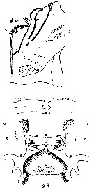 issued from : C. With in The Danish Ingolf-Expedition, Copepoda I, 1915, III, 4. [Pl. IV, Fig.4, b-c]. As Euchirella intermedia. Female: b, labrum (oral view); c, left lobus labialis.
|
 issued from : C. With in The Danish Ingolf-Expedition, Copepoda I, 1915, III, 4. [Pl. VIII, Fig.3]. As Euchirella intermedia. Female: 3, right A2 (anterior view).
|
 issued from : C. With in The Danish Ingolf-Expedition, Copepoda I, 1915, III, 4. [Text-fig.32, a]. As Euchirella intermedia. Female: a, left P4 (posterior view).
|
 issued from : C. With in The Danish Ingolf-Expedition, Copepoda I, 1915, III, 4. [Text-fig.32, b-d]. As Euchirella intermedia. Male: b, head (lateral); c, P5 (posterior view); d, distal segments of left P5.
|
 Euchirella truncata Euchirella truncata female: 1 - Genital segment asymmetrical. 2 - Crest absent. 3 - Genital segment with lateral side swelled (dorsal view), with large projection in the left half of the segment dorsally. 4 - Coxopodite of P4 with 1 spine. 5- Posterior border of genital segment without projection on the right. The projection of left lateral margin of segment, occupying nearly all segment's length (dorsal view). 6 - Genital segment with ear-like, small, nearly symmetrical projections on the right and on the left in the anterior part of segment (dorsal view).
|
 Euchirella truncata Euchirella truncata male: 1 - P5 uniramous on left P5 and large biramous on right P5. Exopodal segment 2 and endopod of right P5 elongated and sharpened in their distal parts forming tongs. Endopod of right P5 significantly exceeding distal border of exopodal segment 1 of right P5. 2 - Crest absent. 3 - No spine present near place wherefrom exopod of right P5 begins. 4 - Left P5 without rudimentary endopod. When closed tongs of exopod of left P5 compact, rounded, clavate-like. 5 - Proximal part of exopodal segment 1 of right P5 with 1 middle sized tooth it is not larger than the 3 following ones.
|
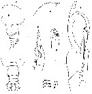
 issued from : E.L. Markhaseva in Proc. Zool. Inst. RAN, St. Petersburg, 1996, 268. [p.172, Fig.134]. Female & Male (from N Atlantic).
|
 issued from : J.C. von Vaupel Klein in Crustaceana, Supplt 9, Studies on Copepoda, III, 1984. [p.66, Fig.7, p]. Euchirella truncata: Structural feature of exopodite 1 and exopodite 2 of A2 (lateral aspects of left appendages and medial views of right antennal parts; compare with Fig.7n which represents left A2 medial face drawn from a lateral view). p, medial. Nota: a single large outgrowth vs moderately well developed as in E. bitumida.
|
 Issued from : J.C. von Vaupel Klein in Crustaceana, Supplt 9, Studies on Copepoda, III, 1984. [p.70, Fig.11, J]. Euchirella truncata: Setal armature of the endopod of Mx1 (detail of left appendage in posterior aspect; setules/spinules omitted). Complement as in E. formosa (Fig.11, i), 4 setae but with a far weaker, 5th seta in addition.
|
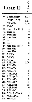 Issued from : J.C. von Vaupel Klein in Crustaceana, (Supplement) 9, 1984. [p.93, Table II]. Euchirella truncata Female: Datamatrix stating observed states of characters from Table I (p.87-90) presently examined; nos. refer to the input nos. used in Table I (see to the family Aetideidae).
|
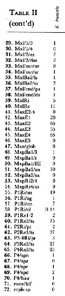 Issued from : J.C. von Vaupel Klein in Crustaceana, (Supplement) 9, 1984. [p.94, Table II (cont' d) ]. Euchirella truncata Female: Datamatrix stating observed states of characters from Table I (p.87-90) presently examined; nos. refer to the input nos. used in Table I (see to the family Aetideidae).
|
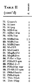 Issued from : J.C. von Vaupel Klein in Crustaceana, (Supplement) 9, 1984. [p.95, Table II (cont' d) ]. Euchirella truncata Female: Datamatrix stating observed states of characters from Table I (p.87-90) presently examined; nos. refer to the input nos. used in Table I (see to the family Aetideidae).
| | | | | Compl. Ref.: | | | Sewell, 1948 (p.329, 520, 531, 548, 556); Grice & Hulsemann, 1968 (tab.2); Roe, 1972 (p.277, tabl.1, tabl.2); Björnberg, 1973 (p.324, 386); Deevey & Brooks, 1977 (p.256, Table 2, station "S"); Seguin & al., 1993 (p.23); Suarez-Morales & Gasca, 1998 a (p108); Holmes, 2001 (p.48); Ikeda & al., 2006 (p.1791, Table 2); Schnack-Schiel & al., 2010 (p.2064, Table 2: E Atlantic subtropical/tropical); Medellin-Mora & Navas S., 2010 (p.265, Tab. 2); in CalCOFI regional list (MDO, Nov. 2013; M. Ohman, comm. pers.); Benedetti & al., 2016 (p.159, Table I, fig.1, functional characters); El Arraj & al., 2017 (p.272, table 2, spatial distribution); | | | | NZ: | 17 | | |
|
Distribution map of Euchirella truncata by geographical zones
|
| | | | | | | | | | | |  Issued from : J.M. Bradford & J.B. Jillett in New Zealand Ocean. Inst. Memoir, 86, 1980. [p.88-89, Figs.65-67]. Issued from : J.M. Bradford & J.B. Jillett in New Zealand Ocean. Inst. Memoir, 86, 1980. [p.88-89, Figs.65-67].
Distribution of several species of Euchirella in the Tasman Sea and around New Zealand.
Nota: Euchirella indica (= Euchirella massinensis indica). |
| | | | Loc: | | | South Africa SE (Durban), off E Tristan da Cunha, G. of Guinea (off Lagos), off E St. Paul Is., off Cape Verde Is., Morocco-Mauritania, Canary Is., off Madeira, off Portugal, Brazil, off Amazon, Caribbean Sea, Caribbean Colombia, G. of Mexico, Sargasso Sea, off Cape Cod, off Bermuda: Station "S" (32°10'N, 64°30'W), off Acadia, Faroe Is., W Ireland, Medit. (Alboran Sea, Eastern Basin), Arabian Sea, Indian, Indonesia-Malaysia, China Seas, off Galapagos, Hawaii, Pacif. (N central), Pacif. (tropical), Japan (Izu, NE), California, New Zealand, off Peru, off Juan Fernandez Is., Chile (N-S).
Type locality: San Diego (California). | | | | N: | 45 | | | | Lg.: | | | (1) F: 6,2; M: 5,1; (7) F: 5,66; M: 5,3; (10) F: 6-5,6; (11) F: 5,5-5,231; (37) F: 6,8-5,2; M: 5,6-4,5; (41) F: 6,05-5,55; (56) F: 6,19; (72) F: 6,81-5,4; M: 5,08-4,6; (110) F: 5,35-5,25; M: 4,92; (143) F: 6,6; M: 5,6; (199) F: 4,64-4,41; (201) F: 6,4-5,4; M: 5-4,5; (202) F: 6,2; M: 5,1; (236) F: 5.4-6.8; (1257) F: 5,75-6,25; {F: 4,41-6,83; M: 4,50-5,60}
The mean female size is 5.767 mm (n = 23; SD = 0.6650) and the mean male size is 5.027 mm. The size ratio (male : female) is 0.85 (n = 8; SD = 0.053). | | | | Rem.: | epi-bathypelagic. Sargasso Sea: 500-1500 m (Deevey & Brooks, 1977, station "S");
For Park (1975, p.294) when compared, Esterly's original specimens from the San Diego region and Atlantic specimens of E. truncata were identical, and they were distinguished from E. pseudotruncata, mainly by the genital segment of female and P5 of male. In E. truncata female, the genital segment in dorsal view has a small ear-shaped process on each side close to the proximal end and a large protrusion on the left side which begins immediately behind the ear-shaped process and extends to the distal end of the segment; the posterior region of the protrusion is distinctly hollow; laterally, the hollow area is approximately ovoid extending in a dorsoventral direction, and its surface is of a pitted structure. Judging from the size ranges of the body lengths of E. Truncata and E. pseudotruncata, the Atlantic records of E. intermedia by With (1915: 5.66 mm) and Sars (1925: 6.20 mm) and those of E. gracilis by Wolfenden (1911: 5.6-6.0 mm) all seem to be referable to E. truncata. This latter synonymy is disputable by different authors and not admitted in the World Copepoda database (Walter & Boxshall, 2011). | | | Last update : 19/06/2023 | |
|
|
 Any use of this site for a publication will be mentioned with the following reference : Any use of this site for a publication will be mentioned with the following reference :
Razouls C., Desreumaux N., Kouwenberg J. and de Bovée F., 2005-2024. - Biodiversity of Marine Planktonic Copepods (morphology, geographical distribution and biological data). Sorbonne University, CNRS. Available at http://copepodes.obs-banyuls.fr/en [Accessed April 25, 2024] © copyright 2005-2024 Sorbonne University, CNRS
|
|
|
|


;)
;)
;)
;)
;)
;)
;)
;)
;)
;)
;)
;)
;)
;)
;)
;)
;)
;)
;)
;)
;)
;)
;)
;)
;)
;)
;)
;)
;)
;)
;)

