|
|
 |
|
Calanoida ( Order ) |
|
|
|
Arietelloidea ( Superfamily ) |
|
|
|
Arietellidae ( Family ) |
|
|
|
Metacalanalis ( Genus ) |
|
|
| |
Metacalanalis hakuhoae Ohtsuka, Nishida & Machida, 2005 (F) | |
| | | | | | | Ref.: | | | Ohtsuka & al., 2005 (p.2486, Descr.F, figs.F, Rem.) | 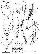 issued from : S. Ohtsuka, S. Nishida & R.J. Machida in J. Nat. Hist., 2005, 39 (27). [p.2487, Fig.1]. Female (from Sulu Sea): A-B, habitus (dorsal and lateral, respectively); C, rostrum (ventral); D, last compound pedigerous somite and genital double-somite (lateral); E, genital double-somite (ventral); F, left A1; J, terminal segments of left A1; H, right A1; I, terminal segment of right A1; J, terminal segments of A2 endopod; K, A2 exopod (terminal segments missing). Scale bars in mm. Nota: Cephalosome slightly asymmetrical in dorsal view. Rostrum strongly curved, with pair of thick filaments. Prosome 2.6 times as long as urosome. Urosome 4-segmented. Genital double-somite with ventral transverse ridge midway; genital system asymmetrical, with right seminal receptacle larger than left; paired gonopores and copulatory pores located at mid-length; each copulatory pore slit-like, located at inner corner of gonopore, connected to curved, thick copulatory pore. Two large, mature eggs visible within prosome. A1 considerably asymmetrical, with left ca 1.5 times longer than right; left A1 21-segmented.
|
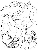 issued from : S. Ohtsuka, S. Nishida & R.J. Machida in J. Nat. Hist., 2005, 39 (27). [p.2488, Fig.2]. Female: A, A2; B, Md; C, Mx1; D, Mx2; E, basal spine of Mx2; F, Mxp; G, 4th endopodal segment of Mxp (largest seta omitted); H, 5th endopodal segment of Mxp (largest seta omitted). Scale bars in mm. Nota: A2: Endopod indistinctly 3-segmented; exopod 10-segmented with setal formula 0, 0, 1, 1, 1, 1, 1, 1, 0, 2; terminal segment lacking vestigial element (as seen in Metacalanus) Coxa unarmed; basis with inner seta half as long as exopod. Md without patch of long setules on gnathobase; cutting edge with 2 patches of minute spinules and 4 teeth, dorsalmost of which tricuspid; endopod rudimentary, 1-segmented, with 2 plumose setae terminally; rxopod 5-segmented, with setal formula 1, 1, 1, 1, 2; terminal setae well developed Mx1 with 1 naked short, and 4 serrate long spines on inner distal corner of praecoxal arthrite; coxal endite with moderately developed, serrate seta; coxal epipodite bearing 6 setae; basal seta absent; endopod 1-segmented, bulbous, with 2 unequal setae; exopod 1-segmented, lamellar, bearing 3 terminal setae. Mx2 stout; 1st praecoxal endite with 2 unequal spinulose setae and vestigial element; 2nd praecoxal to 2nd coxal endites each bearing 2 spinulose setae; basal spine highly sclerotized, with short row of 3 or 4 spinules at mid-length; endopod 4-segmented, with setal formula 1, 3, 2, 2; setae bearing longitudinal row of large spinules. Mxp with syncoxa bearing 1 middle and 2 subterminal serrate setae and terminal patch of short spinules; basis as long as syncoxa, with 2 serrate medial setae, row of setules along proximal half of inner margin, and longitudinal patch of spinules; 1st endopodal segment almost separate from basis, with serrate seta; 2nd to 5th segments bearing 4, 4, 3, and 3 setae, respectively; 6th segment with setae a and b not reduced.
|
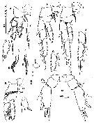 issued from : S. Ohtsuka, S. Nishida & R.J. Machida in J. Nat. Hist., 2005, 39 (27). [p.2489, Fig.3]. Female: A, P1 (posterior); B, 3rd exopodal segment of P1 (posterior); C, outer lateral margin of endopod of P1 (posterior); D, inner basal seta of left P1; E, inner basal seta of right P1; F, right P2 (anterior); G, 3rd exopodal segment of left P2; H, phoront of apostome ciliates (?) on intercoxal sclerite of P2; I, right P3 (anterior); J, 2nd and 3rd exopodal segments of left P3 (anterior); K, P4 (posterior); L, 1st exopodal segment of right P4 (posterior; 2nd and 3rd segments missing); M, P5 (posterior). Phoronts of apostome ciliates (?) indicated by arrowheads. Scale bars in mm. Nota: Assymmetry of the swimming legs 1 to 3 (maybe, leg 4 also). P5 nearly symmetrical; coxae and intercoxal sclerite separate; basis bearing thick, subterminal seta on posterior surface; endopod lacking; exopod 1-segmented, with 2 lateral and 2 terminal spines; acute process present at base of inner terminal spine; outer terminal process more acutely pointed in right leg than in left.
|
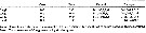 issued from : S. Ohtsuka, S. Nishida & R.J. Machida in J. Nat. Hist., 2005, 39 (27). [p.2490]. Female: Armature formula of swimming legs P1 to P4. Roman numerals = spines; Arabic numerals = setae.
| | | | | NZ: | 1 | | |
|
Distribution map of Metacalanalis hakuhoae by geographical zones
|
| | | | Loc: | | | Sulu Sea (central)
Locality type: 08°52.66'N, 120°25.28'E. | | | | N: | 1 | | | | Lg.: | | | (983) F: 1,55; {F: 1,55} | | | | Rem.: | hyperbenthic (2430-2450 m).
For Ohtsuka & al. (2005, p.2491), asymmetry of the swimming legs may be related to an as yet unknown swimming behaviour of the new genus as observed in Paramisophria spp. that swim with the left lateral side parallel to the bottom and the left A1 extended anteriorly (see in Fosshagen, 1968; Ohtsuka & Mitsuzumi, 1990). Gut content analysis revealed that the species fed upon copepods. Stalked cysts on legs are possibly assignable to phoronts of apostome ciliates. | | | Last update : 17/01/2015 | |
|
|
 Any use of this site for a publication will be mentioned with the following reference : Any use of this site for a publication will be mentioned with the following reference :
Razouls C., Desreumaux N., Kouwenberg J. and de Bovée F., 2005-2024. - Biodiversity of Marine Planktonic Copepods (morphology, geographical distribution and biological data). Sorbonne University, CNRS. Available at http://copepodes.obs-banyuls.fr/en [Accessed April 25, 2024] © copyright 2005-2024 Sorbonne University, CNRS
|
|
|
|


;)
;)
;)
