|
|
 |
|
Calanoida ( Order ) |
|
|
|
Clausocalanoidea ( Superfamily ) |
|
|
|
Aetideidae ( Family ) |
|
|
|
Pseudochirella ( Genus ) |
|
|
| |
Pseudochirella obtusa (Sars, 1905) (F,M) | |
| | | | | | | Syn.: | Undeuchaeta obtusa Sars, 1905 b (p.13, Rem.F); Lysholm & Nordgaard, 1921 (p.17);
Euchirella obtusa : Farran, 1908 b (p.40);
Euchirella dubia A. Scott, 1909 (p.60, figs.F); Sewell, 1948 (p.553);
Chirudina abyssalis With, 1915 (p.147, figs.F); Chirundina abyssalis : Sewell, 1948 (p.500);
Pseudochirella polyspina Brodsky, 1950 (1967) (p.186, figs.F); Arashkevich, 1969 (p.701, figs.F); Tanaka, 1957 b (p.194, figs.F); Grice & Hulsemann, 1967 (p.15); Morris, 1970 (p.2300); Vaupel-Klein, 1970 (p.4, 15, figs.F); Minoda, 1971 (p.25); 1972 (p.326); Morioka, 1972 a (p.314); Deevey & Brooks, 1977 (p.256, Table 2, station "S"); Park, 1978 (p.169, figs.F,M); Arashkevich, 1969 (p.701, figs.F); Gardner & Szabo, 1982 (p.252, figs.F); Hopkins & Torres, 1988 (tab.1); Chihara & Murano, 1997 (p.689, Pl.47: F,M); Ikeda & al., 2006 (p.1791, Table 2); Homma & Yamaguchi, 2010 (p.965, Table 2); Homma & al., 2011 (p.29, Table 2, 3, abundance, feeding pattern: suspension feeders);
Pseudochirella spinifera Brodsky, 1950 (1967) (p.188, figs.M); Tanaka & Omori, 1969 b (p.164, figs.M); Vaupel-Klein, 1970 (p.4, 15); Minoda, 1971 (p.26); 1972 (p.326); Morioka, 1972 a (p.314); Ikeda & al., 2006 (p.1791, Table 2); Homma & Yamaguchi, 2010 (p.965, Table 2); Homma & al., 2011 (p.29, Table 2, abundance);
no Pseudochirella polyspina (M): Tanaka & Omori, 1969 a (p.160, figs.M);
no Pseudochirella obtusa (M): Vervoort, 1949 (p.37, figs.M); 1952 g (n°48, p.3, fig.M); Bradford & Jillett, 1980 (p.71, fig.M);
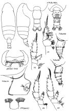
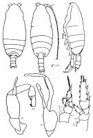
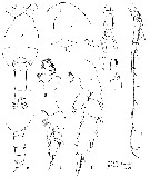
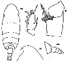
Pseudeuchirella obtusa: Björnberg, 1973 (p.324, 388) | | | | Ref.: | | | Sars, 1925 (p.83, figs.F); Rose, 1929 (p.24); Sewell, 1929 (p.131, figs.F, Rem.); Rose, 1933 a (p.110, figs.F); Jespersen, 1934 (p.65, Rem.F); Lysholm & al., 1945 (p.19); Sewell, 1947 (p.101, Rem.F); Davis, 1949 (p.32, figs.F); Vervoort, 1949 (p.37, F, no M); 1952 g (n°48, p.3, figs.F, no M); 1963 b (p.150, Rem.); Bradford & Jillett, 1980 (p.71, figs.F, no M, fig.73, distribution chart); Vaupel Klein, 1984 a (p.52); Markhaseva, 1989 (p.36, 38, 41, figs.F,M); Markhaseva & Razzhivin, 1993 (p.612); Markhaseva, 1996 (p.278, figs.F,M, Rem.); Bradford-Grieve & al., 1999 (p.879, 923, figs.F,M); Vives & Shmeleva, 2007 (p.600, figs.F,M, Rem.) | 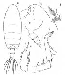 issued from : Tanaka O. in Publ. Seto mar. Biol., Lab., 1957, 6 (2). [Fig.55, p.194]. As Pseudochirella polyspina. Female : a, habitus (dorsal aspect); b, head (lateral aspect); c, last thoracic segment and urosome (lateral aspect); d, Mxp.; e, exopodite of P1; f, inner margin of first basal joint of P4. Nota Female: - Cephalothorax about 3.55 times the abomen length. - Head and 1st pediger segment indistinctly separte, 4th and 5th pediger segments separate. - Posterolateral corners of last thoracic segment rounded. - Rostrum moderately long and strong, directs posteriorly. - Abdomen 4-segmented; segments and caudal rami in proportional lengths 47 : 20 : 13 : 7 : 13 = 100. - Genital segment furnished with hairs on the lateral proximal and distal margin, and also on the ventral side at the base of genital opening, and on the distal ventral margin. - Caudal rami about as long as wide. - A1 23-segmented, extends to distal end of caudal rami. - A2 exopod 3/5 as long as endopod. - Md with a strong biting part, the tooth on outer margin long. - Mx1 with 7 long and 2 short setae on outer lobe; 11 setae on exopod; 15 setae on endopod; 5 setae on 2nd basal segment; 4 setae and a small process on 3rd inner lobe; 5 setae on 2nd inner lobe. - Mxp with 2nd basal segment 1.5 times as long as the 1st; anterior distal margin of the segment with a row of spinules and a double rows of spinules on the proximal margin. - P1 with a slender outer edge spine on the exopodal segments; 1st and 2nd segments incompletely fused. - P4 with 13 spines on inner margin of coxa.
|
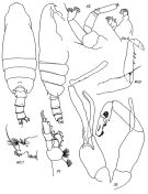
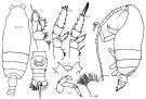
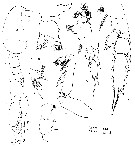
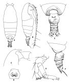 issued from : T. Park in Antarctic Res. Ser. Washington, 1978, 27. [p.170, Fig.45]. As Pseudochirella polyspina. Female: A, habitus (dorsal view); B, idem (right lateral side); C, forehead (lateral); D, genital segment (ventral view); E, posterior part of metasome and urosome (dorsal view); F, idem (right lateral side); G, A2.
|
 issued from : T. Park in Antarctic Res. Ser. Washington, 1978, 27. [p.171, Fig.45]. As Pseudochirella polyspina. Female: A, Mx1; B, P1 (anterior); C, P2 (idem); D, basipod of P4 (posterior).
|
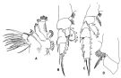
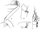
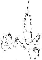
 issued from : T. Park in Antarctic Res. Ser. Washington, 1978, 27. [p.172, Fig.47]. As Pseudochirella polyspina. Male: A, forehead (lateral); B, posterior part of metasome and urosome (dorsal view); C, last metasomal and first two urosomal segments (right lateral); D, A2; E, P1 (anterior); F, P5 (anterior); G, two distal exopodal segments of left P5 (anterior); H, idem (posterior)
|
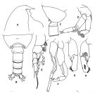
 issued from : E.L. Markhaseva in Proc. Zool. Inst. RAN, St. Petersburg, 1996, 268. [p.282, Fig.225]. Female (from N Atlant.): CP4: coxopod of fourth legs. Nota: Cephalothorax about 3-4 times longer than urosome. Posterior corners of last thoracic segment rounded, sometimes covered by hairs, symmetrical. Urosomal segments 1-3 covered with hairs to different degree in different specimens. Length of A1 varying: possibly slightly longer than cephalothorax, or reaching the end of caudal rami. Exopodite of P1 3-segmented with indistinctly separated segments 1 and 2. Endopodite of P2 2-segmented. Coxopodite of P4 with 8-15 spines.
|
 issued from : E.L. Markhaseva in Proc. Zool. Inst. RAN, St. Petersburg, 1996, 268. [p.283, Fig.226]. Male (from NW Pacif.: Kuril Trench). Nota: Cephalothorax slightly less than 3 times longer than urosome. Cephalon and thoracic segment 1, as thoracic segments 4 and 5 fused (line of fusion sometimes visible dorsally). Posterior corners of last thoracic segment rouded, with spines removed to the back side of specimen (lateral view). Oral parts with reduced setation. Exopodite of P1 3-segmented; external spine not exceeding the midlength of segment 2. Endopodite of P2 2-segmented. Exopodal segment 2 of left P5 with 2 teeth; segment 3 more than 3 times longer than wide.
|
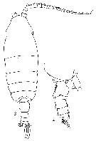
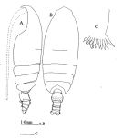 issued from : J.M. Bradford & J.B. Jillett in Mem. N.Z. oceanogr. Inst., 86, 1980. [p.73, Fig.49]. Female: A, habitus (lateral left side); B, idem (dorsal); C, inner edge of basipod 1 of P4. Nota: The basipod 1 of P4 has 10 teeth.
|
 Issued from : J.C. von Vaupel-Klein in Zool. Verh. Leiden, 1970, 110. [p.16, Fig.4,f]. As Pseudochirella polyspina. Female: f, left P1 (anterior surface). Nota: This specimen resembles Brodsky's (1950, p.185, fig.102) description and figures but for P1; the external marginal spines of 1st and 2nd exopodal segment are serrate, the terminal spine has a serrated outer edge, while the inner edge has long hairs; the enopodite bears a row of fine spinules (not figured by Brodsky).
|
 Issued from : G.O. Sars in Résult. Camp. Scient. Prince Albert I, 69, pls.1-127 (1924). [Pl.XXIV, figs.1-4]. Female: habitus (dorsal); 2, idem (lateral left side); 3, forehead, with labrum and labium (lateral); 4, basipodal segment of P4. Head and thoracic segment 1 imperfectly separate (a suture medial visible) on the dorsal surface), Th4 and 5 separate. Rostrum relatively strong, pointed art apex and slightly curved. A1 extending little after the cephalosome. Corners of Th5 (in lateral view) short and blunt. Coxopodite of P4 with comb of about 8 strong spines across mediodistal portion of posterior surface. Genital segment symmetrical; as long as the followingt two urosomal segments. Anal segment shorter than the preceding segment. Caudal rami comparatively short bearing 4 plumose setae of length almost equal. Md strong with masticatory blade large with outer tooth extended as a claw.
|
 Issued from : K.A. Brodskii in Calanoida of the Far Eastern Seas and Polar Basin of the USSR. Opred. Fauna SSSR, 1950, 35 (Israel Program for Scientific Translations, Jerusalem, 1967) [p.188, Fig.104]. As Pseudochirella spinifera. Male (from NW Pacif.): habitus (dorsal and lateral left side); S1, P1; S2, P2; S5, P5 (Le = left leg; Ri = right leg). Nota: Head poorly defined from thorax; two terminal thoracic segments fused. Rostrum simple, well defined. A1 reaching middle or end of 2nd abdominal segment. Endopod of A2 equal to 4/5 length of exopod or slightly longer. Dorsal surface of last thoracic segment bearing 1 long, slightly curved spine on each side. Exopod of P1 3-segmented, with 2 outer spines and rudimentary 3rd spine on proximal segment. Endopod of P2 2-segmented. P5 relatively simple in structure. Right endopodite larger than the left, widened, distally unciform. Penultimate segment of left exopodite with 2 triangular processes.
|
 Issued from : K.A. Brodskii in Calanoida of the Far Eastern Seas and Polar Basin of the USSR. Opred. Fauna SSSR, 1950, 35 (Israel Program for Scientific Translations, Jerusalem, 1967) [p.186, Fig.102]. As Pseudochirella polyspina. Female (from NW Pacific): habitus (dorsal and lateral left side); R, forehead (ventral); A2; urosome (ventral); S1, P1; S2, P2; S3, P3S4B1, inner distal edge of basipodal segment of P4. Nota: Head without crest. Rostrum present. 4th and 5th thoracic segments clearly separate. Abdomen 1/4 length of cephalothorax. Genital segment symmetrical. Abdominal segments densely hairy on ventral side. Caudal rami not divergent, not longer than anal segment. A1 only slightly longer than body length. A2 endopod slightly more than half length of exopod. Exopod of P1 2-segmented, outer spines equally long, distal spine of 1st segment ending slightly before base of spine on 2nd segment. Endopodite of P2 1-segmented. Endopod of P3 with poorly defined 2nd and 3rd segments. First basipodite of P4 armed with 11-15 large lamellar spines inserted into large process of basipodite.
|
 issued from : R.B.S. Sewell in Mem. Indian Mus., 1929, X. [p.132, Fig.50]. Female (from S India): a, Mxp; b, Md (biting edge); c, P1; d, basal portion of P4.
|
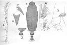 issued from : A. Scott in Siboga-Expedition, 1909, XIX a. [Plate XIV, Figs.1-7]. As Euchirella dubia. Female (from Manipa Strait): 1, habitus (dorsal); 2, forehead (lateral); 3, last thoracic and genital segment (left side); 4, rostrum; 5, A1; 6, Mx1; 7, P4 (basal portion only).
|
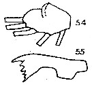
 issued from : O. Tanaka & M. Omori in Publ. Seto Mar. Biol. Lab., 1969, XVII (3). [p.165, Fig.4). As Pseudochirella spinifera. Male (from Izu Region, Japan): a-b, forehead (dorsal and lateral, respectively); c-d, last thoracic segment and urosome (dorsal and left la-side, respectively); e, rostrum (frontal view); f, A1; g, Mx1; i, P1; i, P2; j, P5; k, 2nd and 3rd segments of exopod of left P5.
|
 issued from : O. Tanaka & M. Omori in Publ. Seto Mar. Biol. Lab., 1969, XVII (3). [p.161, Fig.2). As Pseudochirella polyspina. With doubt in this species. Male (from Izu Region, Japan): a,head (lateral); b, last thoracic segment and urosome (lateral, right side); c, Md; d, Mx1; e, Mx2; f, Mxp; g, P1; h, P2; i, P5; j, 2nd and 3rd segments of exopod of left P5. Nota Male : - Prosome and urosome iratio 66 to 34. - Head and 1st pediger segment fused. - Posterolateral margin of the last thoracic segment is rounded, devoid of any lateral spines. - Urosomal segments and furca in proportional lengths 16, 30, 21, 18, 2, 13 = 100. 2nd, 3rd and 4th urosomal segments fringed with fine spinules on the distal margin. Caudal ramus as long as wide. A1 extends about to the distal margin ofthe 4th urosomal segment ; proportional lengths of the segments : 1 (65), 2 (55), 3 (34), 4 (28), 5 (31), 6 (28), 7 (30), 8-10 (69), 11 (28), 12-13 (69), 14 (39), 15 (48), 16 (48), 17 (55), 18 (45), 19 (67), 20 (63), 21 (41), 22 (51), 23 (53, 24-25 (53). A2 exopod 7-segmented which is 1.5 times the length of the endopod ; 1st exopod segment without setae, 2nd segment with a small process carrying a minute seta ; endopod with 7 setae on the outer lobe, and 8 setae on the inner lobe. Md palp well developed, cutting blade is small, without teeth. Mx1 has the following numbers of setae on the various lobes : 5 long and 2 small setae on the outer lobe ; 11 setae on the exopod ; 6+4+4 setae on the endopod ; 4 setae on the 2nd basal segment ; 1 apical seta on the reduced 3rd inner lobe ; the 2nd inner lobe is rudimentary ; the 1st inner lobe has 4 rudimentary setae. Mx2 reduced, with on the various lobes : 6long setae on the endopod ; 2 setae and 1 small process on the 1st to 3rd lobes, respectively ; 3 setae on the 4th lobe ; 1 strong and 2 short setae on the 5th lobe. Mxp well developed, 2nd basal segment 1.4 times the length of the 1st one. P5 resembles that of P. spinifera.
|
 issued from : G.P. Farran in Fish. Ire. Sci. Invest., 1906, II [1908]. [Pl. II, Figs.20-21]. As Euchirella obtusa. Female (from W Ireland): 20, A2; 21, P4.
|
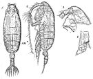
 issued from : G.P. Farran in Fish. Ire. Sci. Invest., 1906, II [1908]. [Pl. IV, Figs.1-2]. Female: 1, last thoracic segment and urosome (lateral); 2, habitus (dorsal).
|
 issued from : C.C. Davis in Univ. Wash. Publs Biol., 1949, 14. [Pl.4, Figs.49-53]. Female (from NE Pacific): 49, habitus (dorsal); 50, exopod of P1; 51, basipod of P4; 52, forehead (lateral); 53, last thoracic segment and urosome (lateral). Nota : Rostrum short, one-pointed. Head rather vaulted, but with no sign of a crest. Head and 1st thoracic segment feebly separated, 4th and 5th segments distinctly separated. Lateral corners very short and rounded. Genital segment as long as the three following segments combined. Caudal rami slightly broader than long and bear terminally 4 plumose setae. The genital swelling is not paricularly large, its greatest thickness is in the anterior part, anda long the posterior ventral edge there is a tuft of hairs. The 2nd and 3rd urosomal segments bear short hairs. A1 23-segmented (11th and 12th nearly fused), reach slightly the posterior border of the prosome. Endopod of A2 about 7/10 the length of exopod. The 1st basal segment of P4 with on its inner posterior margin a row of 8 to 13 spines.
|
 issued from : C.C. Davis in Univ. Wash. Publs Biol., 1949, 14. [Pl.5, Figs.54-55]. Female: 54, endopod of P1; 55, Md (masticatory blade)
|
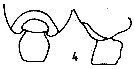 Issued from : E.L. Markhaseva in USSR Acad. Sci., Zool. Inst., Explor. Fauna Seas. Marine Plankton, 1989, 41 (49). [p.52, Fig.4]. Female: Thoracic segment 5 and genital segment. Nota from the key: 1 - Genital segment symmetrical. 2 - Thoracic posterior corners symmetrical. 3 - Exopodite segment 2 of A2 with 1 seta. 4 - Thoracic posterior corners segment 5 without point. 5 - Thoracic posterior corners segment 5 (in lateral view) protruded into lobes, rounded with a concave part.
|
 Pseudochirella obtusa Pseudochirella obtusa Male: 1 - Exopodite 2 of left P5 with tooth. 2 - Exopodite 2 of left P5 with 2 teeth distally. 3 - Posterior corners of last thoracic segment with spines (sub-dorsally). 4 - Exopodite 3 of left P5 more than 3 times longer than wide.
|
 issued from : Tanaka O. in Publ. Seto mar. Biol., Lab., 1957, 6 (2). [p.195]. As Pseudochirella polyspina. Female A1: proportional lengths of segments - A1 23-segmented, extends to distal end of caudal rami. The 1st segment has aswelling on the posterior margin.
| | | | | Compl. Ref.: | | | Sewell, 1948 (p.331, 500, 520, 531, 545); C.B. Wilson, 1950 (p.317); Roe, 1972 (p.277, tabl.1, tabl.2); Kovalev & Schmeleva, 1982 (p.83); Grice & Hulsemann, 1967 (p.15); Ehrhardt, 1967 (p.738, geographic distribution, Rem.); Vives, 1982 (p.291); Roe, 1984 (p.357); Lozano Soldevilla & al., 1988 (p.58); Razouls & al., 2000 (p.343, Appendix); Holmes, 2001 (p.51); Kosobokova & al., 2007 (p.929: Tab.7); Gaard & al., 2008 (p.59, Table 1, N Mid-Atlantic Ridge); Galbraith, 2009 (pers. comm.); Park & Ferrari, 2009 (p.143, Table 4, Appendix 1); Mazzocchi & Di Capua, 2010 (p.423); Bode & al., 2015 (p.268, Table 1, chemical components, trophic level, geographic zone); Belmonte, 2018 (p.273, Table I: Italian zones) | | | | NZ: | 18 | | |
|
Distribution map of Pseudochirella obtusa by geographical zones
|
| | | | | | | | | | | | 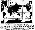 Issued from : E.L. Markhaseva in Issled. Fauny Morei, 1989, 41 (49). [p.55, Fig.9]. Issued from : E.L. Markhaseva in Issled. Fauny Morei, 1989, 41 (49). [p.55, Fig.9].
Geographical distribution of P. obtusa.
1: from literature; 2: literature as P. polyspina; 3: present occurences. |
| | | | Loc: | | | Antarct. (Weddell Sea, SW & SE Atlant., Indian, S & SE Pacif., Drake Passage), sub-Antarct. (SW Atlant., SE Pacif.), Atlant. (temperate & tropical), Angola, off Ghana, Canary Is., Canary-Azores, off W Cabo Finisterre, Sargasso Sea, off Bermuda (Station "S"), off SE Cape Cod, Labrador Sea, S Davis Strait, S Iceland, off W Scotland, off W Ireland, Medit. (Ligurian Sea, Tyrrhenian Sea), Arabian Sea, S Laccadive Is., Indian, Indonesia-Malaysia, Philippines, Japan, off Hokkaido, Station Knot (44°N, 155°E), Okhotsk Sea, Kuril-Kamchatka, Bering Sea , S Aleutian Basin, Aleutian Trench, S Aleutian Is., Alaska, Station "P" (50°N, 145°W), Cap Flattery, Vancouver Is., Pacif. central subarct., New Zealand, N Marquesa Is., S Pacif., off Peru, Chile.
Type locality: North Atlantic. | | | | N: | 44 | | | | Lg.: | | | (1) F: 5,8; (5) F: 7; (7) F: 5,4; (11) F: 5,75; (14) F: 6,05-5,55; M: 5,5-5,45; (22) F: 5,8; M: 5,2-4,7; (37) F: 6,5-5,25; M: 5,5-4,8; (41) F: 6,45-5,25; (49) F: 5,8-5,6; (56) F: 6,28; (111) F: 6,4-5,7; M: 5,2-5,1; (199) F: 5,92-5,17; (201) F: 5,9; (208) F: 6,7-6,28; M: 5,3; (866) F: 5,66-6,5; M: 4,83-5,33; (1257) F: 5,5-6,0; {F: 5,17-7,00; M: 4,70-5,50} | | | | Rem.: | Meso-bathypelagic. Sargasso Sea: 1000-2000 m (Deevey & Brooks, 1977, station "S");
Sampling depth (Antarct. sub-Antarct.) : 0-1000 m.
Characteristics female from Sewell (1947, p.101): The proportional lengths of the cephalothorax and abdomen as 78 to 22. The proportional lengths of the various segments of the body (cephalon to caudal rami) as 397 : 135 : 88 : 73: 50 : 31 : 91 : 50 : 35 : 21 : 29 = 1000. The line of fusion of the head and 1st pediger segment is clearly visible in stained specimens and the fusion appears to be incomplete dorsally; 4th and 5th thoracic segments separate. Forehead devoid of any crest. Genital segment symmetrical, on either side about the middle of its length there is a slight rounded protuberance. All abdominal segments are hairy. Caudal rami as broad as long. A1 24-segmented (segments 8-9 fused, 24-25 partially separate) reaches back to the anal segment. In A2 the proportional lengths of endopod and exopod as 28 to 47; segments 1 and 2 of the endopod separate, there is a row of hairs on the 1st basal segment. In Md the biting blade with the most anterior tooth elongate and sickle-shaped; the 2 nd basal segment bears 3 setae, of which the proximal is much the stoutest. In Mx1 the 2nd inner lobe bears 5 setae, 3rd with 4 setae; the 2nd basal segment with 4 setae; the endopod with 15 setae (4 + 4 + 7); exopod with 11 setae; outer lobe with 9 setae, the proximal two being small and delicate. In Mxp the proportional lengths of the two basal segments as 5 to 8; the anterior margin of the 1st basal segment produced distally in a bluntly rounded eminence that is covered with small spinules; on the distal border is a nipple-like prominence (similar to that present in C. notacantha).
For Vervoort (1963b, p.150) the Atlantide specimens differ from specimens described by A. Scott as Euchirella dubia by the naked condition of the urosome, A. Scott's specimens being distinctly haired.
After Markhaseva (1996, p.281) the first description of P. obtusa by Sars (1905) and subsequent redescription (Sars, 1924-1925) are brief and inadequately documented. This misled Brodsky (1950) in his assumption to originate P. polyspina from the north-western part of the Pacific Ocean. After examination of the material, the author concludes on the identity between P. obtusa and P. polyspina. Likewise the male identified by Vervoort (1949) as P. obtusa should be attributed to the male of P. dubia (Sars, 1905) and the male described under the name P. spinifera Brodsky (1950) and later synonymized with P. polyspina (Park, 1978) should probably be considered as a male of P. obtusa.
All the authors are not in accordance between the synonymy of P. obtusa Sars, 1905 and P. polyspina Brodsky, 1950; as well as between P. obtusa and P. spinifera Brodsky, 1950.
: R. Stephen, 2007 : Data sheets of NIO, Kochi, India (on line). | | | Last update : 23/01/2021 | |
|
|
 Any use of this site for a publication will be mentioned with the following reference : Any use of this site for a publication will be mentioned with the following reference :
Razouls C., Desreumaux N., Kouwenberg J. and de Bovée F., 2005-2024. - Biodiversity of Marine Planktonic Copepods (morphology, geographical distribution and biological data). Sorbonne University, CNRS. Available at http://copepodes.obs-banyuls.fr/en [Accessed April 25, 2024] © copyright 2005-2024 Sorbonne University, CNRS
|
|
|
|


;)
;)
;)
;)
;)
;)
;)
;)
;)
;)
;)
;)
;)
;)
;)
;)
;)
;)
;)
;)

;)
