|
|
 |
|
Calanoida ( Order ) |
|
|
|
Epacteriscoidea ( Superfamily ) |
|
|
|
Ridgewayiidae ( Family ) |
|
|
|
Ridgewayia ( Genus ) |
|
|
| |
Ridgewayia delfine Figueroa & Hoefel, 2008 (F,M) | |
| | | | | | | Ref.: | | | Figueroa & Hoefel, 2008 (p.138, figs.F,M, Table 2, 3, Rem.) | 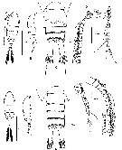 issued from : D.F. Figueroa & K.L. Hoefel in J. Crustacean Biol., 2008, 28 (1). [p.139, Fig.1]. Female (from 00°45.426', 90°18.932'W): A-B, habitus (dorsal and lateral, respectively); C, urosome (ventral); D, A1 (segments 1-19 [I-XX]); E, A1 (segments 20-26 [XXI-XXVIII]). Male: F-G, habitus (dorsal and lateral, respectively); H, urosome (ventral); I, A1 (segments 1-19 [I-XX], with reverse detail of segments 14-18 [XV-XIX]); J, A1 (segments 20-24 [XXI-XXVIII], with reverse detail of segment 20 [XXI-XXIII]. Nota Female: Prosome 6-segmented; cephalosome clearly separate from 1st pedigerous somite. - Posteriolateral angles of prosome rounded and extending 1/3 length along genital double-somite. - Large eye present in anterior cephalosome, red pigmented in fresh specimens. - Rostrum a simple process produced ventrally with rounded tip; pair of secretory gland openings present near tip. - Urosome 4-segmented. - Genital double-somite symmetrical: genital operculum somewhat offset to right of midventrum; seminal receptacle present on left side only. - All urosomal segments, except for anal, have row of denticles along posterior borders. In genital double-somite, row of denticles has a gap along border directly behind genital operculum. - Surface of all urosomal segments, except for area arounfd genital opening, entirely covered with small spinules. - Caudal rami symmetrical with their surfaces covered with minute setules, and bearing 6 setae. Small blade-like seta sited on outer distal corner, followed medially by longer naked seta striated with helical lines, then 2 long , plumose median setae jointed basally. Small plumose seta extends dorsally, followed by longer plumose seta on distal inner margin. Distal 3rd of inner margin bearing row of long setules. - A1 barely reaches the tip of caudal ramus. Variation in length among individuals, with few specimens with A1 extending slightly beyond caudal ramus. A1 26-segmented, segments 13 to 22 each with distal transverse row of spinules. Nota Male: Body as in female. - Urosome 5-segmented. - Caudal rami similar to those of female, except lacking row of setules on distal inner margins. - Left A1 as in female. - Right A1 24-segmented, weakly geniculate with 4 segments beyond geniculation. Rows of spinules present on segments XV, XVI, XVII, XVIII, XIX, and XXI-XXIII. Segments XI and XIV constricted.
|
 issued from : D.F. Figueroa & K.L. Hoefel in J. Crustacean Biol., 2008, 28 (1). [p.140, Fig.2]. Female: A, A2; B, Md; C, Mx2; D, Mx1; E, Mxp. Nota: A2 with plumose seta on coxa adjacent to patch of setules; basis with 2 setae of unequal length; exopod indistinctly 8-segmented, setal formula 1, 1, 1, 1, 1, 1, 1, 4; endopod 2-segmented with 1st bearing 2 subterminal setae, 2nd segment bilobed, subterminal lobe with 9 setae, terminal lobe with 7 setae. 2 rows of spinules/setules along inner margin of 2nd segment. - Md gnathobase bearing 9 teeth; with row of 10 smaller teeth at distal inner edge of mandible blade forming a saw-like element; tuft of fine setules present on gnathobase, near attachment of palp; basis with 4 setae along inner margin (1 large, 2 medium and 1 small); exopod indistinctly 4-segmented with setal formula 1, 1, 1, 3; endopod 2-segmented, 1st segment with 4 setae, 2nd segment with 11 setae. - Mx1 well developed; praecoxal arthrite bearing 6 spiniform and 3 spinulose distal setae and 1 anterior and 4 slender posterior setae. With tuft of fine setules present along insertion of posterior seta. Coxa with 9 setae on epipodite and 5 setae on endite. Basis with 1 seta on exite, 4 setae on 1st endite and 5 setae on 2nd endite. Exopod 1-segmented, with 3 + 8 setae. Endopod 2-segmented with setal formula 4 + 4, 7. - Mx2 with 1st praecoxal endite with 1 proximally directed seta at base and 5 long distal setae; 2nd praecoxal endite and 1st and 2nd coxal endites with 3 setae each. Basis with 1 sclerotized seta and 3 long setae. Endopod 4-segmented with the distal 3 segments partly fused, bearing 3, 2, 2, 3 setae. With tuft of fine setules present around distal edge of 1st and 2nd praecoxal endites and 1st and 2nd coxal endites. - Mxp with syncoxal endites bearing 1, 2, 4, 3 setae, respectively. Several setules on each of first three endites, and large patch of fine setules present on 4th endite. Basis with 3 setae, with patch of long setules along inner margin. Endopod 6-segmented, with setal formula 2, 4, 4, 3, 3 (inner) + 1 (outer), 4. 1st endopod segment distinctly separate from basis.
|
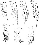 issued from : D.F. Figueroa & K.L. Hoefel in J. Crustacean Biol., 2008, 28 (1). [p.141, Fig.3]. Female: A-E, P1 to P5, respectively (anterior). Male: F, right P5 (anterior); G, left P5 (anterior). Nota Female: P1 with von Vaupel Klein organ for grooming (see Barthelemy & al., 1998), consisting of curved inner basal setae and a distally serrated process originating anteriorly from 1st endopodal segment and extending halfway along 2nd segment. With tuft of setules present on surface of this process. With row of fine setules present on distal edge of 1st exopod segment. Segment 2, with lamellate, serrated process at distal margin, between outer setae and insertion point of 3rd segment. With seta-like process in between outer setae a,d this lamellae process. - P2, P3 and P4 with serrated distal margins on 1st and 2nd endopodal segments; with hairy protuberances on center distal margin of 2nd exopodal segment. - P5 3-segmented exopod and 2-segmented endopod. Inner coxal seta absent, basal seta present. Surface of leg covered with minute setules and/or denticles. Nota Male: P1-P4 as in female. - P5 biramous, asymmetrical, strongly modified.Coxa without setae. Basis of right leg bears single seta on posteriolateral surface, near distal outer margin. Distal half of inner margin serrated and covered with minute setules. Right leg with 2-segmented exopod and 1-segmented, elongate, endopod. Exopod segments without setae. 1st exopodal segment with outer spine reaching base of 1st outer spine on 2nd exopodal segment. 2nd exopodal segment with 2 outer spines. Distal end of this segment modified, with 2 processes on anterior surface. 1st process, next to base of 2nd spine, consisting of curved flange with brush-like elements on outer surface. 2nd process, just distal to 1st, protruding anteriorly and covered with brush-like elements. Exopod ends with terminal projection, outer surface of which covered with brush-like elements. Endopod elongate, reaching tip of exopod. Seat present on anterodistal margin, before mid-length of segment, otherwise unarmed. Left leg with single seta on anterolateral surface of basis, near distal outer margin. Exopod 3-segmented. 1st segment with outer spine reaching distal edge of 2nd segment. 2nd segment with small acute process on distal outer margin. Outer spine on 2nd segment long and straight, rather conical in shape, with thick base tapering slightly distally. Tip of spine very thin and curved inward. Brush-like setules along posterior and anterior inner margins of spine.3rd segment complex and strongly modified, with 5 discernable elements. In order from anterior to posterior: 1st element as thin naked seta near outer margin; 2nd element leaf-like, with anteriorly directed marginal folds; 3rd element slender, lamellae and protruding from middle of 2nd element, with several thin, finger-like extensions at tip; 4th element consists of 2 segments, 1st thick and rectangular along inner edge, 2nd stems from strong distal joint, slender and curved towards outer spine and then projects distally and inwards with forked tip and small, petal-like process; 5th element as another leaf-like process with anteriorly directed marginal folds, and tapering distally into very thin tip.
|
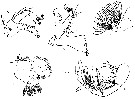
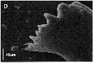 issued from : D.F. Figueroa & K.L. Hoefel in J. Crustacean Biol., 2008, 28 (1). [p.142, Fig.4, D]. Female (SEM image): D, Md (anterior).
|
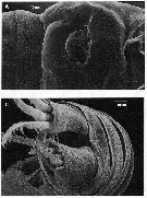 issued from : D.F. Figueroa & K.L. Hoefel in J. Crustacean Biol., 2008, 28 (1). [p.142, Fig.4, A, C]. Female (SEM images): A, genital double somite (ventral); C, caudal rami (posterior-dorsal).
|
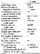 issued from : D.F. Figueroa & K.L. Hoefel in J. Crustacean Biol., 2008, 28 (1). [p.145, Table 2]. Characters female of R. define.
|
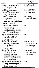 issued from : D.F. Figueroa & K.L. Hoefel in J. Crustacean Biol., 2008, 28 (1). [p.145, Table 3]. Characters male of R. define.
| | | | | NZ: | 1 | | |
|
Distribution map of Ridgewayia delfine by geographical zones
|
| | | | Loc: | | | E Pacific (Island of Santa Cruz, Galapagos) | | | | N: | 1 | | | | Lg.: | | | (1073) F: 0,91-0,98; M: 0,82-0,94; {F: 0,91-0,98; M: 0,82-0,94} | | | | Rem.: | In a swimming hole, near from the shore, pools influenced by tidal current. | | | Last update : 17/08/2021 | |
|
|
 Any use of this site for a publication will be mentioned with the following reference : Any use of this site for a publication will be mentioned with the following reference :
Razouls C., Desreumaux N., Kouwenberg J. and de Bovée F., 2005-2024. - Biodiversity of Marine Planktonic Copepods (morphology, geographical distribution and biological data). Sorbonne University, CNRS. Available at http://copepodes.obs-banyuls.fr/en [Accessed April 18, 2024] © copyright 2005-2024 Sorbonne University, CNRS
|
|
|
|


;)
;)
;)
;)
;)
;)
;)
