|
|
 |
|
Calanoida ( Order ) |
|
|
|
Diaptomoidea ( Superfamily ) |
|
|
|
Acartiidae ( Family ) |
|
|
|
Acartia ( Genus ) |
|
|
|
Euacartia ( Sub-Genus ) |
|
|
| |
Acartia (Euacartia) forticrusa Soh, Moon, Park & Maran, 2013 (F,M) | |
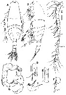
| | | | | | | Ref.: | | | Soh & al., 2013 (p.719, Descr.F,M, figs.F,M, table 2: molecular mtCOI sequences). | 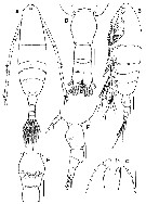 Issued from : H.Y. Soh, S.Y. Moon, E.O. Park & B.A.V. Maran in J. Crustacean Biol., 2013, 33 (5). [p.720, Fig.1]. Female (from 35°01'06.01''N, 127°47'06.77''E): A-B, habitus (dorsal and lateral, respectively); C, forehead with rostral filaments (ventral); D, urosome (dorsal); E, genital double-somite (ventral); F, posterior part of metasome and urosome (lateral). Scale bars: 100 µm (A, B); 50 µm (C-F). Nota: Prosome:Urosome ratio 4.05;1. Prosome 5-segmented; cephalosome and 1st pedigerous somite completely separate, 4th and 5th pedigerous somites fused. Posterior corners of 5th pedigerous somite rounded, naked. Urosome 3-segmented. Proportional lengths of urosomites and caudal rami 47 : 19 : 12 : 22 = 100. Genital double-somite most swollen anterolaterally, with paired copulatory and gonopores ventromedially.
|
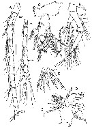 Issued from : H.Y. Soh, S.Y. Moon, E.O. Park & B.A.V. Maran in J. Crustacean Biol., 2013, 33 (5). [p.721, Fig.2]. Female: A, A1; B, A2; C, Md; D, Mx1; E, precoxa arthrite of Mx1. Scale bars: 50 µm (A-D; 25 µm (E). Nota: A1 20-segmented, extending to midlength of genital double-somite; anxestral segments XVIII and XIX with row of setules, XXI with 3 rows of setules. A2: basis and 1st endopodal segment fused to form elongated allobasis , 2nd endopodal segment elongated, 3rd endopodal segment short. Exopod short, 4-segmented (setation formula 1, 2, 2, 3). Md: coxa well developed gnathobase bearing 9 large a,d 3 small sharp teeth. Endopd 2-segmented. Exopod 4-segmented. Mx1: praecoxa and coxa incompletely fused, praecoxal arthrite with 9 elements; coxal endite with 3 setae, coxal epipodite with 9 setae; basal endite 1 with 1 seta; basal exite with 1 seta; endopod (with 5 terminal setae) and exopod (with 2 outer setae) fused with hairs along inner margin.
|
 Issued from : H.Y. Soh, S.Y. Moon, E.O. Park & B.A.V. Maran in J. Crustacean Biol., 2013, 33 (5). [p.722, Fig.3]. Female: A, Mx2; B, Mxp; C, P1 (anterior); D, P2 (anterior); E, P3 (anterior); F, P4 (anterior); G, 3rd exopodal segment of P4 (anterior); H, P5. Scale bars: 50 µm (A-F); 25 µm (G, H). Nota: Mx2: praecoxa and coxa incompletely fused, setation formula of endites 4, 2, 2, 3; basis with 1 long seta and row of setules on distal margin; endopod 3-segmented, last two segments ibcompletely fused, with setation formula 2, 2, 3. Mxp: praecoxa and coxa incorporated into syncoxa bearing 6 setae; basis with 1 spiniform seta; endopod 2-segmented, 1st and 2nd endopodal segments with 3 and 2 spiniform setae, respectively. P5 symmetrical, 3-segmented, both coxae completely incorporated in intercoxal sclerite; basis oblong-ovate, with outer seta inserted at about midlength; exopod tapering, distally short, thick, bent at midlength, distal portion serrated, base slightly swommen.
|
 Issued from : H.Y. Soh, S.Y. Moon, E.O. Park & B.A.V. Maran in J. Crustacean Biol., 2013, 33 (5). [p.719]. Female & Male: Spine (Roman numeral) and seta ( (arabic numeral) for the swimmeing legs P1 to P4.
|
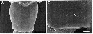 Issued from : H.Y. Soh, S.Y. Moon, E.O. Park & B.A.V. Maran in J. Crustacean Biol., 2013, 33 (5). [p.726, Fig.6]. Scanning electon micrographs of variants of Acartia forticrusa female: A-B, genital double-somite (dorsal). Arrows indicate rows of setules. Scale bars: 20 µm (A); 5 µm (B)
|
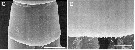 Issued from : H.Y. Soh, S.Y. Moon, E.O. Park & B.A.V. Maran in J. Crustacean Biol., 2013, 33 (5). [p.726, Fig.6]. Scanning electon micrographs of variants of Acartia forticrusa female: C-D, first urosomite (dorsal). Arrows indicate rows of setules. Scale bars: 20 µm (C); 5 µm (D).
|
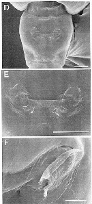 Issued from : H.Y. Soh, S.Y. Moon, E.O. Park & B.A.V. Maran in J. Crustacean Biol., 2013, 33 (5). [p.725, Fig.5]. Scanning electon micrographs of Acartia forticrusa female: D, genital double-somite (ventral); E-F, genital field (ventromedially) with paired copulatory and gonopores, each gonopre covered with epicuticule. Scale bar: 10 µm (D).
|
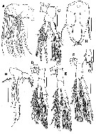
 Issued from : H.Y. Soh, S.Y. Moon, E.O. Park & B.A.V. Maran in J. Crustacean Biol., 2013, 33 (5). [p.724, Fig.4]. Male (from Beolgyo Sream estuary): A-B, habitus (dorsal and lateral, respectively); C, right A1; D-F, 13-15 segments of right A1; G, P5 (anterior view; L = left leg; R = right leg); H, 3rd segment of left P5. Scale bars: 100 µm (A, B); 50 µm (C); 25 µm (D-H). Nota: Prosome:Urosome ratio 3.47 : 1. Posterior corners of 5th pediger somite naked. Urosome 5 somites, each usually unarmed, but sometimes 2nd somite furnished with rows of setules. Caudal rami nearly as broad as long. Proporsions of urosomites and caudal rami 11 : 34 : 22 : 8 : 11 : 14 = 100. Right A1 18-segmented, geniculate between segments XX and XXI; ancestral segments IX-XI and XIX with row of trnasverse spinules, XX and XXI-XXIII with longitudinal row of denticles. Other mouthparts and P1 to P4 as in female. P5 asymmetrical: intercoxal sclerite completely fused to both coxae. Left leg 3-segmented, basis armed with outer seta and round lobe on posterior surface, basis slightly longer than outer seta; exopod 2-segmented, 1st segment short, unarmed; distal segment furnished with proximal tuft of hair, 1 long medial spine with 2 rows of teeth along its inner margin and blunt projection at its base; distal segment terminates in one short process and 1 terminal spine. Right leg 4-segmented: basis short, with outer seta longer than its segment; exopod 3-segmented, 1st segment with long slender seta posteromedially, 2nd segment with oblong inner lobe, grooved terminally, bearing 1 spine on distal border, 3rd segment with very short spine on medial margin and curved thick terminal spine.
|
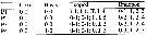
 Issued from : H.Y. Soh, S.Y. Moon, E.O. Park & B.A.V. Maran in J. Crustacean Biol., 2013, 33 (5). [p.727, Table 1]. Comparison of morphological features between A. forticrusa and its congeners A. southwelli and A. sarojus.
| | | | | NZ: | 1 | | |
|
Distribution map of Acartia (Euacartia) forticrusa by geographical zones
|
| | | | | | 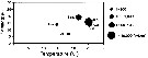 Issued from : H.Y. Soh, S.Y. Moon, E.O. Park & B.A.V. Maran in J. Crustacean Biol., 2013, 33 (5). [p.729, Fig.8]. Issued from : H.Y. Soh, S.Y. Moon, E.O. Park & B.A.V. Maran in J. Crustacean Biol., 2013, 33 (5). [p.729, Fig.8].
Temperature-salinity-abundance diagram for A. forticrusa.
Nota: The species occurrs in salinities ranging from 5 to 20 in the estuaries of Beolgyo Stream and Seomjin River (sothern Korea), between June and November. However, its peak abundance was in salinity of ca. 15 in June and ca.20 in September. When the water temperature is less than 20°C, it is replaced by A. hudsonica. Also A. forticrusa generally occurs together with A. ohtsukai and Tortanus dextrilobatus in the Seomjin River estuary (southern Korea). |
| | | | Loc: | | | China Seas ( South China Sea, East China Sea, Bohai Sea), S Korea (Seomjin River estuary, Beolgyo Stream estuary) | | | | N: | 1 | | | | Lg.: | | | (1145) F: 0,79-0,88; M:0,67-0,80; {F: 0,79-0,88; M: 0,67-0,80} | | | | Rem.: | For Soh & al. (2013, p.723) this species closely resembles A. southwelli and A. sarojus, but it differs from the latter two species (see Table 1, p.727). | | | Last update : 22/09/2014 | |
|
|
 Any use of this site for a publication will be mentioned with the following reference : Any use of this site for a publication will be mentioned with the following reference :
Razouls C., Desreumaux N., Kouwenberg J. and de Bovée F., 2005-2024. - Biodiversity of Marine Planktonic Copepods (morphology, geographical distribution and biological data). Sorbonne University, CNRS. Available at http://copepodes.obs-banyuls.fr/en [Accessed April 19, 2024] © copyright 2005-2024 Sorbonne University, CNRS
|
|
|
|


 forticrusa - Plate 1 of morphological figures',750,500);)
 forticrusa - Plate 2 of morphological figures',750,500);)
 forticrusa - Plate 3 of morphological figures',750,500);)
 forticrusa - Plate 7 of morphological figures',750,500);)
 forticrusa - Plate 8 of morphological figures',750,500);)

 forticrusa - Plate 1 of morphological figures)
 forticrusa - Plate 2 of morphological figures)
 forticrusa - Plate 3 of morphological figures)
 forticrusa - Plate 4 of morphological figures)
 forticrusa - Plate 5 of morphological figures)
 forticrusa - Plate 6 of morphological figures)
 forticrusa - Plate 7 of morphological figures)
 forticrusa - Plate 8 of morphological figures)
 forticrusa - Plate 9 of morphological figures)
 forticrusa - Distribution map 2)
