|
|
 |
|
Calanoida ( Order ) |
|
|
|
Clausocalanoidea ( Superfamily ) |
|
|
|
Clausocalanidae ( Family ) |
|
|
|
Clausocalanus ( Genus ) |
|
|
| |
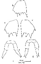
Clausocalanus farrani Sewell, 1929 (F,M) | |
| | | | | | | Syn.: | Clausocalanus arcuicornis minor Brodsky, 1962 c (part., p.116, figs.F); Grice, 1962 (p.189, pl.7, figs.5,6: F);
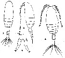
Clausocalanus pergens : Mori, 1937 (1964) (pl.14, figs.2-5); Fleminger, 1964 a; 1967 a (tab. I); Chen & Zhang, 1965 (p.49, figs.F); Kos, 1972 (Vol.1, figs. partim) | | | | Ref.: | | | Sewell, 1929 (p.94, Descr.F, figs.F); Farran, 1936 a (p.81, Rem.); Sewell, 1947 (p.55); Frost & Fleminger, 1968 (p.52, Descr.M, figs.F,M, Rem.); Silas, 1972 (p.646); ? Chen & Zhang, 1974 (p.103, 111, figs.M); Dawson & Knatz, 1980 (p.7, figs.F,M); Chahsavar-Archad & Razouls, 1982 (p.37, fig.F); van der Spoel & Heyman, 1983 (p.44, fig.56); Chihara & Murano, 1997 (p.776, Pl.89,94: F,M); Bucklin & al., 2003 (p.335, tab.2, fig.1, Biomol) | 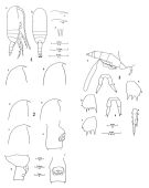 issued from : B. Frost & A. Fleminger in Bull. Scripps Inst. Oceanogr. Univ. California, San Diego, 1968, 12. [Pl.34, p.168-169; Pl.35, p.170-171; Pl.6, p.172-173]. Female: 1 a, habitus (right lateral view); 1 b, idem (dorsal view); 1 c-d, rostrum (right lateral); 1 e, rostrum (anteroventral); 1 f-g, frontal region (right lateral; 1 a-c taken from different specimens; 1 d-e from a fourth; 1 f and g from fifth and sixth specimens respectively; 2 a-c, frontal region (right lateral); 2 d, Th.3 (posterior part), Th.4-5 and genital segment (right lateral); 2 e, genital segment (right lateral); 2 f, genital segment (ventral view); 2 a-f taken from different specimens; 3 a, Th.4-5 (posterior part) and urosome with spermatophore attached; 3 b, B2 of P2; 3 c-d, B2 of P3; 3 e, Re3 and St of P3; 3 f-g, P5; 3 a from one specimen; 3 b,c,e from another; 3 d, f, g from third, fourth and fifth specimens, respectively.
|
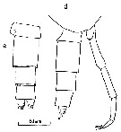
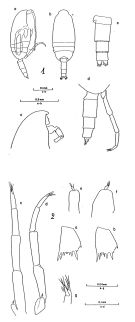 issued from : B. Frost & A. Fleminger in Bull. Scripps Inst. Oceanogr. Univ. California, San Diego, 1968, 12. [Pl.37, p.174-175; Pl.38, p.176-177]. Male: 1 a, habitus (right lateral view); 1 b, idem (dorsal view); 1 c, frontal region (right lateral); 1 d, Th.4-5 (posterior part) and urosome (right lateral); 1 e, urosome (dorsal view); 1 a-e taken from different specimens; 2 a, B2 of P2; 2 b, B2 of P3; 2 c, P5 (posterior); 2 d, P5 (right lateral); 2 e, right P5 (anterior); 2 f, right P5 (right lateral); 2 g, 4P5 (distal part) and 5P5 (posterior); 2 a-b taken from one spegimen; 2 c,e from another; 2 d,f,g from a third.
|
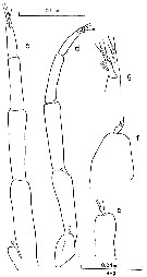
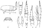 issued from : R.B.S. Sewell in Mem. Indian Mus., 1929, X. [p.94, Fig.38]. Female (from S India): a, habitus (lateral left side); b, urosome (ventral); c, genital opening; d, P2; e, P3; f, P4; g, P5; g', the end of distal segment of P5 (enlarged).
|
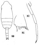 issued from : Q.-c Chen & S.-c. Zhang in Studia Marina Sinica, 1974, 9. [Pl.7, Figs.80-823]. Doubtful. Male (from South China Sea): 80, habitus (dorsal); 81, basipodite 2 of P2; 82, P5.
|
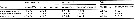
 issued from : B. Frost & A. Fleminger in Bull. Scripps Inst. Oceanogr. Univ. California, 1968, 12. [p.46, Table 3a]. Clausocalanus farrani Females: Measurements and ratios . TL = total body length ; SL = spermatophore length ; P :U = ratio of prosome length to urosome length ; U :U1 = ratio of total urosome length to length of 1st urosomal segment (genital segment) ; S :U1 = ratio of spermatophore sac length to U1 length of female on which spermatophore is attached ; r = sample range; m = sample mean; n = number of specimens measured; s = sample standard deviation.
|
 issued from : B. Frost & A. Fleminger in Bull. Scripps Inst. Oceanogr. Univ. California, 1968, 12. [p.47, Table 3b]. Clausocalanus farrani Males: Measurements and ratios . TL = total body length; P :U = ratio of prosome length to urosome length ; P:U2 = ratio of prosome length to length od 2nd urosomal segment; U2:2P5 = ratio of U2 length to length of 2nd segment of longer ramus of P5; U2:3P5 = ratio of U2 length of 3rd segment of longer ramus of P5; 3P5:2P5 = ratio of length of 3P5 of longer ramus to length of 2P5 of longer ramus; r = sample range; m = sample mean; n = number of specimens measured; s = sample standard deviation.
|
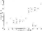 issued from : B. Frost & A. Fleminger in Bull. Scripps Inst. Oceanogr. Univ. California, 1968, 12. [p.25, Fig.8]. Ratio of length of A1 fused segments 20-21 to length of A1 segment 22 (ordinate) plotted against length of A1 fused segments 20-21 (abscissa) for males of Clausocalanus farrani and C. minor.
|
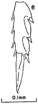 issued from : B. Frost & A. Fleminger in Bull. Scripps Inst. Oceanogr. Univ. California, 1968, 12. [p.173, Pl.36, e]. Female: e, exopodal segment 3 and terminal seta of P3.
|
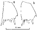
 issued from : B. Frost & A. Fleminger in Bull. Scripps Inst. Oceanogr. Univ. California, 1968, 12. [p.173, Pl.36, b, c-d, f-g]. Female: b, basipodite 2 of P2; c-d, basipodite 2 of P3, f-g, P5 (from different specimens).
|
 issued from : B. Frost & A. Fleminger in Bull. Scripps Inst. Oceanogr. Univ. California, 1968, 12. [p.175, Pl.37, d, e]. Male: d, posterior part of last thoracic segment and urosome (right lateral); e, urosome (dorsal).
|
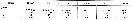
 issued from : B. Frost & A. Fleminger in Bull. Scripps Inst. Oceanogr. Univ. California, 1968, 12. [p.177, Pl.38, c-d, e-f, g]. Male: c, P5 (posterior); d, P5 (right lateral); e, right P5 (anterior); f, right P5 (right lateral); g, 4th segment of P5 (distal part) and 5th segment of P5 (posterior).
|
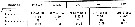
 issued from : B. Frost & A. Fleminger in Bull. Scripps Inst. Oceanogr. Univ. California, 1968, 12. [p.177, Pl.38, a, b]. Male: a, basipodite 2 of P2; b, basipodite 2 of P3.
|
 Issued from : T. Mori inThe pelagic Ciopepoda from the neighbouring waters of Japan, 1964 (1937). [Pl. 14, Figs. 2-5] As Clausocalanus pergens. After Frost & Fleminger, 1968 (p.52) : Syn. from Clausocalanus farrani. Female (from Japan ): 2, habitus (dorsal); 3, same (lateral); 5, P5. Male: 4, habitus (dorsal).
| | | | | Compl. Ref.: | | | Grice & Hulsemann, 1967 (p.14); Fleminger, 1967 a (tabl.1); Björnberg, 1973 (p.310, 385); Fleminger & Hulsemann, 1973 (p.340, carte); Timonin, 1976 (p.79, vertical distribution); Timonin & Voronina, 1977 (p.288, fig.6); Carter, 1977 (1978) (p.35); Star & Mullin, 1981 (p.1322, abundance); Smith S.L., 1982 (p.1331, abundance, monsoon effect); Almeida Prado Por, 1983 (p.141, tab.); Guangshan & Honglin, 1984 (p.118, tab.); Binet, 1984 (tab.3); 1985 (p.85, tab.3); Almeida Prado Por, 1985 (p.250); Brinton & al., 1986 (p.228, Table 1); Madhupratap & Haridas, 1986 (p.105, tab.1); Jimenez-Perez & Lara-Lara, 1988; Echelman & Fishelson, 1990 a (tab.2); Hirakawa & al., 1990 (tab.3); Yoo, 1991 (tab.1); Shih & Young, 1995 (p.72); Noda & al., 1998 (p.55, Table 3, occurrence); Suarez-Morales & Gasca, 1998 a (p109); Lavaniegos & Gonzalez-Navarro, 1999 (p.239, Appx.1); Hsiao & al., 2004 (p.325, tab.1); Lan & al., 2004 (p.332, tab.1); Lo & al., 2004 (p.89, tab.1); Kazmi, 2004 (p.229); Smith & Madhupratap, 2005 (p.214, tab.4); Cornils & al., 2007 (p.1261, feeding); Cornils & al., 2007 (p.57, Table I, fig.2, Rem: reproduction; abnormality); Valdés & al., 2007 (p.103: tab.1); Dur & al., 2007 (p.197, Table IV); McKinnon & al., 2008 (p.844: Tab.1, p.846: Tab.II,); Cabal & al., 2008 (289, Table 1); Lan Y.C. & al., 2008 (p.61, Table 1, % vs stations); C.-Y. Lee & al., 2009 (p.151, Tab.2); Lan Y.-C. & al., 2009 (p.1, Table 2, % vs hydrogaphic conditions); Cornils & al., 2010 (p.2076, Table 3); Hsiao S.H. & al., 2011 (p.475, Appendix I); Hsiao & al., 2011 (p.317, Table 2, indicator of seasonal change); Pillai H.U.K. & al., 2011 (p.239, Table 3, vertical distribution); Tutasi & al., 2011 (p.791, Table 1, 3, fig.9, abundance distribution vs La Niña event, Rem. p.797, fig.11);in CalCOFI regional list (MDO, Nov. 2013; M. Ohman, comm. pers.); Tachibana & al., 2013 (p.545, Table 1, seasonal change 2006-2008); Tseng & al., 2013 (p.507, seasonal abundance); Hirai & al., 2013 (p.1, Table I, molecular marker); Lidvanov & al., 2013 (p.290, Table 2, % composition); Zaafa & al., 2014 (p.67, Table I, occurrence); El Arraj & al., 2017 (p.272, table 2, spatial distribution); Palomares-Garcia & al., 2018 (p.178, Table 1: occurrence); | | | | NZ: | 13 | | |
|
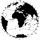
Distribution map of Clausocalanus farrani by geographical zones
|
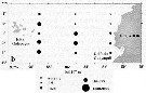
| | | | | | | | | | | | 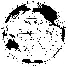 issued from : B. Frost & A. Fleminger in Bull. Scripps Inst. Oceanogr. Univ. California, San Diego, 1968, 12. [p.54, Chart 6, a]. issued from : B. Frost & A. Fleminger in Bull. Scripps Inst. Oceanogr. Univ. California, San Diego, 1968, 12. [p.54, Chart 6, a].
Occurrence of C. farrani in samples examined; closed circles represent samples examined; open circles represent samples in which adults were found; bars through open circles represent samples from which specimens were removed for measurements. |
 issued from : B. Frost & A. Fleminger in Bull. Scripps Inst. Oceanogr. Univ. California, San Diego, 1968, 12. [p.55, Chart 6, b]. issued from : B. Frost & A. Fleminger in Bull. Scripps Inst. Oceanogr. Univ. California, San Diego, 1968, 12. [p.55, Chart 6, b].
Occurrence of C. mastigophorus in samples examined; closed circles represent samples examined; open circles represent samples in which adults were found; bars through open circles represent samples from which specimens were removed for measurements. |
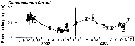
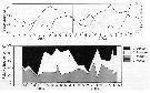 issued from : A. Cornils, B. Niehoff, C. Richter, T. Al-Najjar & B. Schnack-Schiel in J. Plankton Res., 2007, 29 (1). [p.60, Figs.1, 2]. issued from : A. Cornils, B. Niehoff, C. Richter, T. Al-Najjar & B. Schnack-Schiel in J. Plankton Res., 2007, 29 (1). [p.60, Figs.1, 2].
Above: Annual cycle of sea surface temperature and depth -integrated (0-100 m) chlorophyll a (mg/m2)
Below: Relative frequency (%) of the clausocalanid females over the investigation period (March 2002 to December 2003) in the northern Gulf of Aqaba (29°27'87 N, 34°57' 87 E; depth 300 m) for Clausocalanus farrani, C. furcatus, C. minor and Ctenocalanus vanus.
Nota: Collected by vertical hauls from 100 m depth to the surface. Sampling performed between 9 a.m. and 3 p.m. on a monthly basis. |
 issued from : A. Cornils, B. Niehoff, C. Richter, T. Al-Najjar & B. Schnack-Schiel in J. Plankton Res., 2007, 29 (1). [p.65, Fig.8]. issued from : A. Cornils, B. Niehoff, C. Richter, T. Al-Najjar & B. Schnack-Schiel in J. Plankton Res., 2007, 29 (1). [p.65, Fig.8].
Prosome length (mm) displayed with SE bars over the investigation period. |
 issued from : A. Cornils, B. Niehoff, C. Richter, T. Al-Najjar & B. Schnack-Schiel in J. Plankton Res., 2007, 29 (1). [p.65, Table III]. issued from : A. Cornils, B. Niehoff, C. Richter, T. Al-Najjar & B. Schnack-Schiel in J. Plankton Res., 2007, 29 (1). [p.65, Table III].
Percentage of clausocalanid females infected with Blastodinium sp. (dinoflagellate) or showing abnormalities of the P5 (%), found in the preserved females. |
 Issued from : P. Tutasi, S. Palma & M. Caceres in Scienc. Mar., 2011, 75 (4). [p.799, Fig.9 b] Issued from : P. Tutasi, S. Palma & M. Caceres in Scienc. Mar., 2011, 75 (4). [p.799, Fig.9 b]
Geographic distribution of Clausocalanus farrani in September and October 2001, associated with the weak La Niña event of 2001. |
| | | | Loc: | | | South Africa (E) (Natal), off Cape Verde Is. (in Chasavar-Archad & Razouls, 1982), Morocco-Mauritania, off W Tangier (in Zaafa & al., 2014), off Amazon, E Medit. (Lebanon coast)*, Red Sea (G. of Aqaba), Somalia, Arabian Sea, Indian, Andaman Sea (Barren Island) Australia (North West Cape), S Burma, SW Celebes, China Seas (Yellow Sea, East China Sea, South China Sea), Taiwan Strait, Taiwan (SW, E, NW, NE, N, Mienhua Canyon), Japan, Kuchinoerabu Is., Tokyo Bay, off S Baja California, G. of California, La Paz, W Mexico, Hawaii, Pacif. (E & W equatorial), Pacif. (N Central Gyre), Australia (Great Barrier), New Caledonia, Pacif. (sub-tropical), Chile.
* according to Almeida Prado Por, 1983 (C.R.: original document not seen). | | | | N: | 67 | | | | Lg.: | | | (29) F: 1,08-0,87; (30) F:1,2-1,04; M: 0,99-0,87; (34) F: 1,22-1,09; (109) M: 0,85-0,65; (290) F: 1,2-1; (338) M: 0,7-0,65; {F: 0,87-1,22; M: 0,65-0,99}
The mean female size is 1.088 mm (n = 8; SD = 0.1196) and the mean male size is 0.785 mm (n = 6; SD = 0.1394). The size ratio (male : female) is establish on one sample with males and females: 0.83. | | | | Rem.: | For Frost & Fleminger (1968, p.53) the ventral protuberance of the genital segment anterior to the genital pores distinguishes females of C. farrani from all other Clausocalanus species except C. jobei; the latter differs from farrani only in the shape, curvature, and length of the rostrum. Females of C. farrani differ from minor in the form of the P5, and from paululus in body length, in the form and spacing of basipodite 2 spiniform processes 2 and 3 of P3. The C. farrani male differs from jobei in the armature of the distal segment of left P5 and the ratio of left 2nd segment of P5 length to urosomal segment 2 width. The very similar males of C. farrani and minor are distinguished by the length ratio of A1 fused segments 20-21 to segment 22 and usually by the segmentation and appearance of the right P5, particularly the size of the distal segment. Males of C. farrani and paululus are easily separated by body length, the urosomal segment 2/3rd segment of P5 and 3rd segment of P5/ 2nd segment of P5 length ratios, and the form of basipodite 2 spiniform processes 2 and 3 of P3. The authors have not located designated types or original material for C. farrani, but Sewell(s (1929) description and illustration are sufficient to leave little doubt as to the identity of his material.
After Cornils & al (2007, p.63) the percentage of females infected by Blastodinium sp. was 1.9 %, ±0.5); with impact on P5 (abnormal), the left ramus has developed one or two additional segments.
The Atlantic locality records need a confirmation although Calef & Grice (1967) have reported the species off Guyane. | | | Last update : 25/10/2022 | |
| | | | My first time to find several C. farrani (1-1.3 mm) in the SW of El Hierro island (Canary archipelago; Latitude = 27º45.87 N; Longitude=018º10.80 W)in April 2014 in the epipelagic strata (0-200m depth) at mid day | |
|
|
|
 Any use of this site for a publication will be mentioned with the following reference : Any use of this site for a publication will be mentioned with the following reference :
Razouls C., Desreumaux N., Kouwenberg J. and de Bovée F., 2005-2024. - Biodiversity of Marine Planktonic Copepods (morphology, geographical distribution and biological data). Sorbonne University, CNRS. Available at http://copepodes.obs-banyuls.fr/en [Accessed April 24, 2024] © copyright 2005-2024 Sorbonne University, CNRS
|
|
|
|


;)
;)
;)
;)
;)
;)
;)
;)
;)
;)
;)

;)
;)
;)
;)
