|
|
 |
Fiche d'espèce de Copépode |
|
|
Calanoida ( Ordre ) |
|
|
|
Diaptomoidea ( Superfamille ) |
|
|
|
Pontellidae ( Famille ) |
|
|
|
Labidocera ( Genre ) |
|
|
| |
Labidocera johnsoni Fleminger, 1964 (F,M) | |
| | | | | | | Ref.: | | | Fleminger, 1964 (p.2295; in Contribution 1772, vol.34, part II, Scripps Inst. Oceanogr., San Diego; Pilot Register of Zoology, card n° 3A: Descr.F,M, figs.F,M); 1975 (p.401 & suiv.) | 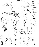 issued from : A. Fleminger in Pilot Register of Zoology, card n°. 3-A, 1964. [Fig.1]. Female (Baja California): a, posterior portion of body (right lateral); b, head (lateral); c, urisome with spermatophore (right lateral); d, habitus (dorsal); e, last thoracic segment and urosome (dorsal); f, same with spermatophore (dorsal); g, right Md (cutting edge of gnathobase); h, genital area; i, genital segment (right ventro-lateral view); j, P5 (lateral; atypical specimen); k, P5 (posterior); l, P5 (lateral view). Copepodite female stage V: m, P5 (posterior); n, P5 (posterior; from unusually large: 2.33 mm). Nota: Urosome 3-segmented; genital segment longest, symmetrical, lacking genital pore; caudal rami about as long as wide Figs. a-j, n taken from northern Gulf of california specimens; figure m taken from southern Gulf of California specimen; figs. g, k and l taken from coastal Pacific specimens.
|
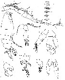 issued from : A. Fleminger in Pilot Register of Zoology, card n°. 3-A, 1964. [Fig.2]. Male: a, right A1 (dorsal view); b, head (lateral); c, posterior portion of body (right lateral); d, habitus (dorsal); e, P5 (posterior); f, chela of right P5 (dorso-lateral view; another specimen); g, last thoracic segment with P5 and urosome (dorsal); h, left P5 (medial view). Copepodite male stage V: i, P5 (posterior). Nota: Right A1 with middle segments weakly tumid, 6th segment from apex lacking spiniform process; urosome with 4 subequal segments; caudal rami about equal in length to urosomal segment 3
| | | | | Ref. compl.: | | | Silas & Pillai, 1973 (1976) (p.774); Brinton & al., 1986 (p.228, fig.11: spatial distribution, Table 1); Hernandez -Trujillo, 1989 (tab.1); 1989 a (tab.1); He, 1994 (tab.1); Suarez-Morales & Gasca, 1998 a (p.110); Hernandez-Trujillo & al., 2010 (p.913, Table 2); Lavaniegos & al., 2012 (p. 11, Appendix); in CalCOFI regional list (MDO, Nov. 2013; M. Ohman, comm. pers.); Hernandez-Trujillo & al., 2013 (p.303, weight vs length); Beltran Castro, 2014 (p.44, Table 1, 2: molecular CO1); Jerez-Guerrero & al., 2017 (p.1046, Table 1: temporal occurrence); Palomares-Garcia & al., 2018 (p.178, Table 1: occurrence)
| | | | NZ: | 2 | | |
|
Carte de distribution de Labidocera johnsoni par zones géographiques
|
| | | | Loc: | | | W Baja California (Bahia Magdalena), Gulf of California, Bahia de La Paz, Bahia de los Angeles, W Mexico (Pacif.), Bahia Cupica (Colombia) | | | | N: | 13 | | | | Lg.: | | | (470) F: 3,26-2,21; M: 3,01-2,12; {F: 2,21-3,26; M: 2,12-3,01} | | | | Rem.: | Coastal.
''Labidocera trispinosa'' Group. (in Brinton & al., 1986).
Diagnosis for L. johnsoni in the 'darwinii' species group after Prusova & Al-Yamani (2014, p.1165) :
Females;
- 1: Urosome of 3 somites.
- 2: Genital somite asymmetrical, caudal rami of approximately equal length.
- 3: Caudal rami symmetrical, or roughly symmetrical, separate from anal somite.
- 4: Exopod of nearly symmetrical P5 with 2 or 3 points, genital pore ventro-lateral
- 5: P5 exopod with 3 terminal points, middle one the largest, a fourth point sometimes present.
Males:
- 1: Urosome of 5 somites.
- 2: Prosome posterior corners asymmetrical, right corner longer or modified.
- 3: Prosome posterior corners strongly asymmetrical, with 3 prominent spiniform prongs on right, lateral-most the largest.
- 4: Lateral-most prosomal prong not curved in lateral view.
- 5: Dorsal-most prosomal prong about 1/2 length of lateral-most prong; P5 exopodal segment 1 with relatively wide rounded process medially. | | | Dernière mise à jour : 13/11/2020 | |
|
|
 Toute utilisation de ce site pour une publication sera mentionnée avec la référence suivante : Toute utilisation de ce site pour une publication sera mentionnée avec la référence suivante :
Razouls C., Desreumaux N., Kouwenberg J. et de Bovée F., 2005-2024. - Biodiversité des Copépodes planctoniques marins (morphologie, répartition géographique et données biologiques). Sorbonne Université, CNRS. Disponible sur http://copepodes.obs-banyuls.fr [Accédé le 24 avril 2024] © copyright 2005-2024 Sorbonne Université, CNRS
|
|
|
|


;)
;)
