|
|
 |
Fiche d'espèce de Copépode |
|
|
Calanoida ( Ordre ) |
|
|
|
Diaptomoidea ( Superfamille ) |
|
|
|
Pontellidae ( Famille ) |
|
|
|
Pontellina ( Genre ) |
|
|
| |
Pontellina morii Fleminger & Hulsemann, 1974 (F,M) | |
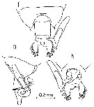
| | | | | | | Syn.: | Pontellina plumata : Mori, 1937 (1964) (p.100, part., Pl;48, figs.1-12); Dakin & Colefax, 1940 (p.99, figs) | | | | Ref.: | | | Fleminger & Hulsemann, 1974 (p.79, figs.F,M, fig.12); Silas & Pillai, 1973 (1976) (p.851, figs.M, Rem.); Hulsemann & Fleminger, 1975 (p.176: figs juv.V F, M; F, M, Rem.); Greenwood, 1979 (p.105, figs.F,M); Hulsemann & Fleminger, 1990 (p.99, 105, figs.F, pores cuticulaires); Ohtsuka & Onbé, 1991 (p.214); Chihara & Murano, 1997 (p.873, Pl.161: F,M); Bradford-Grieve, 1999 b (p.204, figs.F,M, figs.186, 194); Mulyadi, 2002 (p.155, figs.F,M, Rem.); Phukham, 2008 (p.100, figs.F,M) | 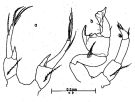 issued from : J.G. Greenwood in Proc. R. Soc. Qd, 1979, 90. [p.106, Fig.7]. Female (from Moreton Bay, E Australia): a, P5. Male: b, P5.
|
 issued from : E.G. Silas & P.P. Pillai in J. mar. biol. Ass. India, 1973 (1976), 15 (2). [p.849, Fig.35, c-f]. Male (from Indian Ocean): c, urosome (dorsal); d, Md (masticatory edge); e, right A1 (geniculate part); f, P5. Scale as in Calanopia minor.
|
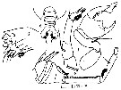
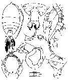
 issued from : A. Fleminger & K. Hulsemann in Fishery bulletin, 1974, 72 (1). [p.80, Fig.9]. Female (from different area): a, Th 4-5 and genital segment (lateral right side); b, habitus (lateral right side; other specimen); c, variation in Th 4-5 (lateral view); d, habiyus (dorsal); e, Th 4-5 and urosome (dorsal view; same as a); f, variation in Th 4-5 (dorsal view); g, P5 (anterior view); h, variation observed in lateral margin of right caudal ramus (dorsal view); i, endopod of P5 in other specimens (first two: right side; three to four: : left side). Nota: In contrast to junction of spine and Th4-5 corner relativelyabrupt in both dorsal and lateral views; spine roughly 1/2 as long as that in its congener from the eastern equatorial Pacific. Genital segment with posterolateral cluster of coarse hairs on both sides, lacking anterolateral cluster found in P. plumata although several fine hairs may occur at this site, posterior margin of segment bordered by fine, long hairs as in P. plumata. Right caudal ramus considerably shorter than in P. plumata relative to prosome, with median ratio of length to width 1.25:1 (range 1.12-1.44:1); lateral edge with small point variable in shape just anterior to base of outemost seta, glandular tissue within ramus as in P. plumata. P5 with exopod bearing hairs along median margin; endopod relatively longer than that in P. plumata; exopod being less than 1.8 times longer than endopod, median 1.45:1, range 1.22-1.76:1; endopod typically with 2 relatively equal apical spines.
|
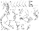 issued from : A. Fleminger & K. Hulsemann in Fishery bulletin, 1974, 72 (1). [p.82, Fig.11]. Male: a, right A1 (dorsal view); b, variation in Th 4-5 spine (lateral view; from different area); c, Th 4-5, P5 and first two segments of urosome (lateral right side); d, P5 (posterior view; same as a); e, P5 chela (lateral view; same as c); f, apex of distal segment of P5 enlarged (same as c); g, aberrant chela showing a weakened subapical spur on distal segment (posterior view); h, aberrant chela (lateral view; same as g). Nota: Th 4-5 ending posteriorly in a small spiniform process similar to female. P5 with chela of plumata-type but both segments showing distinctive features; distal segment short, not reaching opposing disto-lateral digitiform process on proximal segment; Left P5 with exopod longer than that in P. plumata. Right caudal ramus differing from that in P. plumata in having a relatively shorter length, median length-to-width ratio 1.93:1, range 1.80-2.07:1, but overlapping extensively with its congener from the eastern equatorial Pacific. Left P5 with edopod 1 considerably longer than that in P. plumata
|
 issued from : A. Fleminger & K. Hulsemann in Fishery bulletin, 1974, 72 (1). [p.101, Fig.33, f-h]. Female: Th 4-5 and urosome with attached spermatophore (dorsal, lateral and ventral, respectively).
|
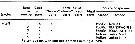 issued from : A. Fleminger & K. Hulsemann in Fishery bulletin, 1974, 72 (1). [p.111, Table 19]. List of identified particles from microscopic analysis of stomach contents in adulte female.
|
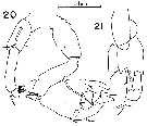 issued from : K. Hulsemann & A. Fleminger in Bull. Mar. Sci., 1975, 25 (2). [p.181, Figs.20-21]. Male: 20, P5 (posterior view); 21, chela in lateral view). Striation on process in fig.21 is assumed to function as friction pad.
|
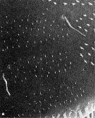
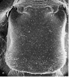 issued from : K. Hulsemann & A. Fleminger in Mar. Biol., 1990, 105. [p.101, Fig.1, c]. Female: c, genital segment (dorsal view). Nota: Scanning electron microscope examination revealed on the genital segment many patches of small spinules ornamenting the integument dorsally and laterally. The spinules are flattened, triangular, non-articulated, rigid structures rising from the integument, usually pointing obliquely posteriad.
|
 issued from : K. Hulsemann & A. Fleminger in Mar. Biol., 1990, 105. [p.101, Fig.2, c]. Female: c, genital segment (dorsal view). Nota: The spinules are arranged in polygonal patches. Within a patch they are of similar size and tend to be arranged in rows.
|
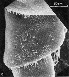
 issued from : K. Hulsemann & A. Fleminger in Mar. Biol., 1990, 105. [p.103, Fig.3, a]. Patches of epicuticular spinules, two peg sensilla and pore of integumental gland in smooth area between patches on genital segment of female (dorsal view), 1800 x)
|
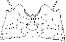 issued from : K. Hulsemann & A. Fleminger in Mar. Biol., 1990, 105. [p.104, Fig.4]. Female: genital segment (dorsal, lateral and ventral patterns separated by dotted line). Integument cut dorsally and laid out flat, showing positions of integumental organs: peg sensilla (o) and pores of integumental glands (filled circle); symbols are larger than organs.
|
 issued from : K. Hulsemann & A. Fleminger in Mar. Biol., 1990, 105. [p.104, Fig.5-7]. Female: genital segment (left to right: dorsal view, right latera viewl and ventral view).Generalized distribution of peg sensilla (o) and pores of integumental glands (filled circle); symbols are larger than organs. Organs shown occurred in at least 40 % of specimens examined (n = 25).
|
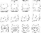 issued from : K. Hulsemann & A. Fleminger in Mar. Biol., 1990, 105. [p.105, Fig.8]. Female: genital segment (left to right: dorsal view, right latera viewl and ventral view) illustrating geographical variation of pore signature pattern; idealized view of integumental organ types, numbers and arrangment. Organs shown are those appearing in at least 40 % of specimens from one regional sample. . Top: dorsal view; middle: right lateral view; bottom: ventral view. Left: types of organs inferred from several intact specimens stained in chlorazole black E and cleared in lactophenol. Symbols: peg sensilla (o); pores of integumental glands (filled circle); symbols are larger than organs. Symbols used for regional samples: (black circle) organs present in 100 % of specimens examined; (clear circle) 80 to 99 %; (black triangle) 60 to 79 %; (white triangle) 40 to 59 %.
|
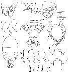
 issued from : Mulyadi in Treubia, 2002, 32. [p.156, Fig.58]. Female (from Celebes Sea): a, habitus (dorsal); b, metasomal somite 5 and urosome (dorsal); c, metasomal somite 5 and genital complex (right lateral); d, P5. Male: e, metasomal somite 5 with P5 and urosomal somites 1-2 (right lateral); f, right A1; g, P5; h, chela of right P5 (lateral); i, apex of distal segment of P5.
|
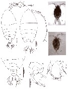 issued from : N. Phukham in Species diversity of calanoid copepods in Thai waters, Andaman Sea (Master of Science, Univ. Bangkok). 2008. [p.184, Fig.58]. Female (from W Malay Peninsula): a, habitus (dorsal); b, P5. Male: c, habitus (dorsal); d, urosome (dorsal); e, P5. Body length after the drawings: F = 1.564 mm; M = 1.283 mm.
| | | | | Ref. compl.: | | | Madhupratap & Haridas, 1986 (p.105, tab.1); Chen Y.-Q., 1986 (p.205, Table 1: abundance %, Table 2: vertical distribution); Othman & al., 1990 (p.561, 564, Table 1); Padmavati & al., 1998 (p.349); Hsiao & al., 2004 (p.326, tab.1); Dur & al., 2007 (p.197, Table IV); Jitlang & al., 2008 (p.65, Table 1);Hsiao S.H. & al., 2011 (p.475, Appendix I) | | | | NZ: | 8 | | |
|
Carte de distribution de Pontellina morii par zones géographiques
|
| | | | | | | | | 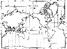 issued from : A. Fleminger & K. Hulsemann in Fishery bulletin, 1974, 72 (1). [p.83, Fig.12]. issued from : A. Fleminger & K. Hulsemann in Fishery bulletin, 1974, 72 (1). [p.83, Fig.12].
Geographical distribution of captures recorded during the present study. |
| | | | Loc: | | | E South Africa, Arabian Sea, Indian, Bay of Bengal, W Malay Peninsula (Andaman Sea), Indonesia-Malaysia (NE Celebes: Manado Bay), Philippines, China Seas (South China Sea), Taiwan (SW & E), Japan, Australia (G. of Carpentaria, Great Barrier Reef, Moreton Bay, New South Wales), Pacif. (tropical), off Baja California, off Peru
Type locality: Varuna (western Pacific) | | | | N: | 14 | | | | Lg.: | | | (256) M: 1,6-1,28; (275) F: 1,88-1,38; M: 1,68-1,26; (1087) F: 1,65-1,70; M: 1,5-1,55; {F: 1,38-1,88; M: 1,26-1,68} | | | | Rem.: | épipélagique.
Voir aussi les remarques en anglais | | | Dernière mise à jour : 08/12/2020 | |
|
|
 Toute utilisation de ce site pour une publication sera mentionnée avec la référence suivante : Toute utilisation de ce site pour une publication sera mentionnée avec la référence suivante :
Razouls C., Desreumaux N., Kouwenberg J. et de Bovée F., 2005-2024. - Biodiversité des Copépodes planctoniques marins (morphologie, répartition géographique et données biologiques). Sorbonne Université, CNRS. Disponible sur http://copepodes.obs-banyuls.fr [Accédé le 18 avril 2024] © copyright 2005-2024 Sorbonne Université, CNRS
|
|
|
|


;)
;)
;)
;)
;)
;)
;)
;)
;)
;)
;)
;)
;)

;)
