|
|
 |
Fiche d'espèce de Copépode |
|
|
Calanoida ( Ordre ) |
|
|
|
Clausocalanoidea ( Superfamille ) |
|
|
|
Scolecitrichidae ( Famille ) |
|
|
|
Pseudoamallothrix ( Genre ) |
|
|
| |
Pseudoamallothrix laminata (Farran, 1926) (F,M) | |
| | | | | | | Syn.: | Scolecithrix laminata Farran, 1926 (p.265, Descr.F, figs.F); Bradford, 1973 (p.145, Rem.); Bradford & al., 1983 (p.103);
Scolecithricella lamellifer Tanaka, 1962 (p.78, figs.F);
Scolecithricella laminata : Grice & Hulsemann, 1965 (p.224, 239, figs.F); 1967 (p.16); Park, 1970 (p.476); Roe, 1972 (p.277, tabl.1, tabl.2); 1975 (p.320, figs.F,M, Rem.); Vives, 1982 (p.292); Lozano Soldevilla & al., 1988 (p.59); Markhaseva & Kobosokova, 1998 (p.51, figs.F); Kosobokova & Hirche, 2000 (p.2029, tab.2); Holmes, 2001 (p.59); Vives & Shmeleva, 2007 (p.795, figs.F, Rem.);
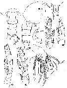
? Amallothrix profunda Brodsky, 1950 (1967) (p.263, figs.M). | | | | Ref.: | | | Vyshkvartzeva, 1999 (2000) (p.228, Rem.); Vyshkvartzeva, 2003 (p.43: Rem.) | 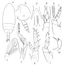 issued from : O. Tanaka in Publ. Seto Mar. Biol. Lab., 1962, X (1). [p.79, Fig.146]. As Scolecithricella lamellifer. Female (Izu region): a, dorsal); b, forehead (left lateral side); c, last thoracic segment and urosome (left lateral side); d, rostrum (dorsal view); e, Mx2; f, Mxp; g, P1; h, P2; i, proximal part of terminal spine of exopod of P2; j, P3; k, proximal part of terminal spine of exopod of P3; l, P4; m, P5. Nota Female: - Cephalothorax about 3.8-4 times the length of abdomen. - Head and 1st pediger segment fused, 4th and 5th pedigers incompletely fused, line of fusion faintly visible both from the dorsal and lateral sapects. - Posterolateral corners of the last thoracic segment broadly rounded, but slightly produced at the distal margin in lateral aspect. - Forehead slightly produced in dorsal aspect. - Rostrum composed of a short basal portion to which strong spines attached, length of spine about 3 times the depth of excavation. - Abdominal segments and caudal rami in the proportional lengths 33 : 19 : 23 : 9 : 16 = 100. - Genital segment sinuate ventrally. - Abdominal segments 1-3 striated with fine teeth on the distal border. - Caudal rami 1.5 times as long as wide. - A1 23-segmented on left side, extends to the end of the 3rd abdominal segment. - A2 exopod 1.7 times as long as endopod; 1st exopodal segment with a knob on anterior margin about the middle of the segment (asin Amallothrix indica Sewell (= Pseudoamallothrix indica). - Mx1 with 9 setae on the outer lobe; 9 setae on exopod; 5 setae on exopod, 5 setae on endopodal segments 3-2 and 3 setae on segment 1; 5 setae on the 2nd basal; 3 setae on the 3rd inner lobe; 2 setae on the 2nd inner lobe. - Mx2 with 6 long vermiform filaments and 2 small bud-like on endopod. - Mxp with a small amalliform (bud-like), and 2 long vermiform filaments on the 1st basal segment. - P1 with the outer edge short spine on the 1st exopodal segment, not attaining to the middle of the outer margin of the 2nd segment; there is a group of minute spinules on the inner distal corner of the 1st exopodal segment.. - P2 coxa with a ridge on the outer margin; inner marginal seta located on the well marked projection; inner margin of basis sinuate and furnished with short hairs on the distal half; posterior surface of basal segments densely provided with small spinules. Outer edge spine on 1st exopodal segment short and straight, about 1/2 the length of the outer margin of the 2nd segment. Terminal spine on exopod with about 40 teeth; the teeth not connected with a sort of lamella. The posterior surface of exopod densely furnished with minute spinules. Endopod provided with rather large spinules on the posterior surface. - P3 coxa has also a ridge on the outer margin; basis with a small process and 2 spinules on inner margin; coxa and basis furnished with minute spinules on posterior surface; terminal spine of exopod with 42 teeth (and they make as fenestellae, fig.k). - P4 coxa ha a transparent swelling, oval in shape, on the inner distal margin; inner marginal setae short: basis with a ridge runing distally from the distal angle of the coxa. Coxa and basis furnished with minute spinules; terminal spine of exopod with about 43 teeth which form fenestellae. - P5 2-segmented; distal segment dilated distally, and with a short apical spine and a inner marginal spine which is 4 times as long as the apical one, these two spines closely set. Apical spine finely toothed on either side. The lamellous swelling on the inner margin of the coxa of P4 is very characteristic in the species
|
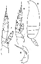
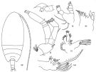 issued from : G.D. Grice & K. Hulsemann in J. Zool., 1965, 146. [p.240, Fig.14, g-m]. As Scolecithricella laminata. Female (from 40°03.5'N, 19°57'W): g, habitus (dorsal); h, forehead (ventral); i, A2; j, Md (mandibular palp); k, Md (cutting edge); l, Mx1; m, Mx2. Nota: The specimens show spines on the 1st exopod segment of P2 (no spines are shown on this segment in Farran's figure); a conspicuous lamella on the inner margin of the 1st basipod segment of P4; the terminal segment of P5 elongate. The smaller specimen (1.63 mm) was similar to the larger specimens in the first two characteristics, but the terminal segment of P5 was less elongate. A smaller specimen (1.63 mm) collected from 32°29'N and 20°09'W was similar to the larger two specimens (above, 2.33 and 2.36 mm) but the terminal segment of P5 was less elongate. Tanaka (1962) used the aforementioned 3 characters to distinguish S. lamellifer from Farran's S. laminata stating that: no spines are present on the 1st exopodal segment of P2 in S. laminata; the lamellous swelling in S. lamellifer is pronounced; the terminal segment of P5 is relatively short compared to its width S. lamellifer. Conclusion: Comparing the 3 characteristics used for separating the two species it appears that S. lamellifer is not specifically distinct from S. laminata.
|
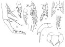 issued from : G.D. Grice & K. Hulsemann in J. Zool., 1965, 146. [p.241, Fig.15, a-j]. As Scolecithricella laminata. Female : a, Mxp; b, P1 (anterior); c, idem (posterior); d, P2; e, details of teeth on terminal spine of P2; f, P3 (2nd and 3rd exopod segments missing); g, basipod segments of P4 (posterior); h, idem (anterior); i, P5 (of 2.33 mm specimen); j, idem (of 1.63 mm specimen).
|
 Issued from : K.A. Brodskii in Calanoida of the Far Eastern Seas and Polar Basin of the USSR. Opred. Fauna SSSR, 1950, 35 (Israel Program for Scientific Translations, Jerusalem, 1967) [p.264, Fig.172]. As Amallothrix profunda. With doubt. Male (from NW Pacif.): habitus (lateral right side); R, rostrum; S5, P5. Remark: The 4th urosomal segment is short comparatively with the one from Roe's male.
|
 issued from : E.L. Markhaseva & K.N. Kosobokova in Zoosyst. Ross., 1998, 7 (1). [p.52, Figs.38-47]. As Scolecithricella laminata. Female (from 81°12'N, 150°14'E): 38-39, pediger segment 5 and urosome (dorsal and lateral, respectively); 40, forehead (lateral); 41, rostrum (frontal view); 42, Mx2; 43, P1; 44, P2; 45, P3; 46, P4; 47, P5. Scale bars: 0.1 mm.
|
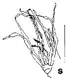
 issued from : G.P. Farran in Biscayan Plankton collected during a Cruise of H.M.S. 'Research', 1900.- Part XIV. The Copepoda. (Linn. Journ. Zoology, XXXVI, 1926). [p.304, Pl.8, Figs.5-8]. As Scolecithrix laminata. Female: 5, habitus (lateral); 6, P2; 7, P3; 8, P5.
|
 issued from : H.S.J. Roe in Bull. British Mus. (Nat. Hist.) Zool., London, 1975, 28 (7). [p.312, Fig.9s]. As Scolecithricella laminata. Female (from NE Atlantic): s, endopod of Mx2. Bar scale 0.1 mm unless indicated. Nota: - Mx1 has 5 large and 2 small setae on the 1st outer lobe, 8 setae on the exopod, 5 on the fused 2nd and 3rd endopodite segment, 2 on the 1st endopodite segment, 6 on the 2nd basipodite, 4 on the 3rd inner lobe, 2 on the 2nd inner lobe and 13 on the 1st inner lobe (of which 4 are on the surface as opposed to the edge). - Mx2 has 8 sensory setae on the endopodite of which only 2 are short and brush-like and 6 are long and worm-like.
|
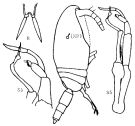
 issued from : H.S.J. Roe in Bull. British Mus. (Nat. Hist.) Zool., London, 1975, 28 (7). [p.312, Fig.9t-v]. As Scolecithricella laminata. Male: t, habitus (dorsal); u, P5; v, 3rd exopodite segment of left P5. Bar scale 0.1 mm unless indicated. Nota: - Cephalothorax 2.3 times as long as the 5-segmented abdomen. - Head and 1st thoracic segment fused, 4th and 5th fused. - Rostrum with2 stout filaments. - The hind margin of the cephalothorax is smoothly rounded. A1 19-segmented, reaches to the end of the caudal rami. - The remaining mouthparts are generally slightly smaller and more weakly chitinized than in the female; their setation patterns are identical to those of the female except: The basipodite of the mandibular palp has no setae; in Mx1 the 1st outer lobe has 6 large and 2 small setae, the exopodite has 9 and the 2nd basipodite has 5 setae; the endopodite of Mx2 is as in the female with 6 long filaments and 2 shorter brush-like setae. - P5 asymmetrical; in the right leg the exopodite has 3 segments, the last of which is roughly triangular in shape; in the left leg the exopodite is much shorter than the 2-segmented endopodite; the 3rd exopodite segment has brunches of fine hairs and 2 large setae. Remarks: Brodskys (1950) description of Amallothrux profunda is incomplete but the stucture of P5, especially the short exopodite of the left leg, is very similar to that of the present specimens.
|
 issued from : O. Tanaka in Publ. Seto Mar. Biol. Lab., 1962, X (1). [p.80]. As Scolecithricella lamellifer. Female: Proportional lengths of segments A1 on the left side. Nota: A1 23-segmented on the left side; segment 10 without seta; segments 12, 14 and 18 with each 2 setae; segments 7, 9, 12, 14, 19 each with a long aesthetasc.
| | | | | Ref. compl.: | | | Kuriyama & Nishida, 2006 (p.299: Tab.II; fig.7, 10, vertical distribution); Kosobokova & al., 2011 (p.29, Table 2, Rem.: Makarov Basin). | | | | NZ: | 7 | | |
|
Carte de distribution de Pseudoamallothrix laminata par zones géographiques
|
| | | | | | | Loc: | | | Cape Verde Is., Canary Is., N Madeira, Haïti, off NE Cape Hatteras, Caribbean Sea, Bay of Biscay, off W Ireland, W Indian, Japan (Izu region, Sagami Bay), Arct. (Makarov Basin) | | | | N: | 13 | | | | Lg.: | | | (8) F: 2,28-1,82; M: 2,43-2,2; ? (22) M: 2,6; (38) F: 2,3; (117) F: 2,4; (226) F: 2,36; 2,33; 1,63; (199) F: 2,28-2,2; M: 2,43; (779) F: 3-2,85; (1000) F: 1,8 ±0,2; {F: 1,60-3,00; M: 2,20-2,43} | | | | Rem.: | bathypélagique, hyperbenthique.
Voir aussi les remarques en anglais | | | Dernière mise à jour : 19/01/2021 | |
|
|
 Toute utilisation de ce site pour une publication sera mentionnée avec la référence suivante : Toute utilisation de ce site pour une publication sera mentionnée avec la référence suivante :
Razouls C., Desreumaux N., Kouwenberg J. et de Bovée F., 2005-2024. - Biodiversité des Copépodes planctoniques marins (morphologie, répartition géographique et données biologiques). Sorbonne Université, CNRS. Disponible sur http://copepodes.obs-banyuls.fr [Accédé le 25 avril 2024] © copyright 2005-2024 Sorbonne Université, CNRS
|
|
|
|


;)
;)
;)
;)
;)
;)
;)
;)

