|
|
 |
Fiche d'espèce de Copépode |
|
|
Calanoida ( Ordre ) |
|
|
|
Clausocalanoidea ( Superfamille ) |
|
|
|
Scolecitrichidae ( Famille ) |
|
|
|
Scaphocalanus ( Genre ) |
|
|
| |
Scaphocalanus longifurca (Giesbrecht, 1888) (F,M) | |
| | | | | | | Syn.: | Scolecithrix longifurca Giesbrecht, 1888; 1892 (p.266, 285, 775, figs.F); Giesbrecht & Schmeil, 1898 (p.45, Rem. F);
Scolecithricella longifurca : A. Scott, 1909 (p.90, Rem.F); Sewell, 1948 (p.553, 562);
? Scaphocalanus insignis Brodsky, 1950 (1967) (p.255); Vives, 1982 (p.292);
? Scaphocalanus temporalis (M) Tanaka, 1953 (p.132); 1961a (p.177, Rem.);
no S. glacialis (M) Tanaka, 1953 (p.132);
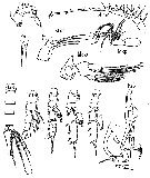
? Scolecithricella vittata (M n°4): Rose, 1942 (p.159, figs.M) | | | | Ref.: | | | Farran, 1929 (p.249, Rem.); Tanaka, 1960 (p.45: Rem.); 1961 a (p.177, figs.F,M, Rem.); Grice, 1962 (p.213, figs.F,M, Rem.); Vervoort, 1965 (p.64, 65: Rem.); Park, 1970 (p.507, figs.F, Rem.); Bradford, 1973 (p.143); Park, 1982 (p.77); Bradford & al., 1983 (p.98, figs.F,M, Rem.) | 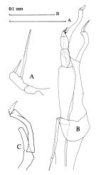 Issued from : J.M. Bradford, L. Haakonssen & J.B. Jillett in Mem. N. Z. oceanogr. Inst., 1983, 90. [p.99, Fig.58]. Female: A, P5. Male: B, P5; C, terminal part of exopod segment 1 and segment 2 of right P5. Nota Female: - Posterolateral corners of last thoracic segment triangularly produced, rounded at apex. - Genital segment produced ventrally, as long as following two urosomal segments. - A1 22-segmented. - P2 exopod segment 2 with curved spine half as long as exopodal segment 2. - Posterior surfaces of basal segments of P2-P4 covered with small triangular teeth. - P5 with 2 distal segments fused, carrying 2 to 3 spines; terminal spine half as long as distal segment; inner edge spine 3 times as long as terminal spine and finely serrated on each side. Nota Male: - Posterolateral corners of last thoracic segment rounded and not produced. - A1 18-segmented, extends to distal end of prosome. - P1 exopod without spine. - P2 exopodal segment 2 with small straight spine. - Left P5 endopodal segment 1 swollen at middle, segment 2 very small; exopod short, 3-segmented, last segment short, covered with hairs and some setae, 2 of which are long. Right P5 endopod does not reach distal margin of exopodal segment 1, which is greatly expanded terminally.
|
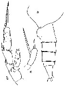
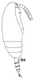 Issued from : T. S. Park in Bull. Mar. Sc., 1970, 20 (2). [p.506, Fig.154]. Female (from Caribbean Sea & G. of Mexico): 154, habitus (lateral right side). Nota: This form is similar to Scaphocalanus major (T. Scott, 1894) in the armature of P2 to P4, and the structure of P5, but can be distinguished from it by the outline of the posterolateral corner of the metasome.
|
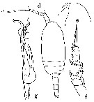
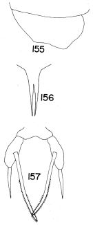 Issued from : T. S. Park in Bull. Mar. Sc., 1970, 20 (2). [p.508, Figs.155-157]. Female: 155, posterior part of metasome (lateral right side); 156, rostrum (anterior view); 157, P5.
|
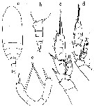
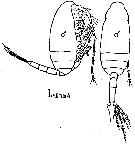 issued from : M. Rose in Annls Inst. océanogr., Monaco, 1942, XXI (3). [p.160, Fig.53]. As Mâle n°4. Male (from Alger bay, Algeria): habitus (lateral and dorsal, respectively). L: Body length = 1.4 mm.
|
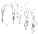
 issued from : M. Rose in Annls Inst. océanogr., Monaco, 1942, XXI (3). [p.161, Figs.54, 55]. As Mâle n°4. Male: Ur, urosome (dorsal); P1 to P5.
|
 issued from : O. Tanaka in Publ. Seto Mar. Biol. Lab., 1961, IX (1). [p.178, Fig.121, a-c]. Female (from Japan: Izu Region): a, last thoracic segment and urosome (lateral, left side); b, P2; c, P5. Nota : Cephalothorax and urosome in proportional lenths 78 :22. Last thoracic segment with lateral distal corner triangularly produced, but narrowly rounded at the apex. Urosomal segment and caudal rami in proportional lengths 37 :20 :17 :9 :18 = 100. Caudal rami 2 times as long as wide. Endopod of A2 1.25 times as long as the exopod. Terminal spine of the exopod with 24 teeth.. Basal segment of P3 coarsely covered with small triangular spines on the anterior surface. P5 2-segmented ; Distal segment delated at the distal part ; terminal spine rather short, about half the length of the distal segment ; inner marginal spine about 3 times as long as the terminal one, and finely serrated one each side of the spine.
|
 issued from : O. Tanaka in Publ. Seto Mar. Biol. Lab., 1961, IX (1). [p.178, d-g]. With doubt. Male (from Izu Region): d, habitus (dorsal); e, forehead (lateral); f, P2; g, P5. Nota : Cephalothorax and abdomen in proportional lengths 69 :31. Head slightly produced anteriorly in dorsal aspect, and obtusely rounded in lateral aspect. Lateral distal corner of the last thoracic segment rounded. Rostrum strong, gradually attenuates to a fine point. Urosomal segments and caudal rami in proportional lengths 13 :28 :23 :23 :2 :12 = 100. Caudal rami slightly divergent. A1 19-segmented on the left side, 18-segmented on the right side (proportional lengths of segments of the right side 65 :98 :47 :33 :33 :22 :33 :142 :38 :33 :49 :49 :49 :49 :65 :33 :77 :83 = 1000) , extend to the posterior margin of the 3rd thoracic segment . Exopod of A2 slightly longer than the endopod (22 :19). Terminal spine of exopod of P2 with about 30 teeth. P5 extends to the distal margin of the 3rd urosomal segment ; endopod of left leg swollen at the middle section ; exopod 3-segmented, short ; endopod of the right leg long, reaches the distal margin of the 1st segment of the exopod of the left leg. Rem. : This male resembles closely S. subbrevicornis except : A1 short, only reaching the distal margin of the thoracic segment, and the segments have the proportional lengths which differ from those found in S. subbrevicornis. Distal segment of exopod of P5 furnished with only 3 stiff hairs on the inner margin, whereas, it is furnished in S. subbrevicornis with, beside 2 strong spines, fine hairs on the inner margin. For Tanaka the present male has been listed in 1953 under S. glacialis iand S. temporalis
|
 issued from : O. Tanaka in Publ. Seto Mar. Biol. Lab., 1961, IX (1). [p.180, Fig.122]. S. longifurca, with doubt. Female (from Izu Region): a, habitus (dorsal); b, last thoracic segment and urosome (lateral, left side); c, P2; d, P2; e, P5. Nota: This specimen measuring 1.76 mm had P5 furnished with a small outer marginal spine near the base of the terminal spine. The specimen has a straight outer edge spine on the 1st segment of the exopod of P2, and the terminal spine of the exopod is more finely serrated on the outer margin. Cephalothorax and urosome in proportional lengths 95:41. Urosomal segments and caudal rami in proportional lengths 7:38:23:23:2:7 = 100. A1 19-segmented on the left (proportional lengths of segments 79:107:51:28:32:23:28:144:37:28:46:42:46:46:28:41:42:69:83 = 1000), 18-segmented on the right side, extend to the end of the 3rd thoracic segment. The author had a doubt concerning the identification as longifurca
|
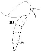 Issued from : W. Giesbrecht in Systematik und Faunistik der Pelagischen Copepoden des Golfes von Neapel und der angrenzenden Meeres-Abschnitte. Fauna Flora Golf. Neapel, 1892. Atlas von 54 Tafeln. [Taf. 37, Fig. 16]. As Scolecithrix longifurca. Female: last thoracic segment and urosome (lateral).
|
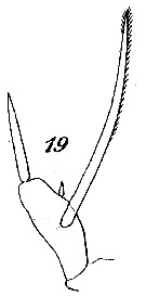 Issued from : W. Giesbrecht in Systematik und Faunistik der Pelagischen Copepoden des Golfes von Neapel und der angrenzenden Meeres-Abschnitte. Fauna Flora Golf. Neapel, 1892. Atlas von 54 Tafeln. [Taf. 13, Fig.19 ]. As Scolecithrix longifurca. Female: 19, P5.
|
 issued from : G.D. Grice in Fish. Bull. Fish and Wildl. Ser., 1962, 61. [p.212, Pl.19, Figs.8-11]. Female (from equatorial Pacific): 8, posterior part of thorax and urosome (lateral, right side); 9, P5. Male: 10, P5; 11, left P5.
| | | | | Ref. compl.: | | | Furuhashi, 1966 a (p.295, vertical distribution in Oyashio/Kuroshio transitional area, Table 7, 8, 9, 10); Grice & Hulsemann, 1965 (p.224); 1967 (p.16); Björnberg, 1973 (p.331, 389); Harding, 1974 (p.141, tab. 3, gut contents); Guangshan & Honglin, 1984 (p.118, tab.); Wiebe & al., 1988 (tab.7); Gopalakrishnan & Balachandran, 1992 (p.167, figs.1, 7, Table 1, 2); Shih & Young, 1995 (p.73); Padmavati & al., 1998 (p.347); Lo & al., 2004 (p.89, tab.1); Kazmi, 2004 (p.229); Kuriyama & Nishida, 2006 (p.300: Tab.II; p.309: Tab.III, fig.7, 10, vertical distribution); Fernandes, 2008 (p.465, Tabl.2); C.-Y. Lee & al., 2009 (p.151, Tab.2); Sano & al., 2013 (p.11, Table 9, food habits) . | | | | NZ: | 11 + 2 douteuses | | |
|
Carte de distribution de Scaphocalanus longifurca par zones géographiques
|
| | | | | | | | | | | | | Loc: | | | NE Atlant. (30-60°N), ? Canary Is., off Amazon, Caribbean Sea, G. of Mexico, off N Bermuda, ? Medit., Indian (Arabian Sea, W Indian, Bay of Bengal, 25°N - 10°S (in Gopalakrishnan & Balachandran, 1992), Indonesia-Malaysia, China Seas (East China Sea, South China Sea), Taiwan (N: Mienhua Canyon, NW), Japan, Sagami Bay, ? NW Pacif., Guaymas Basin, Pacif. (W equatorial), New Zealand, Pacif. equatorial, N Peru, Chile | | | | N: | 26 | | | | Lg.: | | | (9) F: 1,3; 1,25; M: 1,35-1,2; (47) F: 1,75; (101) F: 1,94-1,87; M: 1,53; (108) F: 1,67-1,48; M: 1,35-1,3; ? (147) M: 1,9-1,7; (1000) F: 1,8 ± 0,2; {F: 1,25-2,00; M: 1,20-1,53} | | | | Rem.: | mésopélagique. In vertical tow 4000-3000 m (Harding, 1974, Rem.: possible contaminant species from higher up the water column).
Distributional range (m) from Grice & Hulsemann (1965): 200-1000 (in Kuritama & Nishida, 2006).
L'identification du mâle a souvent provoqué des confusions.
? Cf. Scaphocalanus subbrevicornis.
Voir aussi les remarques en anglais | | | Dernière mise à jour : 19/01/2021 | |
|
|
 Toute utilisation de ce site pour une publication sera mentionnée avec la référence suivante : Toute utilisation de ce site pour une publication sera mentionnée avec la référence suivante :
Razouls C., Desreumaux N., Kouwenberg J. et de Bovée F., 2005-2024. - Biodiversité des Copépodes planctoniques marins (morphologie, répartition géographique et données biologiques). Sorbonne Université, CNRS. Disponible sur http://copepodes.obs-banyuls.fr [Accédé le 25 avril 2024] © copyright 2005-2024 Sorbonne Université, CNRS
|
|
|
|


;)
;)
;)
;)
;)
;)
;)
;)
;)
;)
;)

