|
|
 |
Fiche d'espèce de Copépode |
|
|
Calanoida ( Ordre ) |
|
|
|
Clausocalanoidea ( Superfamille ) |
|
|
|
Scolecitrichidae ( Famille ) |
|
|
|
Scottocalanus ( Genre ) |
|
|
| |
Scottocalanus securifrons (T. Scott, 1894) (F,M) | |
| | | | | | | Syn.: | Scolecithrix securifrons T. Scott, 1894 b (p.47, figs.F, no M); Giesbrecht & Schmeil, 1898 (p.49, Rem.F); ? Cleve, 1904 a (p.197); van Breemen, 1908 a (p.76);
Lophothrix securifrons : Wolfenden, 1904 (p.120, figs.F); 1911 (p.268);
Scottocalanus acutus Sars, 1905 c (p.7, Rem.F);
Amallophora securifrons : Wolfenden, 1904 (p.145);
Scolecithrix cuneifrons Willey, 1918 (1919) (p.194, figs.F,M); Tremblay & Anderson, 1984 (p.6); Scottocalanus securifer : De Decker, 1968 (p.45);
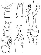
Scottocalanus securiformis: Sameoto & Lewis, 1980; ? Scottcalanus securifrons : Tanaka, 1937 (p.259, figs.F) | | | | Ref.: | | | Farran, 1908 b (p.57, Rem.); A. Scott, 1909 (p.104, figs.F,M); With, 1915 (p.220, figs.F,M); Lysholm & Nordgaard, 1921 (p.21); Sars, 1925 (p.160, figs.F,M); Farran, 1926 (p.267, Rem.); 1929 (p.209, 251); Rose, 1929 (p.26); 1933 a (p.144, figs.F,M); Jespersen, 1940 (p.36); Lysholm & al., 1945 (p.26); Sewell, 1947 (p.143); Brodsky, 1950 (1967) (p.243, figs.F,M); Marques, 1953 (p.103); 1959 (p.211); Tanaka, 1961 a (p.141, Rem.F,M, figs.M); Grice, 1962 (p.213, figs.F); Paiva, 1963 (p.45, figs.F); Vervoort, 1965 (p.36, Rem.); Owre & Foyo, 1967 (p.63, figs.F,M, Rem.: p.65); Ramirez, 1969 (p.66, figs.F,M, Rem.); Tanaka, 1969 (p.275); Gopalakrishnan, 1974 (p.274, figs.F,M, Rem.); Björnberg & al., 1981 (p.637, figs.F); Park, 1983 (p.197, 200, Redescr. F, M, figs.F,M); Bradford & al., 1983 (p.116, figs.F,M, Rem.); Roe, 1984 (p.357); Campaner, 1989 (p.229, figs.F,M, Rem.); Schnack, 1989 (p.137, tab.1, fig.6: Md); Nishida & al., 1991 (p.527, midgut structure, feeding); He & al., 1992 (p.250); Chihara & Murano, 1997 (p.902, Pl.177: F,M); Mulyadi, 1998 a (p.389, Redescr.M, figs.M); Bradford-Grieve & al., 1999 (p.881, 931, figs.F,M); Boxshall & Halsey, 2004 (p.189, figs.F); Mulyadi, 2004 (p.31, figs.M, Rem.); Vives & Shmeleva, 2007 (p.811, figs.F,M, Rem.) | 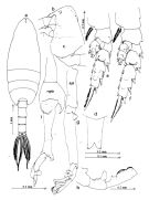 issued from : Mulyadi in The Raffles Bull. Zool., 1998, 46 (2). [p.390, Fig.6]. Male (from 07°29'S, 121°05'E): a, habitus (dorsal); b, forehead (left lateral side); c, last thoracic segment and genital somite (left lateral side); d, rostrum (frontal view); e, P2; f, P3; g, left P5; h, distal segment of right P5; i, right P5.
|
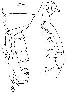
 Issued from : J.M. Bradford, L. Haakonssen & J.B. Jillett in Mem. N.Z. oceanogr. Inst., 1983, 90. [p.118, Fig.73]. Female: A, habitus (lateral right side); B, genital segment (dorsal); C, genital segment and P5 (lateral right side); D, rostrum; E, endopod of P2; F, P5. Male: G, habitus (lateral left side); H, P5. Female: - Posterolateral corners of the last thoracic segment produced posteriorly into triangular expansion ending in sharp point. - Each ramus of rostrum ending in short point. - Genital segment swollen ventrally at mid-length, ventral posterior margin overlaps following segment, posterolateral margins of genital segment with spines. - A1 23-segmented, reaches end of caudal rami. - P5 slightly asymmetrical, with subterminal spine slightly thicker on left. Male: - Posterolateral corners of last thoracic segment ending in small spine - A1 20-segmented. - Right P5 endopod very short; exopod segment 1 long, widened terminally and bearing several processes; segment 2 short, curved with terminal expansion; segment 3 short, rounded with single spine. Left P5 basipod 2 with proximal rounded process; endopod 2-segmented, segment 1 short with distal process, segment 2 with bifurcate tip; exopod segment 1 short, segment 2 enlarged distally, with inner margin densely spinulated, and distal laminous process divided into 2 spines, segment 3 with few proximal teeth and longer pointed spine-like part. Remarks: The south-west Pacific specimen shows some variation in the shape of the posterior metasome borders in females. Brodsky (1950) suggests that specimens which have straight posterior metasome points (see Tanaka, 1937) are a separate species. The south-west Pacific specimens have posterior metasome points which are directed straight back, unlike those illustrated by Sars (1925), which diverge. The male P5 appears to not have as many articulated segments in some branches as described by With (1915). The right P5 exopod appears to be 2-segmented; the terminal segment bears a pair of characteristic semi-circular processes on the distal half of the segment. The left P5 endopod does not seem to have a moveable articulation and bears 1 pointed and 1 semi-circular process at about mid-length.
|
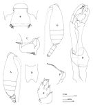
 issued from : T. Park in Antarct. Res. Ser. Washington, 1983, 38 (3). [p.201, Fig.19]. Female: a, habitus (dorsal); b, idem (lateral left side); c, forehead (lateral; d, rostrum (anterior); e, urosome (ventral): f, last thoracic segments and urosome (lateral left side); g, forehead (dorsal); h, A2; i, Md. Nota: Cephalosome and 1st matasomal segment fused, indicated laterally by a fine line, 4th and 5th separate on dorsal side. Urosome about 20/100 length of prosome. Spermatheca with long, somewhat inflated vesicle.
|
 issued from : T. Park in Antarct. Res. Ser. Washington, 1983, 38 (3). [p.202, Fig.20]. Female: a, Mx1; b, distal part of Mx2; c, Mxp; d, P1 (posterior); e, P2 (posterior); f, P3 (posterior); g, P4 (posterior); h, P5 (anterior).
|
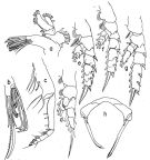
 issued from : T. Park in Antarct. Res. Ser. Washington, 1983, 38 (3). [p.203, Fig.21]. Male: a, forehead (lateral); b, last thoracic segments and urosome (lateral left side); c, forehead (dorsal); d, last thoracic segments and urosome (dorsal); e, rostrum (anterior); f, A2; g, Md; h, Mx1; i, Mx2. Nota: Urosome about 30/100 length of prosome. A1 reaching close to distal end of 3rd urosomal segment.
|
 issued from : T. Park in Antarct. Res. Ser. Washington, 1983, 38 (3). [p.204, Fig.22]. Male: a, Mxp; b, P1 (anterior); c, P2 (posterior); d, P3 (posterior); e, P4 (posterior); f, P5 (anterior); g, P5 (posterior); h, distal part of right P5 exopod; i, left P5 (medial); j, distal part of left P5 exopod (posterior).
|
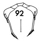 issued from : F.C. Ramirez in Contr. Inst. Biol. mar., Buenos Aires, 1969, 98. [p.64, Lam. XII, figs.92 ]. Female (from off Mar del Plata): 92, P5 Scale bar in mm: 0.05.
|
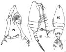 issued from : F.C. Ramirez in Contr. Inst. Biol. mar., Buenos Aires, 1969, 98. [p.62, Lam. XI, figs.79, 81, 82 ]. Female (from off Mar del Plata): 82, habitus (dorsal). Male: 79, P5; 81, habitus (lateral right side). Scale bars in mm: 2 ( (81, 82);
|
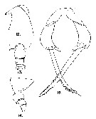
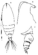
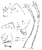
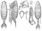 Issued from : G.O. Sars in Résult. Camp. Scient. Prince Albert I, 69, pls.1-127 (1924). [Pl.XLV, figs.1-8]. Female: 1, habitus (dorsal); 2, idem (lateral left side); 3, forehead (lateral); 4, rostrum (frontal view); 5, P5; 6, P5 (lateral view). Male: 7, habitus (dorsal); 8, P5. :
Nota: Female: - Forehead very sallent in dorsal view and with crest. - Rostrum with points slightly divergent. - Posterior angles of the last thoracic segment salient and ending in points. - Urosome short, about ¼ length of prosome. - Genital segment swollen ventrally, and pilose on the posterior edge. - A1 reaching about the middle of the genital segment. - P5 with terminal segment stretched bearing an apical relatively strong spine lesser long than in S. persecans in Sars, (1924-1925). Male : - Posterior angles of the last thoracic segment ending in acute process.. - P5 build as in S. persecans with difference in details. In securifrons the inner ramus of the right leg is reduced like a little rudiment, whereas the oneo of the left le gis well developed and with a complex structure. The apical claw on the right le gis different from S. persecans (see figures).
|
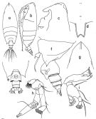
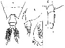
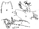
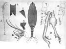 issued from : A. Scott in Siboga-Expedition, 1909, XIX a. [Plate XXVIII, Figs.1-9]. Male (from Indonesia-Malaysia): 1, habitus (dorsal); 2, forehead (lateral); 3, last thoracic and genital segments (left side); 4, rostrum; 5, A1; 6, Mx2 (distal portion); 7, P2; 8, P4; 9, P5.
|
 issued from : A. Scott in Siboga-Expedition, 1909, XIX a. [Plate XXV, Figs.1-9]. Female: 1, habitus (dorsal); 2, forehead (lateral); 3, last thoracic and genital segments (left side); 4, rostrum; 5, A1; 6, Mx2 (distal portion); 7, P2; 8, P4; 9, P5.
|
 issued from : S. Ohtsuka & R. Huys in Hydrobiologia, 2001, 453/454. [p.452, Fig.7, F]. Male (from Japan): F, terminal exopodal segment of left P5. Scale bar = 0.2 mm.
|
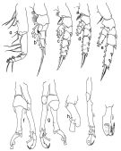
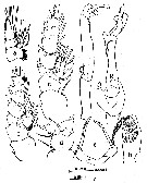
 issued from : O. Tanaka in Publ. Seto Mar. Biol. Lab., 1961, IX (1). [p.142, Fig.106]. As Scottcalanus securifrons. Male: a, last thoracic segment (lateral, right side); b, rostrum (frontal view); c, P2 (posterior view); d, P5; e, distal segments of right P5. Male: - Last thoracic segment terminates one each side into a small spine at the apex. - Urosme 5-segmented; abdominal segments and caudal rami in the proportional lengths 15 : 29 : 21 : 19 : 6 : 10 = 100; 2nd to 4th segments stiated with fine teeth on the distal margin. A1 extends to the distal end of the 3rd urosomal segment (segments 8-9, 20-21 fused on the left side. - Mouth parts and swimming legs as those of the female. - P5 as figured by A. Scott (1909), but the small teeth on the inner margin of the proximal segment of the endopod of the left leg not seen. Female: - Head and pedigerous segment 1 fused, 4th and 5th segments fused. - Head with a high median crest. - Last thoracic segment produced posteriorly into triangular expansions with a sharp point at the apex. Distal lateral margin of the segment not so outwardly curved as figured by A. Scott or by Sars. - Rostrum bifurcate, with a very minute spine at the apex. - Urosome 4-segmented; proportional lengths of the segments and caudal rami 64 : 14 : 5 : 3 : 14 = 100. - Abdominal segments 2 to 4 striated with fine teeth on the distal margin. - Genital segment swollen distally in dorsal aspect; ventral surface swollen at the middle; distal ventral margin of the segment overlaps the following segment; the lateral distal margin of the segment is furnished with rows of spinules on either side, but absent on the distal dorsal and ventral surfaces. - Distal margin of the 2 nd and 3rd abdominal segments are furnished with denticles. - Anal segment very short, almost concealed beneath the preceding. - Caudal rami about as long as wide , furnished with ordinary 4 setae and a small seta. - A1 extends to the distal margin of the caudal rami. - A2 exopod about 1.4 times as long as the endopod (61 : 43). - Md exopod longer than endopod (15 : 12); cutting blade with 8 teeth. - Mx1 with 9 setae on the outer lobe; 8 setae on the exopod; 7 setae on the 2 nd basal; 3 setae on the 3rd inner lobe; 2 setae on the 2 nd inner lobe; 12 setae on the 1st inner lobe (= praecoxal arthrite). - Mx2 has on the endopod 2 small bud-like , and 6 long vermiform filaments; the 5th lobe has a long spine, a ordinary 1 seta, and 2 vermiform filaments; the 4th, 3rd and 2 nd lobe has each 3 setae; the 1st lobe has 4 setae. - Mxp with 3 sensory filaments, of which 1 is short and bud-like, on the 1st basal segment. - P1 3-segmented exopod and 1-segmented endopod, the outer edge spine of the segments of the exopod is fairly long; the distal outer margin of the 1st segment ends in a broadly rounded process. - P2 exopod 3-segmented, endopod 2-segmented; 1st basal segment with a small spine on the outer margin about the middle of segment; 2 nd basal segment with a sharp spine on the inner distal corner; 2 nd and 3rd segments of the exopod furnished with coronas of spinules on the posterior surface ; the 2 nd segment of endopod with several scattened long spinules on the posterior surface; the anterior surface of the 2 nd segment of the endopod with 2 spinules along the mid-line of the segment. - P3 exopod and endopod 3-segmented each. The 2 nd and 3 rd segments of the endopod furnished with spinules on the posterior and anterior surfaces. Terminal spine of the exopod with 27 serrations. - P4 has no marginal spine on the 1st basal segment. The 2 nd and 3rd segments of the endopod have each small spines on the anterior surfaces. - P5 asymmetrical. Subapical spine of the left leg thicker than that of the right. The subapical spine has 2 rows of denticles which diminish in size distally. The apical spine is about 1/3 the length of the 2 nd segment.
|
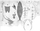
 issued from : G.D. Grice in Fish. Bull. Fish and Wildl. Ser., 1962, 61. [p.212, Pl.19, Figs.12-15]. Female (from 00°03'S, 157°00'E): 12, forehead (lateral); 13, posterior part of thorax and urosome (dorsal); 14, same (lateral, left side); 15, P5.
|
 issued from : C. With in The Danish Ingolf-Expedition, Copepoda I, 1915, III, 4. [p.220, Text-fig. 71]. Female (from 51°32'N, 12°03'W): a, last thoracic segment and urosome (dorsal); b, same (left lateral); c, genital segment (half right); d, segments 8-10 of A1.
|
 issued from : C. With in The Danish Ingolf-Expedition, Copepoda I, 1915, III, 4. [p.221, Text-fig. 72]. Male: a, last thoracic segment and urosome (dorsal); b, segments VII-XIV of A1; c, right P5; exopodal segments 2, 3 of right P5 (right view); e, two first segments of left P5 (dorsal view).
|
 issued from : C. With in The Danish Ingolf-Expedition, Copepoda I, 1915, III, 4. [Pl. VIII, Fig. 13]. Male: last two thoracic segments with P5 and urosome (left lateral); b, left P5; c, same (distal segments).
|
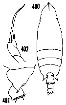 issued from : H.B. Owre & M. Foyo in Fauna Caribaea, 1, Crustacea, 1: Copepoda. Copepods of the Florida Current. [p.62, Figs.400-402]. Female (from 23°31'-25°40'N, 82°32'-79°43'W): 400, habitus (dorsal); 401, last thoracic segment and genital segment (left lateral); 402, P5.
|
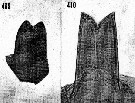 issued from : H.B. Owre & M. Foyo in Fauna Caribaea, 1, Crustacea, 1: Copepoda. Copepods of the Florida Current. [p.63, Figs.409-410]. Female: 409, rostrum. Male: 410, rostrum. Nota: Terminal projections of the bifurcated rostrum minute.
|
 issued from : H.B. Owre & M. Foyo in Fauna Caribaea, 1, Crustacea, 1: Copepoda. Copepods of the Florida Current. [p.65]. Female: Proportional lengths of the segments on A1.
|
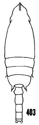 issued from : H.B. Owre & M. Foyo in Fauna Caribaea, 1, Crustacea, 1: Copepoda. Copepods of the Florida Current. [p.62, Fig.403]. Male: habitus (dorsal).
|
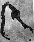 issued from : H.B. Owre & M. Foyo in Fauna Caribaea, 1, Crustacea, 1: Copepoda. Copepods of the Florida Current. 1967. [p.23, Fig.98]. Male: 98, P5.
|
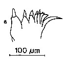 issued from : S.B. Schnack in Crustacean Issue, 1989, 6. [p.143, Fig.6: 8]. 8, Scottocalanus securifrons (from off NW Africa, upwelling region): Cutting edge of Md.
|
 Issued from : O. Tanaka in Japanese J. Zool., 1937, VII, 13. [p.259, Fig.9]. As Scottcalanus securifrons. Female (from coast of Heda, Japan): a, habitus (dorsal); b, forehead (lateral); c, abdomen (lateral). Nota: After Tanaka, the specimen (one female from 4.00 mm length) agrees quite well with Scott's description, except any details. Cephalothorax: 3.28 mm; abdomen: 0.72 mm. Remarks: the posterior end of the last thoracic segment does not curve outwardly as Scott's figure; the length of the cephalothorax is more than 4 times of the combined length of the urosome; the genital segment is longer than the combined length of the following 3 segments. For Brodsky (1950 [1967, p.244]) this specimen differs from S. securifrons, unfortunately the P5 is not shown.
|
 issued from : O. Tanaka in Publ. Seto Mar. Biol. Lab., 1961, IX (1). [p.141]. As Scottcalanus securifrons. Female (from Izu region): Proportional lengths of segments of A1.
|
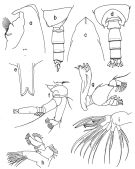
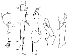
 Issued from : T.C. Gopalakrishnan in Proc. Biol. Soc. Washington, 1974, 87 (28). [p.275, Fig.1]. Female (from 03°29'N, 77°54'E): a, A1; c, forehead (lateral); d, last thoracic segment and genital segment; e, genital segment (enlarged). Male: b, A1; f, forehead (lateral); g, last thoracic segment and urosomites 1 and 2. Nota Female: head and 1st pedigerous somite fused, 4th and 5th fused -Last thoracic segment produced posteriorly into triangular expansion terminating in sharp pointed spine on either side. - Forehead with high median crest. - Rostrum bifid at tip. - Urosome 4-segmented. - Genital segment swollen ventrally at mid-length; its ventral posterior margin overlapping the following segment; posterolateral margins furnished with spines which are absent on dorsal and ventral side. (A Scott 1909 shows spines on the posterior margin , present on the dorsal as well as lateral surfaces). - Posterior margins of 2nd and 3rd abdominal segments with a hyaline fringe. - Anal segment very short. - Caudal rami almost as wide as long, each with 4 setae. - A1 with 23 free segments (when the partly separated 8th and 9th segments are counted as one). Nota Male: General appearance similar to that of female. - Forehead with high median crest. - Last thoracic segment terminating in small spine on either side. - Urosome 5-segmented. - Posterior margin of 2nd to 4th abdominal segments with hyaline fringe. - A1 with 20 segments when fused 8th to 12th segments, partly divided by 2 incomplete sutures between segments 8 and 9 and 11 and 12, are counted as one.
|
 Issued from : T.C. Gopalakrishnan in Proc. Biol. Soc. Washington, 1974, 87 (28). [p.276, Fig.2]. Female: a, rostrum; b, Mx1; c, distal portion of Mx2. Nota: Mx1 with nimbers of setae on different lobes as follow: inner lobe 1 (arthrite) with 12 setae of which 3 are on posterior surface; inner lobe 2 (coxal endite) with 2 setae; inner lobe 3 (basal endite) with 3 setae; basipod 2 (basis) with 5 setae; endopod segment 1 with 3 setae, endopod segments 2 and 3 together with 4 setae; exopod with 8 setae; outer lobe with 9 setae. Exopod segment with fine surface hairs at distal end. - Mx2 endopod with 4 bud-like and 3 vermiform elements<
|
 Issued from : T.C. Gopalakrishnan in Proc. Biol. Soc. Washington, 1974, 87 (28). [p.277, Fig.3]. Female: a, P1; b, P2; c, P3; d, proximal portion of P4; e, P5. Male: f, left P5; g, right P5; h, left P5 (2nd segment of exopodite, enlarged).
Nota Female: P5 asymmetrical; subapical spine of left leg thicker than that of right leg . Subapical spines with 2 rows of teeth.
Nota: Male: The small teeth on the inner margin of the proximal segment of the endopod of the left leg figuresd by A. Scott are not present in Tanaka' specimens, Willey's specimens or in the International Indian Ocean Expedition (1960-65, IIOE) specimens.
| | | | | Ref. compl.: | | | Pearson, 1906 (p.19, Rem.); Sewell, 1948 (p.329, 502, 516, 519, 521, 530, 532, 546); C.B. Wilson, 1950 (p.340); King & Hida, 1955 (p.11); Østvedt, 1955 (p.15: Table 3, p.69); Wickstead, 1962 (p.550, food & feeding); Grice, 1963 a (p.495); Unterüberbacher, 1964 (p.24); De Decker & Mombeck, 1964 (p.14); Furuhashi, 1966 a (p.295, vertical distribution in Kuroshio region, Table 10); Fleminger, 1967 a (tabl.1); Vinogradov, 1968 (1970) (p.258); Morris, 1970 (p.2301); Park, 1970 (p.476); Roe, 1972 (p.277, tabl.1, tabl.2); 1972 b (p.530); Bainbridge, 1972 (p.61, Appendix Table III: occurrence); Vives & al., 1975 (p.43, tab.II, XII); Deevey & Brooks, 1977 (p.256, tab.2, Station "S"); Carter, 1977 (1978) (p.35); Dessier, 1979 (p.205); Vives, 1982 (p.292); Cummings, 1984 (p.163, Table 2); Guangshan & Honglin, 1984 (p.118, tab.); Brenning, 1984 (p.4, Rem.); Tremblay & Anderson, 1984 (p.6, Rem.); Stephen, 1984 (p.161, 169, Distribution vs thermocline & geographic); Brenning, 1985 a (p.28, Table 2); Madhupratap & Haridas, 1986 (p.105, tab.1); Lozano Soldevilla & al., 1988 (p.59); Heinrich, 1990 (p.17); Suarez & Gasca, 1991 (tab.2); Hattori, 1991 (tab.1, Appendix, comme secrifrons); Hernandez-Trujillo, 1991 (1993) (tab.I); Suarez, 1992 (App.1); Gopalakrishnan & Balachandran, 1992 (p.167, figs.1, 6, Table 1, 2); Yen & al., 1992 (p.495, tab.1, mechanoreception); Webber & Roff, 1995 (tab.1); Shih & Young, 1995 (p.74); Kotani & al., 1996 (tab.2); Suarez-Morales & Gasca, 1998 a (p.111); Neumann-Leitao & al., 1999 (p.153, tab.2); Lapernat, 2000 (tabl.3, 4); Razouls & al., 2000 (p.343, Appendix); Lopez-Salgado & al., 2000 (tab.1); Pakhomov & al., 2000 (p.1663, Table 2, transect Cape Town-SANAE antarctic base); d'Elbée, 2001 (tabl. 1); Holmes, 2001 (p.60); Yamaguchi & al., 2002 (p.1007, tab.1); Sameoto & al., 2002 (p.13); Hsiao & al., 2004 (p.326, tab.1); Lo & al., 2004 (p.89, tab.1); Hwang & al., 2007 (p.25); Kuriyama & Nishida, 2006 (p.293, 300: Tab.II, fig.7, 10, vertical distribution, Rem.: p.313); Ikeda & al., 2006 (p.1791, Table 2); Dur & al., 2007 (p.197, Table IV); Neumann-Leitao & al., 2008 (p.799: Tab.II, fig.6); Galbraith, 2009 (pers. comm.); Park & Ferrari, 2009 (p.143, Table 5, Appendix 1, biogeography); Dias & al., 2010 (p.230, Table 1); Medellin-Mora & Navas S., 2010 (p.265, Tab. 2); Hsiao S.H. & al., 2011 (p.475, Appendix I); Teuber & al., 2013 (p.28, Table 1, respiration rates); Teuber & al., 2013 (p.1, Table 1, abundance vs oxygen minimum zone); in CalCOFI regional list (MDO, Nov. 2013; M. Ohman, comm. pers.); Hirai & al., 2013 (p.1, Table I, molecular marker); Sano & al., 2013 (p.11, Table 2, 5, 9, A1, figs.8, 9, feeding habits, vertical distribution); Lidvanov & al., 2013 (p.290, Table 2, % composition); Bonecker & a., 2014 (p.445, Table II: frequency, horizontal & vertical distributions); Bode & al., 2015 (p.268, Table 1, chemical components, trophic level, geographic zone); Bode & al., 2018 (p.840, Table 1, fig.5, respiration & ingestion rates); Dias & al., 2019 (p.1, Table II, IV, occurrrence, vertical distribution); Acha & al., 2020 (p.1, Table 3: occurrence % vs ecoregions). | | | | NZ: | 20 | | |
|
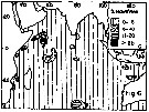
Carte de distribution de Scottocalanus securifrons par zones géographiques
|
| | | | | | | | | | | |  issued from : T.C. Gopalakrishnan & T. Balachandran in Oceanogr. Indian Ocean, 1992. [p.171, fig.6, C]. issued from : T.C. Gopalakrishnan & T. Balachandran in Oceanogr. Indian Ocean, 1992. [p.171, fig.6, C].
Distribution of Scottocalanus securifrons in the Indian Ocean.
Nota: Main occurnces between 10° N - 20° S. |
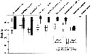 Issued from : M. Sano, K. Maki, Y. Nishibe, T. Nagata & S. Nishida in Progr. Oceanogr., 2013, 110. [p.20, Fig.10]. Issued from : M. Sano, K. Maki, Y. Nishibe, T. Nagata & S. Nishida in Progr. Oceanogr., 2013, 110. [p.20, Fig.10].
Vertical distribution of eight copepod species across 0-1000 m in Sagami bay in 21-27 April 2009.
White and black boxes denote daytime and nighttime distributions. Scaphocalanus echinatus/em> is taken from Kuriyama & Nishida (2006).
Plankton and water samples collected from a fixed station (35°00'N, 139°20'E) using MTD horizontal closing nets (0.33 mm mesh aperture) at 15 depths (0, 50, 95, 100, 150, 195, 200, 295, 300, 395, 400, 500, 600, 800, 1000 m) during both day and night.
Atomic C:N (mean ± SD) = 4.5 ±0.4, n = 22. |
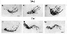 Issued from : M. Sano, K. Maki, Y. Nishibe, T. Nagata & S. Nishida in Progr. Oceanogr., 2013, 110. [p.19, Figs.8, 9]. Issued from : M. Sano, K. Maki, Y. Nishibe, T. Nagata & S. Nishida in Progr. Oceanogr., 2013, 110. [p.19, Figs.8, 9].
Second maxilla (Mx2) and maxilliped (Mxp) from Scaphocalanus echinatus (e), Scottocalanus helenae (f), and Scottocalanus securifrons (f).
Scale bars: Mx2 = 100 µm (e, f); 200 µm (g); Mxp = 100 µm (e); 200 mm (f, g).
Nota: The second maxillae setae in the three species are similar in shape to specialized chemosensory setae; among these, the setae in Sca. echinatus are relatively fine. The maxillipeds of Sco. helenae and Sco. securufrons have short, stout setae; Sca. echinatus have fine plumose setae (as in Calanus sinicus).
The feeding appendages of Sco. helenae and Sco. securifrons do not appear suitable for feeding on fine particulate organic matter (POM), whereas the maxillipeds of Sca. echinatus do. (Compare figures of setal armament from three species in the two genera). |
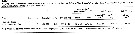 Issued from : M. Bode, R. Koppelmann, L. Teuber, W. Hagen & H. Auel inGlobal Biogeochemical Cycles, 2018, 32. [p.844, Table 1). Issued from : M. Bode, R. Koppelmann, L. Teuber, W. Hagen & H. Auel inGlobal Biogeochemical Cycles, 2018, 32. [p.844, Table 1).
Cf. explanations of these measures in Calanoides natalis from the same authors. |
| | | | Loc: | | | Antarct. (Indian), sub-Antarct. (Indian, SW Pacif.), South Africa (E & W), off E Tristan da Cunha, Namibia, Angola, G. of Guinea, off Lagos, Argentina, Brazil (off Macaé, The Campos Basin, off Natal), SE Atlantic, Cape Verde Is., off Mauritania-NW Cape Verde, off Morocco-Mauritania, Canary Is., off Madeira, off W Cabo Finisterre, Ibero-moroccan Bay, S Brazil, off Rio de Janeiro, off Amazon, Caribbean Sea, Caribbean Colombia, Jamaica, G. of Mexico, Florida, Sargasso Sea, off Bermuda: Station S (32°10N, 64°30W), Azores, Bay of Biscay, off W Ireland, off W Scotland, off E Nova Scotia, Flemish Cape, S Iceland, Faroe Is., Norwegian Sea, N North Sea, Arabian Sea, Natal, Madagascar (Nosy Bé), Indian, Indonesia-Malaysia, Flores Sea, Philippines, China Seas (East China Sea, South China Sea), Taiwan (S, E, Mienhua Canyon), Japan (Izu, Sagami Bay, Onagawa, off Sanriku), off SE Japan, Station Knot, Pacif. (W equatorial), New Zealand, Pacif. (equatorial central, off N Hawaii, Oregon (rare), California, W Baja California, off NE Marquesas Is., off NE Tuamotu Is., Galapagos, Ecuador, Peru, Pacif. (SE tropical) | | | | N: | 112 | | | | Lg.: | | | (1) F: 4,5; (5) F: 4,3; M: 4,75; (6) F: 4,08-3,66; M: 5,33-4,75; (7) F: 4,49; M: 4,98; (9) F: 4,6-3,38; M: 5,3-4,5; (16) F: 4,4-4,05; M: 4,9-4,55; (22) F: 4,9-4,3; M: 4,9-4,7; (35) F: 4,5-3,88; (38) M: 4,9-4,8; (47) F: ± 4; (54) F: 4,01; (73) F: 4,57-4,31; M: 4,99; (75?); (101) F: 4,08; (105) F: 4,07; M: 3,79; (108) F: 4,29; M: 4,81; (128) F: 4; (199) F: 4,53-4,1; M: 4,96-4,48; (254) F: ±4,3; M: 5; (432) F: 4; (539) F: 4,2; M: 4,57; (777) M: 4,55; (800) F: 4,69-4,53; (1000) F: 4,5 ± 0,1; M: 4,8 ± 0,1; (1111) F: 4,2; 4,7; M: 5,09; (1122) M: 4,55; {F: 3,38-4,90; M: 3,79-5,33} | | | | Rem.: | méso-bathypélagique, mais pouvant migrer vers la surface (Roe, 1972; 1972 b, p.530).
Vervoort (1965, p.36, 37) ne partage pas lopinion de Brodsky (1950) concernant l'identification de la femelle de Tanaka (1937).
Sars (1925, p.161) a antérieurement dénommé cette espèce comme S. acutus, en l'identifiant avec celle mentionnée par T Scott. Farran a cependant remarqué que quelques figures de la femelle données par Scott s'accordent mieux avec l'espèce présente, au moins les deux figures 54 et 56. Mais il est apparent que Scott a confondu les deux espèces car les autres figures qu'il donne, en particulier celles du mâle ne sont pas en accord avec la présente espèce, mais se réfèrent à S. persecans. L'espèce présente est distinguable de S. persecans par la forme différente des lobes latéraux du dernier segment pédigère et par la structure du segment génital chez la femelle.
Voir aussi les remarques en anglais | | | Dernière mise à jour : 19/01/2021 | |
|
|
 Toute utilisation de ce site pour une publication sera mentionnée avec la référence suivante : Toute utilisation de ce site pour une publication sera mentionnée avec la référence suivante :
Razouls C., Desreumaux N., Kouwenberg J. et de Bovée F., 2005-2024. - Biodiversité des Copépodes planctoniques marins (morphologie, répartition géographique et données biologiques). Sorbonne Université, CNRS. Disponible sur http://copepodes.obs-banyuls.fr [Accédé le 26 avril 2024] © copyright 2005-2024 Sorbonne Université, CNRS
|
|
|
|


;)
;)
;)
;)
;)
;)
;)
;)
;)
;)
;)
;)
;)
;)
;)
;)
;)
;)
;)
;)
;)
;)
;)
;)
;)
;)

;)
;)
;)
