|
|
 |
Fiche d'espèce de Copépode |
|
|
Cyclopoida ( Ordre ) |
|
|
|
Oncaeidae ( Famille ) |
|
|
|
Oncaea ( Genre ) |
|
|
| |
Oncaea frosti Heron, 2002 (F,M) | |
| | | | | | | Syn.: | Oncaea venusta; Ferrari, 1975 (p.225, fig.51); Boxshall, 1977 (p.124, fig.11a);
Oncaea venusta venella: Böttger-Schnack, 2001 (p.37, 45). | | | | Ref.: | | | Heron, 2002 (p.148, figs.F,M); Böttger-Schnack & Huys, 2004 (p.2: Rem.); Elvers & al., 2006 (p.504, 511: Rem.); Böttger-Schnack & Schnack, 2013 (p. 7, Table 2: Rem.) | 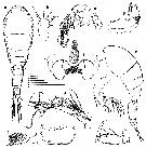 issued from : G.A. Heron in Hydrobiologia, 2002, 480. [p.146, Fig.1]. Female (coast of Liberia: a-b, habitus (dorsal and lateral, respectively; scale bar: u); c, anterior prosome (ventral; scale bar: v); d, right A1 (scale bar: w); e, right A2 (scale bar: x); f, labrum (ventral; scale bar: y); g, left Md (scale bar: y); h, left Mx1 (scale bar: y); i, right Mx2 (scale bar: y); j, left Mxp (scale bar: w). The letter after the explanation of each figure (u-z) indicates the 0.05 mm scale at which the figure is shown. Nota : Body ornamented with refractile granulations and prosome anteriorly with minute grooves. Genital segment length approximately equal to that of remainder of urosome ; gonopore on posterior of upper third of dorsal surface, each lateral area with a setule. Caudal ramus elongate, length variable but usually longer than total of 3 preceding segments, or approximately equal. A1 6-segmented with armature formula 3, 8, 4 + spinule, 3 + 1esthete, 2 + 1 esthete, 7 + 1 esthete. A2 3-segmented, 1st segment with distal inner spinulose seta ; 2nd segment with row of minute dentiform spinules along inner surface, outer rows of setules ; terminal segment with row of setules on posterior surface ; proximal inner surface with a curved spine and 3 setae ; distally 3 setae and 4 long, curved spiniform setae. Labrum posteriorly protuberant ; free margin divided into 2 rounded posteroventral lobes, each with row of dentiform setules inserted on under surface ; lobes separated by a semicircular vertex, from which a thin, hyaline lamella is suspended covered with hyaline, petaloid setules ; Two adjacent conspicuous sclerotized, spiniform protuberances anterior to vertex of labrum. Mx1 flat, bilobed ; 2setae and 1 robust spine on inner lobe ; 4 setae on outer lobe ; sclerotization on inner and outer margins. Mx2 2-segmented ; 1st segment with expanded base and sclerotized bands of support ; 2nd segment produced distally as elongate claw, with 2 inner rows of setules ; outer lateral seta ; 1 seta and 1 curved element with 2 rows of setules medially on proximal inner surface. Mxp 4-segmented ; 2nd segment robust with refractile granules on outer surface, inner surface with 2 spines ; anterior row of setules ; terminal segment consisting of long claw with row of setules on concave surface ; seta near inner base and small outer setule.
|
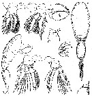 issued from : G.A. Heron in Hydrobiologia, 2002, 480. [p.147, Fig.2]. Female: a-d, P1 to P4 (scale bar: w); e, P5 (scale bar: z). Nota : P5 a short free segment ; 2 terminal setae, distally very fine, the inner slightly longer ; seta on body near leg. P6 probably represented by spiriform setule on each external genital area. Remark : The pediger 2 mid-dorsal dilatation obvious in lateral view, appears to circumscribe a gland-like organ ; a right and left duct originate from the gland, each directed to a small hooded dorsal pore. The gland does not preserve well in formalin, an dits shape and position varies slightly among specimens. Böttger-Schnack (2001) indicates an insignificant dorso-posterior swelling on pediger 2 of the female of Oncaea venusta var. venella, also this form is considerd synonymous with O. frosti. Male: f-g, habitus (dorsal and lateral, respectively; scale bar: v); h, 3rd segment of left A2 (scale bar: x); i, right Mxp (scale bar: w); i, right Mxp (scale bar: w); j, P5 (scale bar: w). Nota : Body with refractile granulations more dense and conspicuous than for female ; scattered hooded pores. Urosome 1st segment with transverse sclerotized ridge dorsoposteriorly. Swimming legs as in female. P5 not delimited from thoracic segment ; lengths of setae similar to those of female ; seta on body near leg. P6 represented by posterolateral flap on ventral surface of genital segment.
|
 issued from : G.A. Heron in Hydrobiologia, 2002, 480. [p.140, Fig.1]. The letter after the explanation of each figure (u-z) indicates the 0.05 mm scale at which the figure is shown.
|
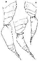 issued from : G.A. Heron in Hydrobiologia, 2002, 480. [p.153, Fig.6]. Female pediger 2-4 and urosome (scale bar: v): a, Oncaea venusta; b, O. frosti; c, O. venella. Nota: Exoskeleton with less dense cuticularization and slightly less conspicuous ornamentation than that of O. venusta. O. frosti displays a urosome flexed less often than that of O. venusta, but the two postgenital segments and anal segment are frequently telescopod; pediger 2 with conspicuous mid-dorsal dilation in lateral view; caudal ramus long, usually longer than sum of preceding three segments. Freshly collected specimens of O. frosti had bright red-orange shading on caudal rami and mouthparts.
| | | | | NZ: | 2 | | |
|
Carte de distribution de Oncaea frosti par zones géographiques
|
| | | | Loc: | | | Atlant. (coast of Liberia, G. of Mexico) | | | | N: | 1 | | | | Lg.: | | | (908) F: 0,95-1,15; M: 0,59-0,7; {F: 0,95-1,15; M: 0,59-0,70} | | | | Rem.: | Cette espèce se distingue de O. venusta et O. venusta venella par ses dimensions intermédiaires à ces deux formes. Sans l'application des méthodes d'analyse génétique, il est difficile de trancher définitivement entre espèces (non reproductibles entre-elles) et simples écophénotypes de populations mélangées a posteriori.
Voir aussi les remarques en anglais | | | Dernière mise à jour : 05/12/2016 | |
|
|
 Toute utilisation de ce site pour une publication sera mentionnée avec la référence suivante : Toute utilisation de ce site pour une publication sera mentionnée avec la référence suivante :
Razouls C., Desreumaux N., Kouwenberg J. et de Bovée F., 2005-2024. - Biodiversité des Copépodes planctoniques marins (morphologie, répartition géographique et données biologiques). Sorbonne Université, CNRS. Disponible sur http://copepodes.obs-banyuls.fr [Accédé le 20 avril 2024] © copyright 2005-2024 Sorbonne Université, CNRS
|
|
|
|


;)
;)
;)
