|
|
 |
Fiche d'espèce de Copépode |
|
|
Calanoida ( Ordre ) |
|
|
|
Diaptomoidea ( Superfamille ) |
|
|
|
Pseudodiaptomidae ( Famille ) |
|
|
|
Pseudodiaptomus ( Genre ) |
|
|
| |
Pseudodiaptomus sulawesiensis Nishida & Rumengan, 2005 | |
| | | | | | | Ref.: | | | Nishida & Rumengan, 2005 (p.27, figs.F,M) | 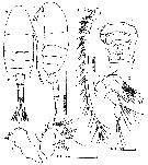 issued from : S. Nishida & I.F.M. Rumengan in Plankton Biol. Ecol., , 2005, 52 (1). [p.28, Fig.1]. Female (from 1°40'48''N, 125°04'12''E): A-B, habitus (dorsal and lateral, respectively); C, genital double-somite (ventral); D, A1; E, A2; F, Md. Nota : Rostrum with paired filaments. Cephalosome and 1st pediger somite separate, 4th and 5th fused withpaired rows of fine hairs along posteromedial margin. A1 symmetical, 22-segmented (segments 6-7 incompletely fused, the former with short spine), each segment except segments 6, 15, 16, 18-20 with aesthetasc ; segment 20 having modified seta with small teeth on medial margin. A2 : endopod 2-segmented ; exopod 4-segmented with 3rd segment inconspicuous and looking like membrane connecting 2nd and 4th segments. . Md endopod 2-segmented, 2nd segment with 9 setae and row of spinules on surface ; exopod 4-segmented ; cutting edge with seta on dorsal margin, 3 cuspidate teeth, smaller cuspidate teeth medially, and blunt molar-like processes ventrally. Proportional lengths of urosomites and caudal ramus 25 : 17 : 17 : 19 : 16 : 23 = 100 ; Genital double-somite, 1st and 2nd abdominal somites with row of triangular spinules on dorsoposterior margin. Ventral surface of genital double-somite with anterior, transverse row of spinules ; paired gonopores covered by broad genital operculum bearing a pair of short, posteriorly directed processes.
|
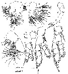 issued from : S. Nishida & I.F.M. Rumengan in Plankton Biol. Ecol., , 2005, 52 (1). [p.29, Fig.2]. Female: A, Mx1; B, Mx2; C, Mxp; D, P1 (posterior); E, P3 (posterior); F, P4 (posterior); G, P5 (posterior). Nota : Mx1 praecoxal arthrite with 9 strong and 6 finer setae ; coxa with 4 setae on endite and 9 setae on epipodite ; basis with 4 and 5 setae on proximal and distal endites, and with 1 seta on exite ; endopod 3-segmented with 4, 4 and 6 setae from 1st to 3rd segments ; exopod with 10 setae. Mx2 with 4 setae on 1st praecoxal endite ; 2nd praecoxal and 2 coxal endites each with 3 setae ; basis with 1 stout seta and 3 thinner setae ; endopod with 9 setae. Mxp praecoxa and coxa completely fused, endites with 0, 2, 3, 4 setae ; basis and 1st endopodal segment nearly fused, with 3 and 2 setae ; 2nd to 6th endopodal segments with 2, 2, 2, 3, 4 setae from proximal to distal ; 2 setae on 2nd segment bifurcated with modified branch. Legs 1-4 biramous with 3-segmented rami ; 1st and 2nd segments of both rami of legs 1-3, except leg 1 endopod, with spinules on inner distal margin. P5 uniramous and symmetrical ; basis with 1 seta, without spinules on distolateral margin ; exopod 3-segmented, 1st segment length ca. 3.0 times width, with distolateral seta ; 2nd segment with distolateral seta and distomedial process with serrate membrane on both margins ; 3rd segment spine-like with medial teeth and short proximomedial process.
|
 issued from : S. Nishida & I.F.M. Rumengan in Plankton Biol. Ecol., , 2005, 52 (1). [p.30]. Female: Seta (Arabic numerals) and spine (Roman numerals) of the swimming legs P1 to P4.
|
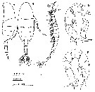 issued from : S. Nishida & I.F.M. Rumengan in Plankton Biol. Ecol., , 2005, 52 (1). [p.30, Fig.3]. Male: A-B, habitus (lateral and dorsal, respectively); C, right A1; D, P5 (anterior); E, P5 (posterior). Nota : Proportional lengths of urosomites and caudal ramus 13 : 22 : 17 : 17 :13 : 18 =100. Genital somite with genital aperture on left posterolateral margin. 1st abdominal somite with ventral row of spinules ; 1st to 3rd abdominal somites with row of triangular spinules on whole of posterior margins. Appendage s similar to those of female , except right A1 and P5. Right A1 geniculate, 21-segmented ; each segment except segments 5-8, 10, 12, 17-20 with aesthetasc ; segments 6 and 7 incompletely fused ; segment 10 with curved spine. P5 asymmetrical, biramous, with two 1-segmented endopods, 2-segmented left exopod and 3-segmented right exopod ; coxa with row of spinules on both surfaces ; basis with distolateral row of spinules ; right leg : 1st segment of exopod with patch of spinules on medialmargin and 2 setae on anterior surface, Y-shaped distal spine with medial fork about two times longer than lateral, and short thick subdistal spinule ; 2nd exopod segment with distolateral serrate spine, with distolateral patch of spinules at base of spine, and posterior-surface seta ; 3rd segment with 1 medial and 1 lateral setae. Endopod bifurcate ; medial branch slender and much longer than lateral, with fine subdistal seta ; lateral branch thick with 5-6 blunt distal spinules, one of which with teeth on tip.Left leg : 1st segment of exopod with short distolateral spine ; 2nd segment with 3 medial and 1 anterior-surface setae, and with small distomedial process and thick distolateral spine extending well beyond distal tip of 2nd segment and curved laterally near tip ; lateral margin distal to this spine fringed with spinules. Endopod with distomedial rows of spinules.
| | | | | NZ: | 1 | | |
|
Carte de distribution de Pseudodiaptomus sulawesiensis par zones géographiques
|
| | | | Loc: | | | Indonesia (N coast of Sulawesi) | | | | N: | 1 | | | | Lg.: | | | (1059) F: 1,19-1,33; M: 0,98-1,06; {F: 1,19-1,33; M: 0,98-1,06} | | | | Rem.: | In Ramosus species group (hickmani subgroup).
For Nishida & Rumengan (2005, p.31) the four species most closely related to P. sulawesiensis, i.e. P. ishigakiensis, P. marinus, P. philippinensis and P.australiensis, all exhibit the Type-III geographic patterns: a confined distribution mainly restricted to the West Pacific (see Walter & al., 2002). The other known species of the hickmani subgroup, i.e. P. ardjuna, P. hickmani, P. hypersalinus and P. jonesi , exhibit the Type-II pattern and are confined mainly to the Indian Ocean (Walter & al., 2002). On the basis of the known occurrence records, the five speciesof Type-III appear to be more-or-less endemic to their respective ranges within the West Pacific: from north to south, the neritic waters of Japan (P. marinus, excluding records of synanthropic introduction and those lacking information in species identifications). | | | Dernière mise à jour : 28/06/2013 | |
|
|
 Toute utilisation de ce site pour une publication sera mentionnée avec la référence suivante : Toute utilisation de ce site pour une publication sera mentionnée avec la référence suivante :
Razouls C., Desreumaux N., Kouwenberg J. et de Bovée F., 2005-2024. - Biodiversité des Copépodes planctoniques marins (morphologie, répartition géographique et données biologiques). Sorbonne Université, CNRS. Disponible sur http://copepodes.obs-banyuls.fr [Accédé le 20 avril 2024] © copyright 2005-2024 Sorbonne Université, CNRS
|
|
|
|


;)
;)
;)
