|
|
 |
Fiche d'espèce de Copépode |
|
|
Calanoida ( Ordre ) |
|
|
|
Hyperbionychidae ( Famille ) |
|
|
|
Hyperbionyx ( Genre ) |
|
|
| |
Hyperbionyx athesphatos Bradford-Grieve, 2010 (M) | |
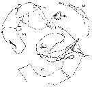
| | | | | | | Ref.: | | | Bradford-Grieve, 2010 (p.2167, figs.M); Blanco-Bercial & al., 2011 (p.103, Table 1, Biol. mol., phylogeny) | 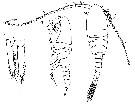 issued from : J.M. Bradford-Grieve in Deep-Sea Res.II, 2010, 57. [p.2168, Fig.1] Male (from 11.522°N, 20.383°W): A-B, habitus (dorsal and lateral, respectively); C, caudal rami (dorsal). Scale bars: 0.1 mm. Nota : Cephalosome and pedigerous somite 1 separate , pedigerous somites 4 and 5 fused. Posterolateral corners of prosome truncate in dorsal view, rounded in lateral view. Rostrum represented by small, blunt ventral extension of margin of head bearing 2 relatively long rostral filaments. Urosome composed of 5 somites ; all urosomites with naked posterior borders. Gonopore on right side of genital somite. Caudal rami asymmetrical, longer and wider on right side, with vestigial seta in position I on left (not visible on right), setae II-VI well developed (on right seta V enlarged), seta II-VI plumose although setae II and VI are plumose on inner border only, seta VII small, situated on dorsal surface directed posteriorly.
|
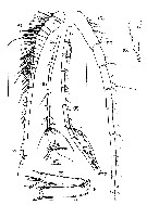 issued from : J.M. Bradford-Grieve in Deep-Sea Res.II, 2010, 57. [p.2168, Fig.2] Male: A, right A1 (ancestral segments I-XX); B, right A1 (ancestral segments XXI-XXVIII); C, right A1 (ancestral segments XXVII-XXVIII); D, left A12 (ancestral segments I-X); E, left A1 (ancestral segments XI-XXVIII); F, left A1 (hinge formed between ancestral segments XX and XXI; G, left A1 (ancestral segments XXVII-XXVIII). Scale bars: 1.0 mm (A-B, E-D); 0.1 mm (C, F, G). Nota : A1 23-segmented on left, geniculate , much longer that whole body ; 25-segmented on right extending as far as middle of urosomite 4.
|
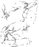
 issued from : J.M. Bradford-Grieve in Deep-Sea Res.II, 2010, 57. [p.2169, Fig.3] Male: A, right A2; B, right A2 (terminal endopod segments); C, right A2 (terminal part of exopod); D, right Md; E, right mx1. Scale bars: 0.1 mm. Nota : Md gnathobase masticatory margin with 2 large terminal teeth, heavily chitinized. Mandibular palp with elongate basis, unarmed ; endopod 2-segmented, segment 1 without setae, segment 2 outer border lined with setules, and 3 terminal setae ; exopod with 6 setae. Mx1 : Praecoxal arthrite with 3 posterior surface setae as well as 7 strong marginal spines and 3 setae ; coxal endite developed with 3 terminal setae ; basal endites 1 and 2 undeveloped and without setae, basis and endopod separate ; endopod segmented, elongate, with 2 small inner setae and 3 terminal setae ; exopod large with 2 strong setae ; basal exite without seta ; coxal epipodite with 2 setae
|
 issued from : J.M. Bradford-Grieve in Deep-Sea Res.II, 2010, 57. [p.2170, Fig.4] Male: A, right Mx2; B, right Mx2 (coxal endite 2); C, right Mx2 (endopod); D, left Mxp (note longitudinal row of spinules on basis on underside as drawn; E, maxilliped terminal part of endopod. Scale bars: 0.1 mm. Nota : Mx2 very well developed. Praecoxal endite 1 in form of elongate lobe with 3 setae, praecoxal endite 2 with 1 seta ; coxal endite 1 with 2 setae, coxal endite 2 lobe-like with 3 setae one of which short and toothed along both borders ; basal endite well developed bearing stout claw, 1 short spine-like seta and 2 other small setae ; endopod segment 1 well-developed bearing strong claw and 3 other small setae, rest of endopod 2-segmented with 6 setae. Mxp : basis with 3 setae placed distally on segment and longitudinal row of densely fine spinules extending almost entire length of segment ; endopod segment 1 separate from basis with 2 setae , endopod segments 2-6 with 4, 4, 3, 3, 4 setae respectively, segment 6 very small but separate, lying almost at right angles to the main axix of endopod and telescoped within terminal part of segment 5, middle seta on endopod segment 5 with well developed teeth along central part of one border.
|
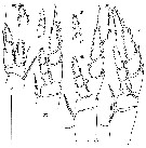 issued from : J.M. Bradford-Grieve in Deep-Sea Res.II, 2010, 57. [p.2171, Fig.5] Male: A, P1 (posterior view); B, anterior view of endopod of P1; C, P2 (posterior view); D, anterior view of endopod segment 2 and 3 of P2; E, P3 (posterior view); F, anterior view of endopod segments 2 and 3 of P3; G, P4 (posterior view); H, anterior view of endopod segments 2 and 3 of P4.Scale bars: 0.1 mm. P1 : endopod segment 1 with large outer swelling bordered by setules, distal and distolateral spinules on anterior surface as well as oblique row of 5 pores, segment 2 and 3 fused (region of fusion visible on posterior surface). P2 to P4 anterior surface of endopod segments 2 and 3 with very fine setules.
|
 issued from : Bradford-Grieve, 2010 [p.2169.]. Male: Segmentation and disposition of spines and setae (Roman numerals) and setae (Arabic numerals) of Swimming legs P1 to P4.
|
 issued from : J.M. Bradford-Grieve in Deep-Sea Res.II, 2010, 57. [p.2171, Fig.6] Male: A, right P5 (posterior view); B, terminal part of right exopod in a different view; C, left P5 (posterior view); D, terminal part of left exopod segment 2 in a different view. Scale bar: 0.1 mm. Cross-scored seta broken. Nota : P5 biramous on both sides with small, 1-segmented endopods articulated to both bases. Right leg with coxa fused to intercoxal sclerite, basis with posterior surface seta ; endopod represented by small knob-like segment articulated to basis ; exopod indistinctly 3-segmented, segment 1 extended at outer distal corner into large rounded lobe bearing stout spine, segment 2 and 3 indistinctly separate, scoop-shaped, with strong outer spine just proximal to midlength and with terminal curved, ear-shaped process on segment 3 with a thickened cuticle (naked spine situated sub-terminally at based of ear-shaped process as well as 3 pores, fig.B) ; inner border in region of division between segments 2 and 3 without setules. Left leg with coxa fused to intercoxal sclerite ; basis with posterior surface seta ; endopod represented by small, tapering, leaf-like segment ; exopod 3-segmented, terminated by complex pincer arrangement of segments 2 and 3 ; segment 1 extended at outer distal corner into large rounded lobe bearing stout spine ; segment 2 curved, complicated with 1 outer spine on process beyond midlength of segment ; with recurved, elongate posterior process and shorter anterior recurved lobe to which articulates long, tapering exopod segment 3 ; posterior process with strong basal part that is wrinkled where it arises from segment 2 and recurves, but then extends into transparent blade with several flanges, milength flange finger-like, small tooth (fig.D) situated opposite finger-like flange and border is serrated both distal and proximal to this spine ; segment 3 articulates to anterior terminal surface of rounded anterior lobe of segment 2, oval in cross-section, tapering and extending parallel to long process of segment 2 although not quite as long, terminal part appears to be articulated.
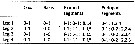
| | | | | NZ: | 1 | | |
|
Carte de distribution de Hyperbionyx athesphatos par zones géographiques
|
| | | | Loc: | | | NE Atlantic (Cape Verde Abyssal Plain) | | | | N: | 1 | | | | Lg.: | | | (1063) M: 8,92; {M: 8,92} | | | | Rem.: | sampled between 4795-4000 m (bottom depth: 4891 m).
For Bradford-Grieve (2010, p.2171) this male is distinguished from H. pluto by the P5. H. athesphatos male P5 has a leaf-shaped endopod (H. pluto has this ramus reduced to a vestigial low process); exopod segment 2 has a very large terminal recurved extension, of hyaline appearance distally, which bears ridges or flanges (no such extension is present in H. pluto), and exopod segment 3 is inserted at midlength having a large, terminal, articulated soft spines extending almost as far as, and opposed to, the large extension of segment 2 (in H. pluto this segment is of similar shape but does not have the large terminal spine). The right P5 appears to be without a tuft of setules on the exopod inner border in the region of the division between segment 2 and 3, unlike H. pluto which has setules in this position. The A2 basis and endopod segment 1 are separate rather than partially fused as in H. pluto. There are also small differences in the disposition of setae on some segments of the A1. For the autor, the species has been characterized by sequences of the nuclear ribosomal genes: 18S, 28S, and the mitochondrial gene Cytochrome B (Blanco-Bercial & Bucklin, in prep.). | | | Dernière mise à jour : 06/01/2015 | |
|
|
 Toute utilisation de ce site pour une publication sera mentionnée avec la référence suivante : Toute utilisation de ce site pour une publication sera mentionnée avec la référence suivante :
Razouls C., Desreumaux N., Kouwenberg J. et de Bovée F., 2005-2024. - Biodiversité des Copépodes planctoniques marins (morphologie, répartition géographique et données biologiques). Sorbonne Université, CNRS. Disponible sur http://copepodes.obs-banyuls.fr [Accédé le 19 avril 2024] © copyright 2005-2024 Sorbonne Université, CNRS
|
|
|
|


;)
;)
;)
;)
;)
;)
