|
|
 |
Fiche d'espèce de Copépode |
|
|
Monstrilloida ( Ordre ) |
|
|
|
Monstrillidae ( Famille ) |
|
|
|
Maemonstrilla ( Genre ) |
|
|
| |
Maemonstrilla okame Grygier & Ohtsuka, 2008 (F) | |
| | | | | | | Ref.: | | | Grygier & Ohtsuka, 2008 (p.482, Descr.F, figs.F, Rem.) | 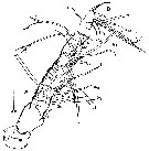 issued from : M.J. Grygier & S. Ohtsuka in Zool. J. Linnean Soc., 2008, 152. [p.484, Fig.28, A-B]. Female (Holotype; from Sesoko Island): A, right A1 (ventral view); B, same, more fully armed tip of left A1 (ventral view; b5-seta seen to be bifid in another specimen). Scale bar = 0.100 mm.
|
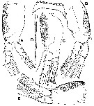 issued from : M.J. Grygier & S. Ohtsuka in Zool. J. Linnean Soc., 2008, 152. [p.486, Fig.20]. Female (Paratype, ovigerous): Right legs P1-P4, setules omitted and setae cut short. A, P1 (anterior; B, P2 and intercoxal sclerite (posterior); C, P3 (anterior); D, outer distal exopodal seta of P3; E, P4 (anterior). Scale bar = 0.100 mm.
|
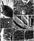 issued from : M.J. Grygier & S. Ohtsuka in Zool. J. Linnean Soc., 2008, 152. [p.483, Fig.17]. SEM. Female (from two non-ovigerous paratypes): A, anterior half of cephalothorax (lateral); B, anteroventral part of cephalothorax (i.e. 'face') between oral papilla (o) and bases of A1 (top corners), showing two pairs of pores and two pairs of scars; C, reticulations of lateral side of cephalothorax; D, reticulations of forehead, showing pair of hair-like sensilla and pair of pores (dorsal to left); E, P2-P4 (lateral view); F, detail of coxal spinulation of a right swimming leg (proximal to left; G, site of absent inner seta (arrow) of 1st endopodal segment of right P1. Scale bars = 0.100 mm (A, E); 0.050 mm (B-D); 0.020 mm (F, H); 0.010 mm in G.
|
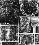 issued from : M.J. Grygier & S. Ohtsuka in Zool. J. Linnean Soc., 2008, 152. [p.485, Fig.19]. SEM. Female (from two non-ovigerous paratypes): A, dorsal surface of free pedigers1 and 2 (anterior at top; arrows indicating obscured pair of large pores); B, dorsal surface of free pediger 3 (anterior at top); C, dorsal surface of free pediger 4 and genital compound somite (g; letter placed on indistinct dorsal suture) (anterior at top); D, telson (t) and caudal rami (f) (dorsal view); E, part of posterior margin of penultimate segment of urosome (posterior to right); F, genital compound somite (ventral view), anterior at bottom, showing copulatory pore (arrow) and small ventral knob on posterior part (above). Scale bars = 0.050 mm (A-C, F); 0.020 (D, E).
|
 issued from : M.J. Grygier & S. Ohtsuka in Zool. J. Linnean Soc., 2008, 152. [p.499, Fig.29]. Female: Dorsal and lateral pore and pit seta patterns, from rear of cephalothorax through genital compound somite. Symbols: dots (three sizes) = pores; larger circles = pits of pit setae. Pattern based on SEM and light microscopical examination.
|
 issued from : M.J. Grygier & S. Ohtsuka in Zool. J. Linnean Soc., 2008, 152. [p.493]. Key to the Ryukyu species of the Maemonstrilla hyottoko species-Group. M. okame Female: 1 - Cuticle of cephalothorax, A1, lateral sides of trunk, dorsum telson, and caudal rami reticulated; outer faces of P1-P4 and dorsum of free pedigers, genital compound somite, and penultimate somite spinulose. Cephalothoracic reticulations comprising ridges with abundant or sparse spinules; simple or complex cuticular ornamentation within at least some meshes. 2 - Oral papilla small, directed more anteriorly than ventral and flanked by 'puffed cheeks'. Genital compound somite lecking dorsal transverse ridge. Compare with M. simplex, M. hyottoko, M. polka, M. spinicoxa.
| | | | | NZ: | 1 | | |
|
Carte de distribution de Maemonstrilla okame par zones géographiques
|
| | | | | | | Loc: | | | S Japan (Sesoko Island)
Type locality: 26°38.2' N, 127°51.8' E. | | | | N: | 1 | | | | Lg.: | | | (1077) F: 1,20-1,67 *
*: sum of lengths of cephalothorax, metasome and urosome in lateral view. | | | Dernière mise à jour : 23/04/2020 | |
|
|
 Toute utilisation de ce site pour une publication sera mentionnée avec la référence suivante : Toute utilisation de ce site pour une publication sera mentionnée avec la référence suivante :
Razouls C., Desreumaux N., Kouwenberg J. et de Bovée F., 2005-2024. - Biodiversité des Copépodes planctoniques marins (morphologie, répartition géographique et données biologiques). Sorbonne Université, CNRS. Disponible sur http://copepodes.obs-banyuls.fr [Accédé le 25 avril 2024] © copyright 2005-2024 Sorbonne Université, CNRS
|
|
|
|


;)
;)
;)
;)
;)

