|
|
 |
Fiche d'espèce de Copépode |
|
|
Monstrilloida ( Ordre ) |
|
|
|
Monstrillidae ( Famille ) |
|
|
|
Maemonstrilla ( Genre ) |
|
|
| |
Maemonstrilla simplex Grygier & Ohtsuka, 2008 (F,M) | |
| | | | | | | Ref.: | | | Grygier & Ohtsuka, 2008 (p.489, Descr. F, figs.F, Rem.) | 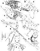 issued from : M.J. Grygier & S. Ohtsuka in Zool. J. Linnean Soc., 2008, 152. [p.490, Fig.22]. Female (from Ishigaki Island): A, habitus (left lateral; reticulations in area marked with asterisk obscured); B, cephalothorax showing full extent of obvoius reticulation (right lateral); C, detail of oral papilla region, showing scars and two pairs of pores (arrowheads); D, anterior end of cephalothorax (dorsal view); E, urosome (lateral view), with P5 and ovigerous spines (one of latter broken), setules of setae omitted, furcal setae cut short; F, urosome (dorsal view), furcal setae cut short and with setules omitted. Scale bars = 0.500 mm (A, B); 0.100 mm in C; 0.200 mm (D-F). Nota: - Forehead transversely wrinkled, with pair of hair-like sensilla (fig.22D); button-like structure present anterodorsally (presumably homologous to wrinkled pit in M. hyokotto), followed by 2 medial pores and 2 pairs of more lateral pores. - Oral papilla conical, nearly straight and protruding ventrally (fig.22A, B), relatively the smallest among species of Maemonstrilla, except that of M. okame. 1 pair of pores close behind oral papilla (fig.22C). 3 cups of naupliar eye well developed, diameters 110-139 µm, lateral ones separated from each other and slightly larger than ventral cup. - P5 as in M. hyottoko. - Dorsum of genital compound somite with transverse partial (fig.22F). Ventrally, posterior part and portion anterior to bases of ovigerous spines each produced into round swelling (fig.22E). Anteriorly directed ovigerous spines cylindrical but tapering to naked tips from slightly swollen, wrinkled region at 2/3 length, reaching perhaps just beyond P1 (fig.22A, E). - Mean diameter of eggs from ovigerous specimen: 32 µm (n = 15) - Cayudal rami armed as M. hyottoko, dorsal seta origunally with setules, but these broken off (fig.22F); ventral pore present.
|
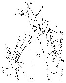 issued from : M.J. Grygier & S. Ohtsuka in Zool. J. Linnean Soc., 2008, 152. [p.484, Fig.18, C, D]. Female: C, left A1 (dorsal view); D, same, more fully armed tip of right A1 (dorsal view). Scale bar = 0.100 mm. Nota: A1 with 3 small sclerites for muscle attachment amidst wrinkled arthropodial membrane at base of 1st segment. 2nd to 4th segments not clearly separated from each other. All setal elements as shown in Grygier & Ohtsuka (1995) present on right A1 (some distal elements lost from left A21). 2 apical 6- greatly differing in size, 1 ind.1 being smaller and 6 ind.2 apparently bearing some setules; 2 ind.v-setae and 3-setae more than 3 times longer than 2 ind.2-setae and 4-setae; 1-seta and 2-setae with 2 rows of minute spinules discernible by light microscopy, 3-seta and 4-setae with even smaller spinules, and 5-seta seemingly smooth.
|
 issued from : M.J. Grygier & S. Ohtsuka in Zool. J. Linnean Soc., 2008, 152. [p.491, Fig.23]. Female: A, P1 and intercoxal sclerite (anterior); B, detail of outer apical exopodal setae (and distal pore) of P1; C, P2 (posterior); D, P3 and intercoxal sclerite (posterior view); E, P4 (anterior). Arrows in A and D indicating vestige of proximal inner endopodal seta. Scale bars = 0.200 mm (A, C-E); 0.100 mm in B.
|
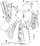
 issued from : M.J. Grygier & S. Ohtsuka in Zool. J. Linnean Soc., 2008, 152. [p.499, Fig.29]. Female: Dorsal and lateral pore and pit seta patterns, from rear of cephalothorax through genital compound somite. Symbols: dots (three sizes) = pores; larger circles = pits of pit setae. Pattern based on SEM and light microscopical examination.
|
 Issued from : M.J. Grygier & S. Ohtsuka in Zool. J. Linnean Soc., 2008, 152. [p.493]. Key to the Ryukyu species of the Maemonstrilla hyottoko species-Group. M. simplex Female: 1 - Only cephalothorax reticulated, and no obviously spinulose regions present. Ridges of cuticular meshwork lacking spinules, and cuticular surface within meshes plain. Compare with M. hyottoko, M. okame, M. polka, M. spinicoxa : 1' - Cuticle of cephalothorax, A1, lateral sides of trunk, dorsum telson, and caudal rami reticulated; outer faces of P1-P4 and dorsum of free pedigers, genital compound somite, and penultimate somite spinulose. Cephalothoracic reticulations comprising ridges with abundant or sparse spinules; simple or complex cuticular ornamentation within at least some meshes.
|
 Issued from : M.J. Grygier & S. Ohtsuka in Zool. J. Linnean Soc., 2008, 152. [p.489]. Maemonstrilla simplex Female (from Ishigaki Island): - Cephalothorax less bulbous and more cylindrical than in other species of this genus; Cephalothorax polygonal reticulation simple and completely non-spinulose. - No reticulation or spinulation noted on other body regions by light microscopy (SEM not attempted). - Articyulations between A1 segments 2-4 not distinct. Spiniform 2v- and 3-setae more than 3 times longer than 4-setae; 1-seta and all 2-setae appearing minutely spinulose even by light microscopy, and 6-setae differing greatly in size. - Oral papimlla relatively smaller than in other species except M. okame, preceded by parallel grooves and pair of tubular pores.
- Basis of P1-P4 slightly protruding on inner side.
- Two large, ronded, ventral lobes on anterior and posterior parts of genital compound somite.
| | | | | NZ: | 1 | | |
|
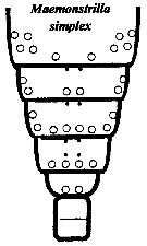
Carte de distribution de Maemonstrilla simplex par zones géographiques
|
| | | | | | | Loc: | | | S Japan (Ishigaki Island)
Type locality: 24°20' N, 124°11' E. | | | | N: | 1 | | | | Lg.: | | | (1077) F: 1,80-1,98 *
*: sum of lengths of cephalothorax, matasome and urosome in lateral view | | | Dernière mise à jour : 24/04/2020 | |
|
|
 Toute utilisation de ce site pour une publication sera mentionnée avec la référence suivante : Toute utilisation de ce site pour une publication sera mentionnée avec la référence suivante :
Razouls C., Desreumaux N., Kouwenberg J. et de Bovée F., 2005-2024. - Biodiversité des Copépodes planctoniques marins (morphologie, répartition géographique et données biologiques). Sorbonne Université, CNRS. Disponible sur http://copepodes.obs-banyuls.fr [Accédé le 18 avril 2024] © copyright 2005-2024 Sorbonne Université, CNRS
|
|
|
|


;)
;)
;)
;)

