|
|
 |
Fiche d'espèce de Copépode |
|
|
Calanoida ( Ordre ) |
|
|
|
Pseudocyclopoidea ( Superfamille ) |
|
|
|
Pseudocyclopidae ( Famille ) |
|
|
|
Pinkertonius ( Genre ) |
|
|
| |
Pinkertonius ambiguus Bradford-Grieve, Boxshall & Blanco-Bercial, 2014 (F,M) | |
| | | | | | | Ref.: | | | Bradford-Grieve & al., 2014 (p.517, Descr.F,M, figs.F;M, genetic catalogued in GenBank); Laakmann & al., 2019 (p.330, fig. 2, 3, phylogenetic relationships) | 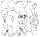 Issued from : J.M. Bradford-Grieve, G.A. Boxshall & L. Blanco-Bercial in Zool. J. Linn. Soc., 2014, 171. [p.511, Fig.2]. Female (44°41.95'S, 173°41.94'E): A-B, habitus (dorsal and lateral, respectively); C, A1; D, rostrum; E, genital double-somite (ventral); F, caudal rami (dorsal); G, same (ventral); H, P5 (anterior); I, detail of joint between exopod segments 2 and 3 of P5, showing (large, unlabelled arrow-heads) location of two pivot points. Selected characters and their states are indicated (e.g for example arrowhead 5.2). Scale bars: A-C, 1.0 mm (A1 at same scale as A and B); D-I, 0.1 mm. Nota : Cephalosome and pedigerous segment 1 separate, 4th and 5th separate. Pedigerous somite 5 produced into pointed lappets extending more than ½ along genital double-somite. Rostrum in form of ventrallydirected rounded plate with pair of filaments. Urosome 4-free somites, fist three bordered posteriorly by unevenly serrated hyaline fringe, largely intact ventrally. Genital double-somite with slight anterior swelling in dorsal view ; in lateral view swollen ventroanteriorly ; in ventral view genital field asymmetrical, gonopores slightly unequally developed, being larger on left, genital operculum kewed to left with hinge aligned at about 45° to anterior-posterior axis of somite, so that right gonopore ant completely covered, right side of genital field bordered by ridge aligned anterior-posteriorly, no such flange on left, copulatory pore and seminal receptacle not obvious, although sac on left apparently linked to left gonopore. Caudal rami slightly asymmetrical, longer on right, with 7 setae each. Seta I vestigial, seta II spiniform, seta V longest, seta IV next longest followed by setae VI and III, seta VII small and spiniform and inserted on dorsoinner distal corner ; left caudal ramus inner border lined with fine setules ; on right ramus row of long setules arranged obliquely on anterior part of ventral surface. A1 27-segmented, extending to posterior border of pedigerous somite 5. P5 articulation of exopodal segment 3 with segment 2, oblique (reminiscent of sepcies of Ridgewayia) ; proximal end of segment 3 narrowing, distance between two pivot points (fig.2H) ensures region of articulation is as wide as exopodal segment 3 at level of proximal inner seta of exopodal segment 3. Outer distal extension of exopodal segment 2 not reaching origin of proximal outer spine of exopodal segment 3.
|
 Issued from : J.M. Bradford-Grieve, G.A. Boxshall & L. Blanco-Bercial in Zool. J. Linn. Soc., 2014, 171. [p.512, Fig.3]. Female: A, A1 ancestral segments I-XII; B, A1 ancestral segments XIII-XIX; C, A1 ancestral segments XX-XXVIII; D, Mx2; E, Mx2 endopod. Scale bars: A-D, 0.1 mm; E, 0.02 mm. Selected characters and their states are indicated (e.g for example arrowhead 20.2). Nota : A1 with 1 aesthetasc present on each of segments I, III, VII, XI, XIV, XVI, XVIII, XXI, XXV, and XXVIII ; small cuticular thickenings found on segmentsII-X, XII, XIII, XV, and XVII ; 1 seta on eight proximal segments, wider than other setae, and attenuated distally into curved narrow tip. Mx2 : Praecoxa, coxa and basis clearly separated, endites 1-4 with 7 (1 very short), 3, 3 and 3 setae, respectively ; basal endite with 3 setae, one of them stout and spiniform ; inner setae on endites 2-5 lined with long spinules ; endopod segment 1 endite with 3 setae, segments 2-4 with 2, 2, 3 setae, respectively.
|
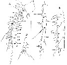
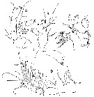 Issued from : J.M. Bradford-Grieve, G.A. Boxshall & L. Blanco-Bercial in Zool. J. Linn. Soc., 2014, 171. [p.514, Fig.4]. Female: A, A2; B, Md; C, Mx1; D, Mxp. Scale bars: 0.1 mm. Selected characters and their states are indicated (e.g for example arrowhead 66:1). Nota : A2 Coxa and basis separate ; endopod 2-segmented with traces of fusion between segments 2 and 3, and between segments 3 and 4 ; segment 1 with 2 inner setae ; segment 2 with 9 plus 7 terminal setae and outer transverse rox of spinules marking boundary between putative endopodal segments 3 and 4. Exopod shorter than endopod, 8-segmented, ancestral segments VIII and IX fused, segments I-VII each with 1 well developed seta, compound distal segment VIII-IX with 3 terminal setae and 1 inner subterminal seta, outermost seta on terminal endopodal segment lined proximally with small spinules. Md : Gnathobase with 7 marginal teeth, ventralmost largest, 5 dorsal teeth bicuspid, small spinulated seta inserted dorsally.Basis with 4 apparently naked setae ; endopod 2-segmented segment 1 with inner lobe and 4 setae, segment 2 with 10 terminal setae, distoinner border with short row of spinules at about midlength. Exopod 5-segmented with setal formula 1, 1, 1, 1, 2. Mx1 : Praecoxal arthrite with 14 spines and setae, including 4 on posterior surface and 1 on dorsal surface ; coxal endite with 4 setae ; basal endites 1 and 2 with 4 and 5 setae, respectively. Endopod segments 1 and 2 fused, segments 2 and 3 separate, with 4, 3, and 7 setae, respectively. Exopod with 11 setae, of which 3 terminal setae short and bordered by fine setules along inner border. Basal exite without seta. Coxal epoipodite with 9 setae, of which 3 proximal setae short. Mxp : 1st syncoxal endite with 1 seta, endites 2 and 3 with 2 and 4 spinulose setae, respectively, crescent-shaped row of fine spinules at base of endite 3 on inner surface ; endite 4 with 3 setae and small peg-like structure, and a few small spinules. Basis with 2 setulose setae and proximal border lined by spinules. Endopod well-developed, longer than basis, endopodal segment 1 apparently separate from basis, endopodal segments 1-6 with 2, 4, 4, 3, 3 plus 1, and 4 spinulose setae, respectively, outer seta of segment 6 wider and longer than adjacent seta, and terminally inserted.
|
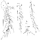
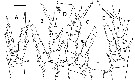 Issued from : J.M. Bradford-Grieve, G.A. Boxshall & L. Blanco-Bercial in Zool. J. Linn. Soc., 2014, 171. [p.515, Fig.5]. Female: A, P1 (posterior); B, P2 (posterior); C, P3 (posterior); D, P3 coxa (anterior); E, P4 (posterior); F, P4 coxa (anterior surface). Scale bar: 0.1 mm. Selected characters and their states are indicated (e.g for example arrowhead 98:1).
|
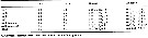 Issued from : J.M. Bradford-Grieve, G.A. Boxshall & L. Blanco-Bercial in Zool. J. Linn. Soc., 2014, 171. [p.519, Table 4]. Setal formulae for swimming legs. Roman numerals indicate spines; Arabic numerals indicate setae. Outer border setation listed first in each segment, with group separated by ;
|
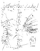
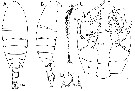 Issued from : J.M. Bradford-Grieve, G.A. Boxshall & L. Blanco-Bercial in Zool. J. Linn. Soc., 2014, 171. [p.516, Fig.6]. Male: A-B, habitus (dorsal and lateral, respectively; C, right A1 (at same scale as A and B); D, caudal rami (dorsal); E0, P5 (posterior); note the deformed left endopod segment 3 that has an extra inner seta - this seta is absent in another specimen. Scale bars: A-C: 1.0 mm; D, E: 0.1 mm. Selected characters and their states are indicated (e.g for example arrowhead 120:1). Nota : Urosome 5 free somites, anterior 4 somites borderd posteriorly by unevenly serrated hyaline fringe. Caudal rami symmetrical, with 7 setae each : seta I vestigial, seta II spiniform, seta V longest, seta IV next longest followede by setae VI and III, weta VII small and spiniform and inserted on dorsoinner distal corner, inner borders of caudal rami lined with fine setules. A2, Md, Mx1, Mx2, Mxp, and P1 P4 identical to those of female. P5 biramous on both sides, with both rami 3-segmented, asymmetrical. Exopod slightly longer on right. Right exopodal segment 2 with triangular inner attenuation, segment 3 in form of claw with terminal spine, rounded distally, fused to segment, with 2 articulated spines (1 medioproximal, other on outer border more distally inserted), with additional outer fused spinule and pore opening. Left leg exopodal segment 2 with inner border swollen, bearing seta modified into scalpel-shaped element thickened along its outer border ; exopodal segment 3 simple,about twice as long as wide, short terminal spine fused to segment at level adjacent to lateral pore opening, 1 articulated inner spine an douter articulated spine more proximally inserted on posterior surface. Posterior surfaces of exopods and endopods ornamented with scattered patches of very small spinules.
|
 Issued from : J.M. Bradford-Grieve, G.A. Boxshall & L. Blanco-Bercial in Zool. J. Linn. Soc., 2014, 171. [p.520, Fig.7]. Male left A1: A, ancestral segments I-XIV; B, ancestral segments XV-XIX; D, ancestral segments XX-XXV; E, ancestral segments XXVI-XXVIII. Scale bar: 0.1 mm. Nota : Left A1 26-segmented.
|
 Issued from : J.M. Bradford-Grieve, G.A. Boxshall & L. Blanco-Bercial in Zool. J. Linn. Soc., 2014, 171. [p.521, Fig.8]. Male right A1: A, ancestral segments I-XIV; B, ancestral segments XV-XIX; C, ancestral segments XX-XXV; D, ancestral segments XXVI-XXVIII. Scale bar: 0.1 mm. Selected characters and their states are indicated (e.g for example arrowhead 39:1). Nota : Right A1 25-segmented, extending just beyond posterior border of 4th pedigerous somite, geniculation between ancestral segments XX and XXI, segments XV-XIX enlarged
| | | | | NZ: | 1 | | |
|
Carte de distribution de Pinkertonius ambiguus par zones géographiques
|
| | | | Loc: | | | SW Pacif. (E New Zealand)
Type locality: 44°41.95'S, 173°41.94'E. | | | | N: | 1 | | | | Lg.: | | | (1156) F: 1,8-2,0; M: 1,8-1,88; {F: 1,8-2; M: 1,8-1,88} | | | | Rem.: | Cette espèce clôt l'essai de synthèse bibliographique mise à la libre disposition de tous ceux qui sont confrontés à l'identification des copépodes pélagiques marins (ou dans des habitats extrêmes), dans l'ensemble des océans et mers du globe, comme des principaux aspects de leur écologie.
Les références citées et analysées sont présentes dans la bibliothèque de l'Observatoire Océanologique de Banyuls-sur-Mer.
Ce travail se veut un hommage rendu à tous ceux qui ont contribué à la connaissance de ce groupe d'organismes marins et à la première synthèse zoogéographique établit par R. B. Seymour Sewell (Lieut.-Colonel, I.M.S. [ret.]).
Voir aussi les remarques en anglais | | | Dernière mise à jour : 02/04/2019 | |
|
|
 Toute utilisation de ce site pour une publication sera mentionnée avec la référence suivante : Toute utilisation de ce site pour une publication sera mentionnée avec la référence suivante :
Razouls C., Desreumaux N., Kouwenberg J. et de Bovée F., 2005-2024. - Biodiversité des Copépodes planctoniques marins (morphologie, répartition géographique et données biologiques). Sorbonne Université, CNRS. Disponible sur http://copepodes.obs-banyuls.fr [Accédé le 25 avril 2024] © copyright 2005-2024 Sorbonne Université, CNRS
|
|
|
|


;)
;)
;)
;)
;)
;)
;)
