|
|
 |
Fiche d'espèce de Copépode |
|
|
Calanoida ( Ordre ) |
|
|
|
Arietelloidea ( Superfamille ) |
|
|
|
Arietellidae ( Famille ) |
|
|
|
Metacalanus ( Genre ) |
|
|
| |
Metacalanus sp.1 Ohtsuka, Boxshall & Roe, 1994 (F,M) | |
| | | | | | | Ref.: | | | Ohtsuka & al., 1994 (p.138, figs.F,M) | 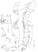 issued from : S. Ohtsuka, G.A. Boxshall & H.S.J. Roe in Bull. nat. Hist. Lond. (Zool.), 1994, 60 (2). [p.139, Fig.21, , B-I]. Female (E China Sea): B, genital double-somite (ventral); E, exopod of A2; F, Md (mandibular palp); G, praecoxal arthrite, coxal endite and endopod of Mx1 (endopod indicated by arrowhead); H, 1st and 2nd praecoxal endites of Mx2; I, basal spine of Mx2. cd = copulatory duct; cp = copulatory pore; g = gonopore; rd = receptacle duct; o, oviduct; s, spermatophore remnant; sr = seminal receptacle. Scales in mm. Nota: Cephalosome partly fused with 1st pedigerous somite. A1 asymmetrical, left longer than right. Exopod of A2 indistinctly 7-segmented (setal formula: 0, 1, 1, 1, 1, 1, 3 (2 setae and vestigial element). Md: Mandibular palp with endopod 1-segmented (almost fused with basis), with 2 setae of unequal lengths; 1st exopod segment with long seta, 5th segment with 2 normal setae of unequal lengths. Mx1 with praecoxal arthrite with 2 slender setae; coxal endite with 1 short seta; coxal epipodite with 5 setae; basal seta absent; endopod (indicated by arrowhead) 1-segmented, bulbous, with short seta terminally. Mx2 with 1st and 2nd praecoxal endites with 1 seta plus 1 vestigial element and 2 setae respectively; basal spine with 2 rows of short spinules proximally. Mxp with 4th and 5th endopod segments with relatively well developed innermost seta, middle-length seta and well-chitinized, long seta; 6th segment with setae (a and b) not reduced. Genital double-somite asymmetrical, left gonopore and copulatory pore absent, right gonopore located near posteroventral margin of genital double-somite, anterior half opening, covered by oval flap (possibly derived from leg 6); copulatory pore small, oval in shape (approximately 0.004 mm in long axis and 0.001 mm in short axis), near inner distal corner of gonopore (copulatory pore blocked by spermatophore remnant); single seminal receptacle large, about half width of somite, located ventromedially; copulatory duct short, curved. Anal operculum triangular (as in M. acutioperculum). Male: C, left A1 (segments I to XVII; D, idem (segments XVIII to XXVIII).
|
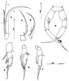 issued from : S. Ohtsuka, G.A. Boxshall & H.S.J. Roe in Bull. nat. Hist. Lond. (Zool.), 1994, 60 (2). [p.144, Fig.26, A-F, G]. Female: A, 4th endopod segment of Mxp (innermost seta indicated by arrowhead); B, 5th endopod segment of Mxp (innermost seta indicated by arrowhead); C, 6th endopod segment of Mxp; D, exopod of P1 (anterior); E, right P5 (posterior); F, left P5 (anterior). P1 3rd exopodal segment with 1 outer lateral spine. P5 coxae separate from intercoxal sclerite and from basis, on both sides; right basis with outer thicker plumose seta than left; endopod represented by large plumose seta; exopod distinctly 1-segmented, with medial and subterminal bipinnate spine along outer lateral margin and spiniform seta terminally. Male: G, P5 (anterior). Nota: Left A1 segments X and XI, XI and XII, and XIV and XV only partly fused near posterior margin; sutures between segments IX and X, and XXI and XXII visible on both surfaces; suture between segments XII to XIV visible only on dorsal surface. P5 with coxae and intercoxal sclerite almost fused; basis separate from coxa; endopod absent; right basal seta thicker and longer than left; right exopod 3-segmented, 2nd segment elongate, with outer seta subterminally and 2 patches of setules and minute seta along inner terminal margin, 3rd segment small, bearing large unipinnate terminal spine fused with segment; left exopod 3-segmented, 2nd segment without inner seta and patches of setules, 3rd segment similar to that of right leg. Scales in mm.
|
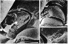 issued from : S. Ohtsuka, G.A. Boxshall & H.S.J. Roe in Bull. nat. Hist. Lond. (Zool.), 1994, 60 (2). [p.140, Fig.22, A-C]. Female (SEM micrographs of genital double-somite): A, copulatory pore indicated by arrow and gonopore (scale bar = 10 µm); B, copulatory pore indicated by arrow (scale bar = 30 µm) ; C, copulatory pore (scale bar = 5 µm)
|
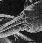 ssued from : S. Ohtsuka, G.A. Boxshall & H.S.J. Roe in Bull. nat. Hist. Lond. (Zool.), 1994, 60 (2). [p.143, Fig.25 A]. Female (SEM micrographs): A, detail of segments IV to VIII of A2 exopod. Scale bar = 10 µm.
| | | | | NZ: | 1 | | |
|
Carte de distribution de Metacalanus sp.1 par zones géographiques
|
| | | | | | | Loc: | | | E China sea (off Okinawa) | | | | N: | 1 | | | | Lg.: | | | (263) F: 0,84-0,81; M: 0,77; {F: 0,81-0,84; M: 0,77} | | | | Rem.: | For Ohtsuka & al., 1994 (p.140) the asymmetry in the genital structures of the female seems to be unique to the new species (M. inaequicornis, M. curvicornis M. acutioperculum have paired genital structures, namely, a pair of gonopores and copulatory pore); as far as the genital structures are concerned, the new species exhibits a more derived state than these species. P5 is similar to that of M. inaequicornis and is the more primitive state in having an endopodal seta and 3 spines on the exopod. | | | Dernière mise à jour : 17/01/2015 | |
|
|
 Toute utilisation de ce site pour une publication sera mentionnée avec la référence suivante : Toute utilisation de ce site pour une publication sera mentionnée avec la référence suivante :
Razouls C., Desreumaux N., Kouwenberg J. et de Bovée F., 2005-2024. - Biodiversité des Copépodes planctoniques marins (morphologie, répartition géographique et données biologiques). Sorbonne Université, CNRS. Disponible sur http://copepodes.obs-banyuls.fr [Accédé le 25 avril 2024] © copyright 2005-2024 Sorbonne Université, CNRS
|
|
|
|


;)
;)
;)
;)

