|
|
 |
Fiche d'espèce de Copépode |
|
|
Calanoida ( Ordre ) |
|
|
|
Arietelloidea ( Superfamille ) |
|
|
|
Heterorhabdidae ( Famille ) |
|
|
|
Neorhabdus ( Genre ) |
|
|
| |
Neorhabdus latus (Sars, 1905) (F,M) | |
| | | | | | | Syn.: | Heterorhabdus latus Sars, 1905 c (p.9, Rem.F);
Hemirhabdus latus : Sars, 1925 (p.232, figs.F,M); Sewell, 1932 (p.306, 307, Rem.); 1948 (p.503); C.B. Wilson, 1950 (p.238); Tanaka, 1964 a (p.28, figs. juv.F); Grice & Hulsemann, 1965 (p.224); Owre & Foyo, 1967 (p.80, figs.F,M); Grice & Hulsemann, 1967 (p.18); Itoh, 1970 a (p.5: tab.1); Roe, 1972 (p.277, tabl.1, tabl.2); Vives, 1982 (p.293); Lozano Soldevilla & al., 1988 (p.59); Suarez & Gasca, 1991 (tab.2); Suarez, 1992 (App.1); Suarez-Morales & Gasca, 1998 a (p.109); Hernandez-Trujillo & Esqueda-Escargera, 2002 (in Appendix); Boxshall & Halsey, 2004 (p.125: fig.F);
Hemirhabdus truncatus: Sewell, 1932 (p.306, figs.F, Rem.); ? 1947 (p.181); 1948 (part., p.330, 530, 533, 547);
Neorhabdus truncatus: Heptner,1972 a (p.58);
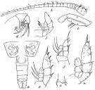
Mesorhabdus truncatus A. Scott, 1909 (p.132, Descr.F, figs.F) | | | | Ref.: | | | Heptner, 1972 a (p.58); Chihara & Murano, 1997 (p.820, Pl.116); Ohtsuka & al., 1997 (p.579, figs.F); Park, 2000 (p.59, Redescr.F,M, figs.F,M, Rem.); Vives & Shmeleva, 2007 (p.320, figs.F,M, Rem.) | 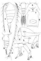 issued from : T. Park in Bull. Scripps Inst. Oceanogr. Univ. California, San Diego, 2000, 31. [p.186, Fig.34]. Female: a, habitus (left side); b, c, urosome (left, dorsal, respectively); d, e, f, forehead (left, dorsal, ventral, respectively); g, genital somite (ventral); h, left A1 (ventral); i, left A2 (posterior); j, left Md (posterior); k, masticatory edge of right Md (posterior). Prosome about 3/4 length of body and about 2.7 times as long as urosome. Rostrum bearing 2 long filaments. Urosome about 3/10 length of body. Anal somite about as long as 3rd urosomal somite. Combined lengths of anal somite and left caudal ramus about as long as genital somite. Dorsal appendicular setae of caudal rami similar in length. A1 reaching about posterior end of caudal ramus; Four lobes of 1st segment with 1+2+2+2 setae and 0+1+1+2 aesthetes; 2nd to 19th segments each with 2 setae (1 middle and 1 dista), number of aesthetes seems variable to a certain extent. A2 basipod a little shorter than 1st endopodal segment. Measured along lateral marginns, 2nd endopodal segment a little longer than 1/2 length of 1st, without posterior appendicular setae on either inner or outer lobe. Exopod slender, much shorter than endopod, with inner marginal seta on last segment reduced to a vestige. Md: 1st tooth from basal spine very small, 2nd and 3rd similar size and shape, tricuspid, and about as long as basal spine. 4th tooth present only on right side (Fig.34-k), is low and more or less triangular. Ventralmost tooth sicklelike, only a little longer than 3rd tooth, initilly diverging somewhat from and then curved toward dorsal teeth. Mandfibular palp about 7/10 length of mandibular blade, with exopod far short of reaching distal end of endopod.
|
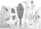
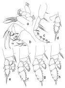 issued from : T. Park in Bull. Scripps Inst. Oceanogr. Univ. California, San Diego, 2000, 31. [p.187, Fig.35]. Female: a, left Mx1 (posterior); b, left Mx2 (posterior); c, right Mxp (anterior); d, P1 (endopod omitted), posterior; e, P1 (anterior); f, P2 (anterior); g, P3 (anterior); h, P4 (anterior). asp = anterior spine; psp = posterior spine; tsp = terminal spine. Mx1 relatively small, with 6 setae on 1st lobe, no setae on 2nd and 3rd inner lobes, 3 or 4 setae on outer lobe, 2 or 3 setae on basis, 6 setae on endopod, and 4 setae on exopod.
|
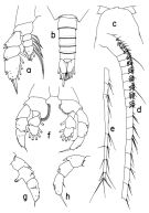 issued from : T. Park in Bull. Scripps Inst. Oceanogr. Univ. California, San Diego, 2000, 31. [p.188, Fig.36]. Female: a, P5 (anterior). Male: b, urosome (dorsal); c, forehead (dorsal); d, segments 1-16 of left A1 (ventral); e, segments 17-25 of left A1 (ventral); f, P5 (anterior); g, exopod of right P5 (posterior); h, exopod of left P5 (posterior). Nota: Coxa in P1 to P4 large, about as wide as long, coxa about as long as endopod and almost twice as long as basis (when measured along medial margins. P5 anteriorly, 1st and 2nd exopodal segments each with 1 pore, 3rd exopodal segment with 4 pores.
|
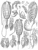 Issued from : G.O. Sars in Résult. Camp. Scient. Prince Albert I, 69, pls.1-127 (1924). [Pl.LXIV, figs.1-16]. As Macrorhabdus latus. Female (from Canary Islands & Madeira Islands): 1, habitus (dorsal); 2, idem (lateral left side); 3, rostrum; 4, A2; 5, left Md; 6, right Md (biting edge); 7, idem (inner part, enlarged); 8, Mx1; 9, Mx2; 10, Mxp; 11, P1; 12, P2; 13, P3; 14, P5. Male: 15, habitus (dorsal); 16, P5.
|
 Issued from : R.B.S. Sewell in Mem. Indian Mus., 1932, X (continued). [p.306, Fig.101]. As Hemirhabdus truncatus. Female (from off SE Sri Lanka): a, last thoracic segment and urosome (dorsal); b, proximal segments of A1; c, Md (biting edge); d, Mx2; e, Mxp (2nd basal segment); f, P1; g, P2; h, P3; j, P5.
|
 issued from : A. Scott in Siboga-Expedition, 1909, XIX a. [Plate XXXIX, Figs.12-21]. As Mesorhabdus truncatus. Female (from Banda Sea): 12, habitus (dorsal); 13, forehead (lateral); 14, last thoracic and genital segments (left side); 15, rostrum; 16, A1; 17, Md; 18, Mx2; 19, Mxp; 20, P1; 21, P5.
|
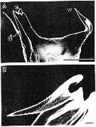 issued from : S. Ohtsuka, H.Y. Soh & S. Nishida in J. Crustacean Biol., 1997, 17 (4). [p.581, Fig.2, A, B]. Female (from S. Japan): A, left mandibular cutting edge; B, terminal part of ventralmost tooth. v = ventral tooh; c = central teeth; d = dorsal tooth. Scale bars : 0.100 mm (A); 0.010 mm (B).
|
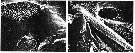 issued from : S. Ohtsuka, H.Y. Soh & S. Nishida in J. Crustacean Biol., 1997, 17 (4). [p.584, Fig.5, C-D]. Female (S Japan): C, left side of labrum (posterior surface) with gland openings (small arrow = type-3 gland opening); D, idem right side of labrum (posterior surface) with gland openings (small arrow = gland type-3; asterisk = small gland opening). Scale bars: 0.010 mm.
|
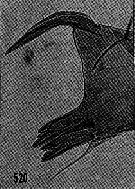 issued from : H.B. Owre & M. Foyo in Fauna Caribaea, 1, Crustacea, 1: Copepoda. Copepods of the Florida Current. 1967. [p.79, Fig.520]. As Hemirhabdus latus. Female (from 23°20'N, 82°55'W): 520, Md (biting edge of gnathobase).
|
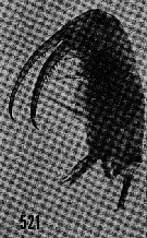 issued from : H.B. Owre & M. Foyo in Fauna Caribaea, 1, Crustacea, 1: Copepoda. Copepods of the Florida Current. 1967. [p.79, Fig.521]. As Hemirhabdus latus. Female: 521, Mx2.
|
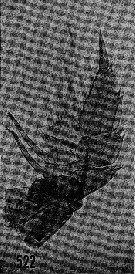 issued from : H.B. Owre & M. Foyo in Fauna Caribaea, 1, Crustacea, 1: Copepoda. Copepods of the Florida Current. 1967. [p.79, Fig.522]. As Hemirhabdus latus. Female: 522, P5.
|
 Neorhabdus latus Neorhabdus latus female and male: 1 - In 3rd lobe of Mx2, anterior spine much smaller than 1st terminal spine (Fig.35-b); Mx1 with 2nd inner lobe missing (Fig.35-a). 2 - 2nd distal seta of Mxp coxa about twice as long as 1st (Fig.35-c); in 3rd lobe of Mx2, 1st terminal spine more than twice length of anterior spine (Fig.35-b).
| | | | | NZ: | 7 | | |
|
Carte de distribution de Neorhabdus latus par zones géographiques
|
| | | | | | 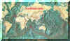 Carte de 1996 Carte de 1996 | |
| | | | Loc: | | | Canary Islands, Madeira Is., off Portugal, Brazil, G. of Mexico, Florida, Indian (SW & W), Banda Sea, Malay Archipelago, Japan (Sagami), central Pacif. (E-W, N-S), off SW Galapagos.
Type locality: North Atlantic (between 27°43'N-34°02'N, 12°21'W-19°09'W. | | | | N: | 13 | | | | Lg.: | | | (1) F: 7,1; (199) F: 8,5; (824) F: 8,91-6,75; M: 8,08-7,16; {F: 6,75-8,91; M: 7,16-8,08} | | | | Rem.: | bathypélagique.
Voir aussi les remarques en anglais | | | Dernière mise à jour : 24/01/2015 | |
|
|
 Toute utilisation de ce site pour une publication sera mentionnée avec la référence suivante : Toute utilisation de ce site pour une publication sera mentionnée avec la référence suivante :
Razouls C., Desreumaux N., Kouwenberg J. et de Bovée F., 2005-2024. - Biodiversité des Copépodes planctoniques marins (morphologie, répartition géographique et données biologiques). Sorbonne Université, CNRS. Disponible sur http://copepodes.obs-banyuls.fr [Accédé le 25 avril 2024] © copyright 2005-2024 Sorbonne Université, CNRS
|
|
|
|


;)
;)
;)
;)
;)
;)
;)
;)
;)
;)

;)
