


|
|
|
|
© copyright 2005-2026 Sorbonne University, CNRS Toute utilisation est soumise à autorisation préalable Any use is subjected to preliminary authorization webmaster |
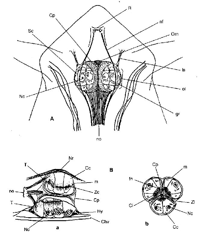 Fig. G15 : Structure de l'oeil nauplien chez Eucalanus elongatus (Calanoïde) (d'après Vaissière, 1961).
A : Vue dorsale. Cp: cellule pigmentaire; gr: granulations réfringentes; ls: ligament suspenseur; Nc: noyau de la cellule cristalline; nf: nerf frontal; no: nerf optique; ol: ocelle latéral; Om: ocelle médian; R: rostre; Sc: sphère cristalline. B : Coupe frontale (a) et longitudinale (b); Cc: cellule cristalline; Chv: chitine de la paroi ventrale; Ci: corps interne; fn: fibre nerveuse; Hy: hypoderme; m: membrane des ocelles; Nr: nerf rostral; T: terminaisons neurofibrillaires; Zc: zone chromophobe; Zl: zone lamellaire. |