|
|
 |
|
Calanoida ( Order ) |
|
|
|
Metridinidae ( Family ) |
|
|
|
Metridia ( Genus ) |
|
|
| |
Metridia curticauda Giesbrecht, 1889 (F,M) | |
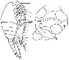
| | | | | | | Syn.: | no Sars, 1920 c (p.10) | | | | Ref.: | | | Giesbrecht, 1892 (p.340, 346, 773, Descr.F, figs.F); Giesbrecht & Schmeil, 1898 (p.108, Rem. F); Wolfenden, 1911 (p.286); Farran, 1929 (p.209, 259); Sewell, 1932 (p.248); Brodsky, 1950 (1967) (p.297, figs.F,M); Vervoort, 1951 (p.121, figs.F,M, Rem.); 1957 (p.123, Rem.); Tanaka, 1963 (p.15, figs.F,M); Vaupel-Klein, 1970 (p.4, 33); Minoda, 1971 (p.36); Bradford, 1971 b (p.23, figs.F,M, Rem.); Gardner & Szabo, 1982 (p.322, figs.F,M); Szabo & Gardner, 1986 (p.1560: Appendix, Rem.); Razouls, 1994 (p.138, figs.F,M); Chihara & Murano, 1997 (p.837, tab.6); Hure & Krsinic, 1998 (p.61, 101); Bradford-Grieve & al., 1999 (p.884, 948, figs.F,M); Bradford-Grieve,1999 b (p.113, figs.F,M, Rem., figs.178, 192); Vives & Shmeleva, 2007 (p.371, figs.F,M, Rem.); Takenaka & al., 2013 (p.201, DNA sequences vs luciferase) | 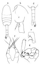 issued from : Tanaka O. in Publs Seto Mar. Biol. Lab., 1963, XI (1). [p.16, Fig.154]. Female: a, habitus (dorsal); b, forehead (left lateral side); c, last thoracic segment and genital somite (left lateral side); d, last thoracic segment and urosome (dorsal); e, 1st segment of endopodite of P2; f, P5. Male: g, P5.
|
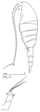 issued from : J.M. Bradford-Grieve in The Marine Fauna of New Zealand: Pelagic Calanoid Copepoda. National Institute of Water and Atmospheric Research (NIWA). NIWA Biodiversity Memoir, 111, 1999. [p.114, Fig.76]. Female (34°32.5'S, 157°31.5'E): A, habitus (lateral left side); B, P5.
|
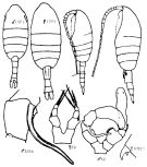 Issued from : K.A. Brodskii in Calanoida of the Far Eastern Seas and Polar Basin of the USSR. Opred. Fauna SSSR, 1950, 35 (Israel Program for Scientific Translations, Jerusalem, 1967) [p.297, Fig.203]. Female (from NW Pacif.): habitus (dorsal and lateral left side); S2En1, hooks on endopodal segment 1 of P2; S5, P5, P5. Male: habitus (dorsal and lateral left side); S5, P5; S5Le, P5 (Le = left leg).
|
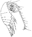 issued from : W. Vervoort in Verh. K. ned. Akad. Wet., Afd. Natuurk., 1951, (Sect. 2) 47 (2). [p.122, Fig.65]. Female (from ± 66°.5S, 11°W): habitus (lateral). Nota: Head and 1st thoracic somite separated, 4th and 5th fused. Postero-lateral margin of the last thoracic somite is not smoothly rounded, but produced into a minor knob, distinctly visible in dorsal and lateral views. A1 reaching the middle of the abdomen (or even slightly longer); the 8th-9th segments completely fused and these segments partly fused with the 10th.
|
 issued from : W. Vervoort in Verh. K. ned. Akad. Wet., Afd. Natuurk., 1951, (Sect. 2) 47 (2). [p.123, Fig.66]. Female: a, urosome (dorsal); b, right P4 (anterior); c, P5 (anterior). Nota: Genital segment formed by fusion of the first 3 urosomal somites, slightly longer than the combined length of the intermediate abdominal segment and anal segment, which two segments have the same length. Caudal rami slightly longer than the anal segment and about twice as long as wide. Exopod of P4 with very short endspine (exactly 1/3 the length of the 3rd exopodal segment); in this respect the females, here, differ from Gisebrecht's account.
|
 issued from : W. Vervoort in Verh. K. ned. Akad. Wet., Afd. Natuurk., 1951, (Sect. 2) 47 (2). [p.124, Fig.67]. Male: a, habitus (lateral); b, P5 (anterior; lt = left foot, rt = right foot). Nota: The males, found in company of the females, strickling resemble the female in external appearance. Clasping A1 on the left side; 21-segmented; right A1 as in female. Mouth parts not reduced. P1 to P4 as in female. P5 resemble those of M. brevicauda, but differ in the proportional length of the segments and other details.
|
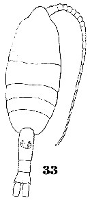 Issued from : W. Giesbrecht in Systematik und Faunistik der Pelagischen Copepoden des Golfes von Neapel und der angrenzenden Meeres-Abschnitte. - Fauna Flora Golf. Neapel, 1892, 19 , Atlas von 54 Tafeln. [Taf.33, Fig.33]. Female: 33, habitus (dorsal).
|
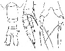
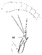 Issued from : W. Giesbrecht in Systematik und Faunistik der Pelagischen Copepoden des Golfes von Neapel und der angrenzenden Meeres-Abschnitte. - Fauna Flora Golf. Neapel, 1892, 19 , Atlas von 54 Tafeln. [Taf.33, Figs.4, 15]. Female: 4, A1 (proximal segments); 15, P5.
|
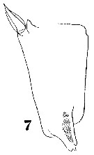 Issued from : W. Giesbrecht in Systematik und Faunistik der Pelagischen Copepoden des Golfes von Neapel und der angrenzenden Meeres-Abschnitte. - Fauna Flora Golf. Neapel, 1892, 19 , Atlas von 54 Tafeln. [Taf.32, Fig.7]. Female: 7, exopodal segment 1 of P2 (anterior view).
|
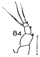 Issued from : J.M. Bradford in N.Z. Oceanogr. Inst., 1971, 206, Part 8, No 59. [p.22, Fig.84]. Female (from Ross Sea): 84, P5. Scale bar: 100 µm. Nota: P5 bears small external spines on all three segments, whereas Vervoort (1951) does not figure the terminal segment spine and Giesbrecht does not figure the penultimate segment spine.
|
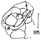 Issued from : J.M. Bradford in N.Z. Oceanogr. Inst., 1971, 206, Part 8, No 59. [p.22, Fig.85]. Male (from Ross Sea): 85, P5. Scale bar: 100 µm. Nota: P5 has more spines on various segments than Vervoort's (1951) figures, but the Ross Sea specimens agree with those figured by Tanaka (1963) from deep water off the Izu region (Japan).
|
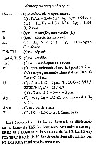 Issued from : C. Razouls in Ann. Inst. océanogr., Paris, 1994, 70 (1). [p.138]. Caractéristiques morphologiques de Metridia curticauda femelle et mâle adultes. Terminologie et abbréviations: voir à Calanus propinquus.
| | | | | Compl. Ref.: | | | Wilson, 1942 a (p.194); Lysholm & al., 1945 (p.32); Sewell, 1948 (p.349, 502, 557, 567, 570, 573, 574); C.B. Wilson, 1950 (p.264); Furuhashi, 1966 a (p.295, vertical distribution in Oyashio/Kuroshio regions, Table 6, 8, 9, 10); Park, 1970 (p.477); Roe, 1972 (p.277, tabl.1, tabl.2); 1972 b (p.543); Björnberg, 1973 (p.337, 387); Deevey & Brooks, 1977 (p.256, tab.2, Station "S"); Kovalev Shmeleva, 1982 (p.84); Vives, 1982 (p.293); Hopkins, 1985 (p.197, Table 1, gut contents); Brinton & al., 1986 (p.228, Table 1); Zmijewska, 1987 (tab.2a); Hopkins & Torres, 1988 (tab.1); Hattori, 1991 (tab.1, Appendix); Ward & al., 1995 (p.195, Table 2); Errhif & al., 1997 (p.422); Suarez-Morales & Gasca, 1998 a (p.110); Voronina & Kolosova, 1999 (p.71); Atkinson & Sinclair, 2000 (p.46, 50, 51, 54, 55, zonal distribution); Lapernat, 2000 (tabl. 3, 4); Razouls & al., 2000 (p.343, tab. 5, Appendix); Ward & Shreeve, 2001 (p.50, tab.3); Yamaguchi & al., 2002 (p.1007, tab.1); Kahle & Zauke, 2003 (p.409, metals concentration); Yamaguchi & al., 2004 (p.480, tab.2); Hsiao & al., 2004 (p.326, tab.1); Ikeda & al., 2006 (p.1791,Table 2); Schnack-Schiel & al., 2008 (p.1045: Tab.2; Fernandes, 2008 (p.465, Tabl.2); Gaard & al., 2008 (p.59, Table 1, N Mid-Atlantic Ridge); Galbraith, 2009 (pers. comm.); Park & Ferrari, 2009 (p.143, Table 4, Appendix 1, biogeography); Homma & Yamaguchi, 2010 (p.965, Table 2); Medellin-Mora & Navas S., 2010 (p.265, Tab. 2); Hsiao & al., 2010 (p.179, Table III, trace metal concentration); Homma & al., 2011 (p.29, Table 2, 3, 5, abundance, feeding pattern: suspension feeders); Michels & al., 2012 (p.369, Table 1, occurrence frequency); Ward & al., 2014 (p.305, Table 6, 7, seasonal and abundance in the ''Discovery'' Investigations in the 1930s); Belmonte, 2018 (p.273, Table I: Italian zones) | | | | NZ: | 18 | | |
|
Distribution map of Metridia curticauda by geographical zones
|
| | | | | | | | | | | | | | | | | | |


;)
;)
;)
;)
;)
;)
;)
;)
;)
;)
;)
;)
