|
|
 |
|
Calanoida ( Order ) |
|
|
|
Clausocalanoidea ( Superfamily ) |
|
|
| |
| | | |
| Stephidae Sars, 1902 ( Clausocalanoidea ) | | Ref.: | Sars, 1902 (1903) (p.60); Gurney, 1931 a (p.85); Rose, 1933 a (p.161); Fosshagen, 1970 (p.37); Andronov, 1974 a (p.1005); Bowman, 1976 (p.189, Rev., Genera Key); Razouls, 1982 (p.389); 1993 (p.311); Bowman & Abele, 1982 (p.9); Brodsky & al., 1983 (p.144, 147); Huys & Boxshall, 1991 (p.419, 467); Bradford-Grieve, 1994 (p.132, Déf.); Chihara & Murano, 1997 (p.913); Ohtsuka & Huys, 2001 (p.461); Boxshall & Halsey, 2004 (p.15; 49; 199: Def.; p.200: Genrera Key); Vives & Shmeleva, 2007 (p.847); Kos, 2016 (p.8, Def.)
Bradford-Grieve J.M., (2002 onwards). Key to calanoid copepod families. Version 1 : 2 oct 2002. http://www.crustacea.net/crustace/calanoida/index.htm  | | Rem.: | 2 (or 4 G.): Miostephos, Parastephos, Speleohvarella, Stephos.
For Kos (2016, p.8, 9) the genera Miostephos Bowman, 1976 and Parastephos Sars, 1902 iare junior synonyms from Stephos.
Hyperbenthic forms, living just above the sea floor in relatively shallow coastal waters or in anchialine habitats, and only occasionally swimming up into the water column. | 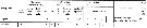 issued from : E.L. Markhaseva & F.D. Ferrari in Invert. Zool., 2005, 2 (2). [p.162, Table 4] Setation of oral parts in females Stephidae (Clausocalanoidea) and ancestral condition of setation. |
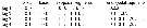 Issued from : G.A. Boxshall & S.H. Halsey in An Introduction to Copepod Diversity. The Ray Society, 2004, No 166, part. I. [p.199]. Armature formula of swimming legs P1 to P4. Nota: Endopod of P4 on right side modified in Parastephos: middle endopodal segment elongate, distal segment with strong spinules on surface and with proximal seta on inner margin slender, denticulated and spiniform. - Female P5 minute ; with coxae and intercoxal sclerite fused to form transverse coxal plate ; each leg tipically uniramous, comprosing basis and single tapering rxopodal process ; basis and process often fused ; process usually ornamented with spinule rows or teeth. - Male P5 powerfully developed and strongly asymmetrical ; right leg slender, uniramous, 4 to 8-segmented, terminal segment often tapering, spiniform ; left leg uniramous, tipically 5-segmented with 4th segment robust and swollen, distal segment variable in form, often spoon-like ; both 4th and 5th segments often with additional processes ; right leg reduced to 2-segmented rudiment in Miostephos- Eggs released into waters. | | | | | (1) Miostephos Bowman, 1976 | |
| | Syn.: | Stephos: Kos (2016, p.8, 9, Rem.) | | Ref.: | Bowman, 1976 (p.185); Razouls, 1982 (p.393); 1993 (p.311); Bradford-Grieve, 1994 (p.132, Def.); Mauchline, 1998 (p.87: F; p.89: M); Bradford-Grieve, 2004 (p.287); Boxshall & Halsey, 2004 (p.200) | | Rem.: | Hyperbenthic forms. Type: Miostephos cubrobex Bowman,1976.
Total: 2 spp.
Diagnosis from Bradford-Grieve (1994, p.95) :
- As for the family definition.
- Urosome female 3-segmented.
- Urosome male 5-segmented.
- Female P5 symmetrical, 3-segmented, last segment small and extending into a point.
- Male P5 very aasymmetrical; left leg slender, elongate, 6-segmented; right leg rudimentary, 3-segmented, similar to the female P5.
For Kos (2016, p.8, 9) the genus Miostephos Bowman, 1976 is a junior synonym from Stephos. | | | | (2) Parastephos Sars, 1902 | |
| | Ref.: | Sars, 1902 (1903) (p.65); van Breemen, 1908 a (p.86); Sars, 1919 (1921) (p.5); Rose, 1933 a (p.163); Razouls, 1982 (p.393); Fleminger, 1988 (p.309); Razouls, 1993 (p.311); Bradford-Grieve, 1994 (p.132, Def.); Mauchline, 1998 (p.87, 89: F; p.89: M); Bradford-Grieve, 2004 (p.287); Boxshall & Halsey, 2004 (p.200) | | Rem.: | Hyperbenthic forms. Type: Parastephos pallidus Sars,1902. Total: 3 spp.
Diagnosis from Bradford-Grieve (1994, p.132) :
- As for the family definition.
- Female urosome powerfully developed with 1 or more of the anterior segments decorated with spines.
- Anterior of head, A1, and mouthparts very similar in the male and female; and similar to those of Stephos except that the distal part of the mandibular blade is considerably more expanded, with the outermost cutting tooth very large and claw-like.
- Swimming legs not very powerful.
- male P4 asymmetrical.
- Female P5 symmetrical or asymmetrical, 3-segmented; with a common basal segment, one globular segment and a terminal claw-like segment with a sharp lateral projection near its base.
Male P5 largely developed and very asymmetrical; right leg slender, terminating in a strong denticulate claw; left leg much coarser, with the antepenultimate segment the largest.
After Kos (2016, p.9, 10) this genus is a junior synonym from Stephos | | | | (3) Speleohvarella Krsinic, 2005 | |
| | Ref.: | Krsinic, 2005 (p.608) | | Rem.: | Type species: Speleohvarella gamulini. Total: 1 sp.
Diagnosis Female:
- Body oval.
- Prosome 5-segmented, cephalosome separate from 1st pedigerous somite, 4th and 5th fused.
- Urosome 4-segmented.
- Genital double-somite slightly longer than wide, with genital area symmetrical.
- Caudal rami asymmetrical, right longer than left, with 4 long terminal setae and 1 inner seta on inner border.
- A1 symmetrical, 24-segmented (ancestral segments II-IV, X-XI and XXVII-XXVIII fused.
- P1-P4 as described for type species.
- 3rd exopodal segment of P4 with outer distal spine exceptionally long, about half the length of terminal spine.
- A2 exopod 7-segmented, 2nd free segment corresponding to ancestral segments II-IV.
- Mouthparts identical in both sexes.
Mx1 displaying 13 armature elements including 4 posyrtior setae on praecoxal arthrite and 2 setae on basal exite.
- P5 symmetrical, uniramous, 3-segmented, terminal segment with spinules in distal half along their margins;
Diagnosis Males:
- Body similar in habitus to female.
- Urosome 5-segmented.
- A1 similar in fusion patterns and armature to that of female but with ancestral segments XII and XIII naked.
- A2, Md, Mx1, Mx2, Mxp and P1-P4 similar to those of female
- P5 asymmetrical, uniramous, right leg short, 3-segmented, with coxa incorporating intercoxal sclerite; left leg elongate and 5-segmented | | | | (4) Stephos T. Scott, 1892 | |
| | Syn.: | Möbianus Giesbrecht, 1892 (p.51, 205); Stephus Giesbrecht & Schmeil, 1898 (p.29); Wolfenden, 1908 (p.21); 1911 (p.204) | | Ref.: | T. Scott, 1892 (p.245); Sars, 1902 (1903) (p.61); van Breemen, 1908 a (p.82, spp. Key); Rose, 1933 a (p.161, spp. Key); Strömgren, 1969 (p.5, Rem.); Fosshagen, 1970 (p.37); Razouls, 1982 (p.389); Huys & Boxshall, 1991 (p.62, 63, 324); Jacoby & Greenwood, 1991 (p.405, species coexisting); Razouls, 1993 (p.311); Bradford-Grieve, 1994 (p.132, Def.); Chihara & Murano, 1997 (p.913); Mauchline, 1998 (p.87, 89: F; p.89: M); Bradford-Grieve, 1999 a (p.13, 25: Rem. M); Bradford-Grieve, 2004 (p.287); Boxshall & Halsey, 2004 (p.200); Vives & Shmeleva, 2007 (p.848, spp. Key) ; Krsinic, 2012 (p.1536, Rem.); Kos, 2016 (p.9, Def., Key F & M); Brylinski & Courcot, 2019 (Rem. p.8-9: the ''hyaline sheath', zoogeography'). | | Rem.: | Hyperbenthic forms. Generally described from specialised habitats, like anchialine caves, aquarium tanks (Naples, Concarneau, Roscoff). Type: Stephos minor T. Scott,1892. Total: 32 spp. (of which 1 doubtful) and 1 juv. unidentified. For Brylinski & Coucot (2019, p.366), the number of species is underestimated, (see Jacoby & Greenwiood (1991) and according to Walter (1986), further studies of epibenthic studies should produce new records, allowing elucidation of phylogenetic relationships and zoogeographical distribution.
Diagnosis from Bradford-Grieve (1999a, p.13):
Female :
Small copepods, usually less than 1mm long.
- Cephalosome and pediger somite 1 may be fused or separate, pedigers 4 and 5 fused or partially fused.
- Posterior prosomal margins may be symmetrical or asymmetrical.
- Rostrum represented by a rounded hnob.
- Caudal rami symmetrical or asymmetrical with 4 subequal terminal setae (III-VI), outer border naked or with 1 minute outer setule (II), inner border with 1 small seta (VII).
- Urosome 4-segmented.
- Genital double-somite symmetrical or asymmetrical; genital aperture may be oriented obliquely (in S. lucayensis and S. hastatus, only a single seminal receptacle is retained); the common operculum may be produced into a posteriorly directed spiniform process. When carrying a spermatophore, the genital double-somite is covered by a hyaline sheath of a complex structure.
- A1 24-segmented (apparently ancestral segment I-II, III-IV and X-XI fused), segments 1 and 2 partly fused, some of proximal segments bearing minute spinules along the posterior border; segment 12-23 sometimes each with a small lamellate plate.
- A2 Coxa and basis separate or partly fused; coxa with 1 seta, basis with 2 setae; exopod 6-7-segmented; segment 1 with 1-2 setae, segment 2 with 2-3 setae, segments 3-6 with 1 seta each, the terminal segment usually with 1 seta proximal to mid-length and 3 terminal setae; endopod segment 1 with 1-2 setae, segment 2 with 13-15 setae.
- Md Gnathobase only slightly expanded with a straight row of moderately incised teeth; basis with 3-4 setae; endopod segment 1 with 4 setae; endopod 2 with a totazl of 9-11 setae; exopod and endopod of approximately equal in length.
- Mx1 praecoxal arthrite with 8-9 terminal spines, and 3-4 posterior setae; coxal endite, and basal endites 1 and 2 with 3, 4, and 5 setae respectively; basis and endopod fused, endopod with 14-16 setae; exopod with 11 setae; coxal epipodite with 9 setae; basal exite without seta.
- Mx2 endites 1-5 with 3-6, 3, 3, 3, and 3-4 setae, respectively; endopod endite 6 with 1 seta and the remaining segments with 4-5 setae.
- Mxp syncoxa with groups of 2, 3, and 3-4 setae from proximal to distal; basis and fused endopod segment 1 with 3 inner setae, 2 distal setae and a group of long spinules; free endopod segments 1-5 with 4, 4, 2-3, 0-1+2-3, 0-1 +3 setae respectively.
- Swimming legs P2-P4 with terminal spine finely serrated on the outer border; spine and seta formula as in table.
- P5 uniramous, symmetrical or asymmetrical, coxae fused with the intercoxal sclerite; basis separate or fused with a single tapering terminal segment, which is usually variously ornamented with setae, spines and teeth.
Male :
- Similar to females in size and general form but may vary in the degree of symmetry or asymmetry of the posterior prosome.
- Urosome of 5 somites, somite 1 with genital opening on left; somite 2 with or without ventral process.
- A1 usually 24-segmented but is equiped with more asethetascs along the anterior margin especially proxymally.
- A2, Md, Mx1, Mx2, Mxp and P1-P4 as in female.
- P5 Powerfully developed as a grasping organ; elongate, uniramous, and very asymmetrical. Right leg slender, usually 4-segmented; coxa and basis sghort, unarmed, segment 3 elongate, unarmed, but may have a distal process, segment 4 either comprising 2 processes set at right angles, or with a proximal process or is simple, spiniform. Left leg 5-segmented; coxa and basis short, unarmed; segment 3 short, unarmed; segment 4 often robust, unarmed or well ornamented with spinules or spiniform processes distally along inner margin; segment 5 complex, with 2-9 elongate, usually articulated, appendages and additional hairs or spines (antarcticum and longipes with segment 5 plain, bifurcate, processes not articulated).
For Brylinski & Courcot (2019) the hyaline sheath in Stephos is a relict structure derived from an ancestral complex spermatophore. It has been suggested that structures can prevent subsequent mating by other males (Ohtuska & Huys, 2001). The efficiency of such a system is uncertain in S. cryptospinus since it does not always cover the genital opening female, and furthermore, the aithors found one specimen carrying two hyaline sheaths. For the authors that is not a general feature of the genus because it is not possible to observe this structure in S. scotti, despite examinating numerus females both bearing spermatophore (s) and without. The origin of this sheat is uncertain but Fosshagen (1970) suggested that it seems yo be secreted from two gland-like structures, one on either syde of the last prosomal segment next of the genital segment; the authors did not find these glandular pores in either sex on any of the species studied using SEM.
The hyaline sheat differs in shape in each species but the precise position can vary from specimen to specimen within a species (Fosshagen, 1970), as also shown in wS. cryptospinus. If the sheath were secreted by the female, it should be a priori located at the same place. Brylinski & Courtot suggest it is produced by the male at the same time as the spermatophore. The variable positioning on the female might reflect variation in copulatory behaviour of males. This structure was suspected to belonging to the spermatophore and to function as an attachment material (Krsinic (2012), but the authors were unable to show a direct connection with the spermatophpre in the five specimens studied by SEM.
Most calanoid copepods prodce simple tubular spermatopjores as in Stephos, but several genera produce complex spermatophores as for example some species of the subgenus Tortanus (Atortus) having highly complex coupling device (Barthélémy & al., 2003). | | Remarks on dimensions and sex ratio: | | The mean female size is 0.905 mm (n = 28; SD = 0.3011) and the mean male size is 0.816 mm (n = 26; SD = 0.2584). The size ratio (Male : Female) is 0.91 (n = 25; SD = 0.0758). The sex ratio (Female : Male is 1,04. | | |
|
|
 Any use of this site for a publication will be mentioned with the following reference : Any use of this site for a publication will be mentioned with the following reference :
Razouls C., Desreumaux N., Kouwenberg J. and de Bovée F., 2005-2025. - Biodiversity of Marine Planktonic Copepods (morphology, geographical distribution and biological data). Sorbonne University, CNRS. Available at http://copepodes.obs-banyuls.fr/en [Accessed November 02, 2025] © copyright 2005-2025 Sorbonne University, CNRS
|
|
|
|


