|
|
 |
|
Calanoida ( Order ) |
|
|
|
Clausocalanoidea ( Superfamily ) |
|
|
|
Phaennidae ( Family ) |
|
|
|
Onchocalanus ( Genus ) |
|
|
| |
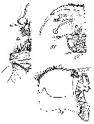
Onchocalanus trigoniceps Sars, 1905 (F,M) | |
| | | | | | | Syn.: | Onchocalanus steueri Pesta, 1920 (p.516, figs.F); Rose, 1933 a (p.136, figs.F); Gamulin,1939; Sewell, 1948 (p.508, 509); C.B. Wilson, 1950 (p.274); Rose & Vaissière, 1952 a (p.118); Mazza, 1966 (p.70); Vives, 1978 a (p.265); 1982 (p.291); Kovalev & Shmeleva, 1982 (p.83); Lozano Soldevilla & al., 1988 (p.58);
O. magnus : With, 1915 (p.225, figs.F);
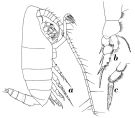
no O. trigoniceps: C.B. Wilson, 1942 a (p.200, fig.F); Fagetti, 1962 (p.23) | | | | Ref.: | | | Sars, 1905 b (p.20, Rem.F); 1925 (p.144, figs.F,M); Rose, 1929 (p.26); Sewell, 1929 (p.176); Rose, 1933 a (p.137, figs.F,M); Jespersen, 1934 (p.83); 1940 (p.33); Sewell, 1947 (p.139, figs. juv.F,M); Vervoort, 1950 a (p.12, figs.F, Rem.); Tanaka, 1960 a (p.115, figs. juv.F); Vervoort, 1965 (p.30, Rem.); Mazza, 1967 (p.178, figs.F,M, juv.); Tanaka & Omori, 1967 (p.250); Bradford & al., 1983 (p.67, Rem., figs.F,M); Park, 1983 a (p.329, figs.F,M, Rem.); Tanaka & Omori, 1992 (p.267, figs.M, as trigonoceps); Chihara & Murano, 1997 (p.854, Pl.140,141: F,M); Bradford-Grieve & al., 1999 (p.880, 929, figs.F,M); Lapernat, 1999 (p.25, Rem.); Vives & Shmeleva, 2007 (p.692, figs.F,M, Rem.) | 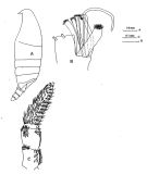 issued from : J.M. Bradford, L. Haakonssen & J.B. Jillett in Mem. N.Z. Oceanogr. Inst., 1983, 90. [p.69, Fig.38]. Female: A, habitus (lateral left side); B, Mx2; C, P5. Nota: The female specimen from the south-west Pacific deviates slightly from Vervoort's (1950) description.
|
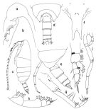
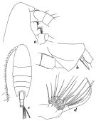
 issued from : T. Park in Antarct. Res. Ser. Washington, 1983, 39 (5). [p.331, Fig.7]. Female: a, forehead (dorsal); b, posterior part of metasomal segments and urosome (dorsal); c, forehead (lateral); d, last thoracic segments of metasome and urosome (lateral right side); e, genital segment (lateral); f, idem (ventral); g, A1; h, A2; i, Md; j, Mx1.
|
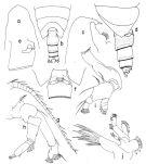
 issued from : T. Park in Antarct. Res. Ser. Washington, 1983, 39 (5). [p.332, Fig.8]. Female: a, Mx2; b, Mxp; c, distal part of Mxp; d, exopod and endopod of P1 (posterior); e, P1 (anterior); f, P2 (posterior); g, P3 (posterior); h, P4 (posterior); i, P5 (posterior).
|
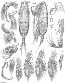
 issued from : T. Park in Antarct. Res. Ser. Washington, 1983, 39 (5). [p.334, Fig.9]. Male: a, forehead (dorsal); b, idem (lateral); c, habitus (lateral left side); d, last thracic segments and urosome (dorsal); e, idem (lateral left side); f, rostrum (anterior); g, Mxp; h, distal part of Mxp; i, P1 (anterior); j, P5 (posterior).
|
 issued from : O. Tanaka in Publ. Sero Mar. Biol. Lab., 1960, VIII (1). [p.116, Fig.95]. As Onchocalanus trigonoceps. immature Female (copepodid stage V from Sagami and Suruga Bays): a, habitus (dorsal); b, forehead (lateral); c, last thoracic segment (lateral left side); d, Mx2; e, P1; f, P2; g, P5. Immature female (copepodid stage IV): h, last thoracic segment and abdomen (lateral left side); i, rostrum (anterior aspect); j, P5. Body size : 4,56 mm ( stage IV); 5394 mm (stage V).
|
 issued from : O. Tanaka & M. Omori in Publs Seto Mar. Biol. Lab., 1992, 35 (4/5). [p.267, Fig.8, a-f]. Male (Izu region, Japan): a, forehead (lateral); b, posterior part of metasome and urosome (dorsal); c, Mx1; d, Mx2; e, right P5; f, left P5.
|
 Issued from : W. Vervoort in Zool. Verh., Leiden, 1950, 10. [p.14, Fig.3]. Female (from Banda Sea): a, habitus (lateral right side); b, left P1 (posterior aspect); c, left P5 (posterior).
|
 Issued from : W. Vervoort in Zool. Verh., Leiden, 1950, 10. [p.15, Fig.4]. Female (from Banda Sea): a, forehead (lateral); b, posterior part of cephalothorax and genital segment (lateral right side); c, habitus (dorsal); d, right Mx2. Nota: Head and 1st thoracic segment separated, 4th and 5th fused; line of fusion visible on the dorsal surface. The abdominal segments and furca in the proportional lengths 30:27:19:4:11 = 100. Anal segment telescoped into 3rd urosomal segment.
|
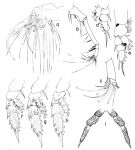
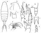
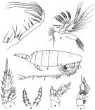
 Issued from : G.O. Sars in Résult. Camp. Scient. Prince Albert I, 69, pls.1-127 (1924). [Pl.XL, figs.1-17]. Female: 1, habitus (dorsal); 2, idem (lateral left side); 3, forehead (lateral); 4, rostrum (frontal view); 5, A2; 6, Md; 7, Mx1; 8, Mx2; 9, Mxp; 10, sensory appendix on basal segment of Mxp; 11, P1; 12, P2; 13, P3; 14, P4; 15, P5. Male: 16, Mx2; 17, P5.
|
 issued from : R.B.S. Sewell in The John Murray Expedition, 1933-34, Scientific Reports, VIII (1), 1947. [p.140, Fig.34]. Female stage V (from Arabian Sea): A, habitus (lateral right side; B, Mx1; C, Mxp; D, P1; E, P2; F, basal portion of P3. male immature: G, P5. Nota: The P5 legs are each composed of 4 segments, instead of 3 as in the female. Remarks female: The proportional lengths of the cephalothorax and abdomen as 77 to 23. The proportional lengths of the various segments of the body (cephalon to caudal rami) as 370:148:91:82:42:20:106:69:42:7:23 = 1000. Head and 1st pediger segment partly fused (the line of separation being visible dorsally, 4th and 5th separate. Rostrum composed of 2 stout spinous processes directed vertically. The posterior thoracic margins prolonged backwards in a triangular flap, at the apex of which is a short spinous projection. A number of small pores, passing through the exoskeleton are present on the cephalon and thoracic segments. On either side of the genital orifice is a flap that is pointed at its anterior end; the genital orifice is surrounded by an area that is profusely covered with short hairs; the posterior segments are hairy.
|
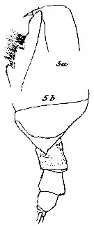 issued from : C. With in The Danish Ingolf-Expedition, Copepoda I, 1915, III, 4. [Pl. VII, Fig. 5, a-b]. As Onchocalanus magnus. Female (from 61°30'N, 17°08'W): a, head (lateral); b, urosome (lateral). Nota: A1 24-segmented, reaching almost to the end of the body. Urosome about 1/3 as long as prosome. the comparative length of the three first abdominal segments and the caudal rami 32, 22, 16 and 11. Anal somite scarcely visible.
|
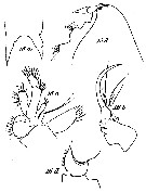 issued from : C. With in The Danish Ingolf-Expedition, Copepoda I, 1915, III, 4. [Pl. VII, Fig. 16, a-d]. As Onchocalanus magnus. Female: a, rostrum (anterior view); b, left Mx1; c, left Mx2; d, P2 (left endopod). Nota: Head with distinct eyes, but without trace of frontal keel or spine. Rostrum bifurcate, with fairly long, slightly divergent spines, one of which, at least, possesses a slender filament.
|
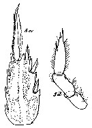 issued from : C. With in The Danish Ingolf-Expedition, Copepoda I, 1915, III, 4. [Pl. VII, Fig. 5, c-d]. As Onchocalanus magnus. Female: c, left P4 (posterior view); d, left P5 (anterior view). Nota: P4 has a marginal setae on the basis and hairs; a glandular pore on the exopodal segment 2; the posterior surface is covered with groups of short spinules or fairly long setae, with all intermediary steps between;
|
 issued from : C. With in The Danish Ingolf-Expedition, Copepoda I, 1915, III, 4. [Pl. VII, Fig. 5, e-g]. ]. As Onchocalanus magnus. Female: e, labrum (anterior view) and labial lobes; f, labrum (oral view); g, serrula 6-dentata and lobus labialis (anterior view). Nota: In front of the prominent labrum (proper a prominent epistoma (fig.5a), which is densely covered with long bristles (fig.5e) (the bristles are neathy arranged into two anterior groups), about three lateral groups of shorter hairs and a single marginal row. The oral surface of the labrum is anteriorly densely covered with a number of short minute prickles; scarcely distinguished from these, two lateral groups of hairs. Almost in the middle, on each side, about three almost completely fused groups of short bristles, and more behind, an oblique group of delicate hairs. In the middle densely placed granules are found in transverse areas. No distinct lamina labialis; in front of the serrula 6-dentata a large inner group of short granules, and an outer longitudinal row of fairly long hairs. Behind, a horse-shoe shaped group of granules on each side and, well separated from this (fig.5 e), irregularly placed short hairs. Along the inner margin of the labial lobes inwards short spines, and more outwards long bristles.
| | | | | Compl. Ref.: | | | Massuti Alzamora, 1942 (p.110); Sewell, 1948 (p.330, 365, 501, 508, 521, 525, 527, 530); C.B. Wilson, 1950 (p.274); Rose & Vaissière, 1952 a (p.118); Mazza, 1966 (p.70); Grice & Hulsemann, 1967 (p.16); Vinogradov, 1968 (1970) (p.261); Roe, 1972 (p.277, tabl.1, tabl.2); Bainbridge, 1972 (p.61, Appendix Table III: occurrence); Björnberg, 1973 (p.331, 388); Deevey & Brooks, 1977 (p.256, tab.2, Station "S"); Vives, 1982 (p.291); Scotto di Carlo & al., 1984 (p.1045); Lozano Soldevilla & al., 1988 (p.58); Madhupratap & Haridas, 1990 (p.305, fig.5: vertical distribution night/day; fig.7: cluster); Padmavati & al., 1998 (p.347); Hure & Krsinic, 1998 (p.101); Voronina & Kolosova, 1999 (p.71); Lapernat, 2000 (tabl. 3, 4); Razouls & al., 2000 (p.343, tab. 5, Appendix); Lapernat & Razouls, 2001 (tab.1); Vukanic, 2003 (139, tab.1); Shimode & al., 2005 (p.113 + poster); Licandro & Icardi, 2009 (p.17, Table 4); Park & Ferrari, 2009 (p.143, Table 4, fig.1, Appendix 1, biogeography); Mazzocchi & Di Capua, 2010 (p.426); Belmonte, 2018 (p.273, Table I: Italian zones) | | | | NZ: | 15 | | |
|
Distribution map of Onchocalanus trigoniceps by geographical zones
|
| | | | | | | | | | | | | | | 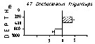 Issued from : M. Madhupratap & P. Haridas in J. Plankton Res., 12 (2). [p.312, Fig.5]. Issued from : M. Madhupratap & P. Haridas in J. Plankton Res., 12 (2). [p.312, Fig.5].
Vertical distribution of calanoid copepod (mean +1 SE), abundance No/100 m3. 47- Onchocalanus trigoniceps.
Night: shaded, day: unshaded.
Samples collected from 6 stations located off Cochin (India), SE Arabian Sea, November 1983, with a Multiple Closing Plankton Net (mesh aperture 300 µm), in vertical hauls at 4 depth intervalls (0-200, 200-400, 400-600, 600-1000 m). |
| | | | Loc: | | | Antarct. (Weddell Sea, Indian, S & SE Pacif.), sub-Antarct. (Indian, SE Pacif.), G. of Guinea (off Ghana, off Lagos), off NW Cape Verde Is., off Mauritania, Canary Is., Portugal, Bay of Biscay, Azores, off Bermuda: Station "S" (32°10'N, 64°30'W), Sargasso Sea, S Strait of Davis, S Iceland, Medit. (Alboran Sea, W Basin., Ligurian Sea, Tyrrhenian Sea, Ionian Sea, off Malta, Adriatic Sea), Arabian Sea, Laccadive Is., Indian, Indonesia-Malaysia, China Seas, Japan (Izu region), off NE Marquesas Is., off NE Easter Is., off Galapagos, New Zealand off Peru, Chile | | | | N: | 32 | | | | Lg.: | | | (1) F: 7,5; (7) F: 8,3; (9) F: 6,7; (11) F: 7,33; (15) F: 9,16-7,83; M: 6,75-6,5; (16) F: 6,91; M: 5,63-5,42; (11) F: 7,33; (64) F: 7,45; M: 4,85; (70) F: 8,9-8,2; (199) F:?; (207) F: 6,24-6,2; (340) F: 5,1; (449) F: 7-6; (867) F: 7,95-8,8; M: 6,6-6,95; {F: 5,10-9,16; M: 4,85-6,95} | | | | Rem.: | meso & bathypelagic.
Sampling depth (Antarct., sub-Antarct.) : 0-2000 m. Sargasso Sea: 1000-1500 m (Deevey & Brooks, 1977, station "S");
The female shows minor variations according to the description of Vervoort (1950 a).
For Park (1983, p.335) this species is closely related to O. paratrigoniceps (See remarks in this species).
Certain locality records in the Pacific are considered as doubtful.
This form resembles a O. paratrigoniceps. | | | Last update : 16/06/2020 | |
|
|
 Any use of this site for a publication will be mentioned with the following reference : Any use of this site for a publication will be mentioned with the following reference :
Razouls C., Desreumaux N., Kouwenberg J. and de Bovée F., 2005-2026. - Biodiversity of Marine Planktonic Copepods (morphology, geographical distribution and biological data). Sorbonne University, CNRS. Available at http://copepodes.obs-banyuls.fr/en [Accessed March 25, 2026] © copyright 2005-2026 Sorbonne University, CNRS
|
|
|
|


;)
;)
;)
;)
;)
;)
;)
;)
;)
;)
;)
;)
;)
;)

