|
|
 |
|
Calanoida ( Order ) |
|
|
|
Clausocalanoidea ( Superfamily ) |
|
|
|
Aetideidae ( Family ) |
|
|
|
Bradyidius ( Genus ) |
|
|
| |
Bradyidius plinioi Campaner, 1978 (F,M) | |
| | | | | | | Syn.: | Bradyidius plinioi plinioi & B. plinioi minor Campaner, 1978 (p.865, figs.F, juv.M, p.871, Descr.F, figs.F). | | | | Ref.: | | | Björnberg & al., 1981 (p.605, 630, figs.F); Alvarez, 1984 (p.94, Descr.M, figs.M, Rem.: p.96); Othman & Greenwood, 1987 (p.1137, Rem.); Bradford-Grieve & al., 1999 (p.879, 918, figs.F, as plinoi) | 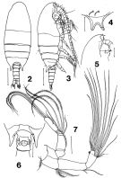 issued from : A.F. Campaner in Ciência e Cultura, 1978, 30 (7). [p.866, Figs. 2-7]. As Bradyidius plinoi plinoi. Female (from continental shelf, Brazil): 2, habitus (dorsal); 3, idem (lateral right side); 4, rostrum (frontal ciew); 5, forehead (lateral); ; 6, last thoracic segment and genital segment (ventral); 7, A2. Scale bars: 0.500 mm (2, 3); 0.200 mm (5); 0.100 mm (6, 7); 0.050 mm (4). Nota: Head and 1st thoracic segment separate (with a very thin suture).
|
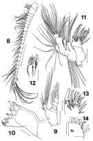 issued from : A.F. Campaner in Ciência e Cultura, 1978, 30 (7). [p.867, Figs. 8-14). As Bradyidius plinoi plinoi. Female: 8, A1; 9, Md (mandibular palp); 10, Md (cutting edge); 11, Mx1; 12, 3rd inner lobe of Mx1; 13, 1st inner lobe of Mx1 (dorsal); 14, idem (ventral). Scale bars: 0.500 mm (8); 0.100 mm (9, 11); 0.050 mm (10, 12-14).
|
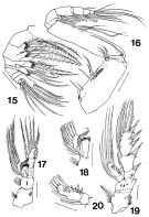 issued from : A.F. Campaner in Ciência e Cultura, 1978, 30 (7). [p.868, Figs.15-19). As Bradyidius plinoi plinoi. Female: 15, Mx2; 16, Mxp; 17, P1; 18, endopodite of P1; 19, P2; 20, endopodite of P2. Scale bars: 0.100 mm. Nota: Occurrence of a group of large spines on endopodite 2 of P2.
|
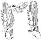 issued from : A.F. Campaner in Ciência e Cultura, 1978, 30 (7). [p.869, Figs. 21-23). As Bradyidius plinoi plinoi. Female: 21, P3; 22, endopodite of P3; 23, P4. Scale bars: 0.100 mm (21-23). Scale bars: 0.100 mm (21-23). Nota: Occurrence of a group of large spines on endopodite 2 of P3.
|
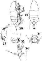 issued from : A.F. Campaner in Ciência e Cultura, 1978, 30 (7). [p.867, Figs. 29-34). As Bradyidius plinoi minor. Female: 29, habitus (dorsal); 30, idem (lateral right side); ; 31, postero-lateral corners of last thoracic segment and genital segment (ventral); 32, exopodite of A2 (partial); 33, terminal part of 1st basipodite of Mxp; 34, Md (exopodite). Scale bars: 0.500 mm (29-30; 0.050 (31-34).
|
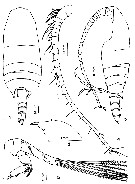 Issued from : M.A. Jimenez Alvarez inBolm. Zool. Univ,. S. Paulo, 1984, 8. [p.95, Figs.1-5] Male (28°36'S, 47°55'W): 1-2, habitus (dorsal and lateral, respectively); 3, forehead with rostrum (lateral); 4, A1; 5, A2. Nota: Cephalon and 1stpedigerous segment partially fused (dorsal visible suture almost complete, 4th and 5th thoracic segments almost completely fusd, ending in potero-lateral projections, acutely pointed and slightly asymmetrical ; the left, a little longer, overreaches the posterior margin of genital segment. Urosome 5-segmented. Proportional length of segments and caudal rami 21.7 : 20.0 : 16.7 : 15.0 : 26.7 = 100. Genital segment slightly asymmetrical. Anal sefment wery reduced and only ventrally visible. Caudal rami with 1 short lateral seta and 3 terminal setae. Left A1 24-segmented, reaching the caudal rami ; segments bearing thick sensory setae up to 13th segment included, and, the thinner ones up to the end and other very thin setules. Right A1 lost. A2 2nd basipod with 2 setae ; exopod and endopod almost equal in length ; 2nd endopodal segment with 5 subterminal and 4 terminal setae on the lateral margin ; externally there is a group of very small spines.
|
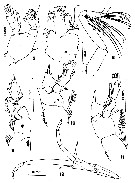 Issued from : M.A. Jimenez Alvarez inBolm. Zool. Univ,. S. Paulo, 1984, 8. [p.103, Figs.6-12] Male; 6, Md (palp); 7, Mx1; 8, Mxp; 9, P1; 10, P2; 11, P3; 12, P5. Mandibular palp with well developed basipod and 1 internal marginal seta. The two-segmented endopod is short. The exopod has 1 long thick seta on each of its four first segments, the last has 2 setae. The gnathobase was not found in this specimen. Mx1 reduced with 3 setae on the 1st external lobe, 9 setae on the exopod ; no setae on the 1st internal lobe, 2setae on the other internal lobe and 6 fine setae plus a very long one on the fused 2nd basipod and endopod. 1st basipod of Mxp without setae ; 2nd basipodal segment with 2 long plumose setae and 1 very shot.. The 1st to 5th endopodal segments bear respectively 5 : 3 : 4 : 3 : 4 setae. P5 asymmetrical and very big. Left leg, shorter than the right with the basipods and the 3 segments of the exopod long, the last segment the shortest and bearing some setae ; endopod reduced, 1-segmented in both legs.. The 1st segment of the exopod of the right leg is much longer than the second, and this is curved with a bulge on the lateral internal margin.
| | | | | Compl. Ref.: | | | Markhaseva, 1998 (p.250, Fig.2) | | | | NZ: | 1 | | |
|
Distribution map of Bradyidius plinioi by geographical zones
|
| | | | Loc: | | | S Brazil | | | | N: | 3 | | | | Lg.: | | | (218) F: 2,52-2,45 (plinioi plinioi); 1,95-1,85 (plinioi minor); (217) M: 1,5; {F: 1,85-2,52; M: 1,50} | | | | Rem.: | hyperbenthic.
For Campaner (1978, p.871) the most interesting morphological feature of this species is the occurrence of a group of large spines on each endopodal segment 2 of P2 and P3, found in most of the Phaennidae and Scolecitrichidae.
After Alvarez (1984, p.96) , Campaner established two subspecies distinguishing one from the other mostly by their body lengths. The two groups also showed differences in the relatve proportions of the prosome : urosome and length : width of the genital segment, in the morphology and length of the metasome margins, in the length and width of the last segment of the exopod of A2 and in the length of the median-terminal seta of the 1st basipod of Mxp (a little longer in B. plinioi minor).
Concerning the body length, Gurney (1931, p.36) and successors (see in the database) wrote that differences in the length of the specimens have little value or none at all for the distinction of species, because their lengths may be due to varying retractions of the body segments during fixation and conservation, and the environmental physical (mainly temperature during the development from nauplius to adult), and biological conditions (food quanlity and quantity). | | | Last update : 30/12/2014 | |
|
|
 Any use of this site for a publication will be mentioned with the following reference : Any use of this site for a publication will be mentioned with the following reference :
Razouls C., Desreumaux N., Kouwenberg J. and de Bovée F., 2005-2025. - Biodiversity of Marine Planktonic Copepods (morphology, geographical distribution and biological data). Sorbonne University, CNRS. Available at http://copepodes.obs-banyuls.fr/en [Accessed August 30, 2025] © copyright 2005-2025 Sorbonne University, CNRS
|
|
|
|


;)
;)
;)
;)
;)
;)
;)
