|
|
 |
|
Calanoida ( Order ) |
|
|
|
Diaptomoidea ( Superfamily ) |
|
|
|
Pontellidae ( Family ) |
|
|
|
Calanopia ( Genus ) |
|
|
| |
Calanopia aurivilli Cleve, 1901 (F,M) | |
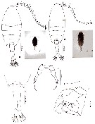
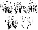
| | | | | | | Syn.: | Calanopia aurivillei : Rose, 1956 (p.461) | | | | Ref.: | | | Cleve, 1901 (p.37, figs.F); Thompson & Scott, 1903 (p.235, 251); A. Scott, 1909 (p.181, figs.F,M); Sewell, 1912 (part. p.368); 1914 a (p.232, Rem.); Früchtl, 1924 b (p.57); Farran, 1929 (p.210, 273); Sewell, 1932 (p.341); Farran, 1936 a (p.115); Kasturirangan, 1963 (p.47, 48, figs.F,M); Saraswathy, 1966 (1967) (p.85); Silas & Pillai, 1976 (p.784, figs.F,M, Rem.); Bradford-Grieve, 1999 b (p.183, figs.F,M, figs.184, 194); Mulyadi, 2002 (p.36, figs.F,M, Rem.); Othman & Toda, 2006 (p.306, F,M); Phukham, 2008 (p.73, figs.F,M); Lacuna & al., 2013 (p.64, figs.F,M, Rem.: p.77, morphological variation) | 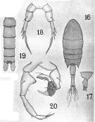 issued from : A. Scott in Siboga-Expedition, 1909, XIX a. [Plate XLVIII, Figs.16-20]. Female (from off Galle, Ceylon): 16, habitus (dorsal); 17, last thoracic and genital segments (left side)18, P5. Male: 19, urosome (dorsal); 20, P5.
|
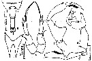 issued from : E.G. Silas & P.P. Pillai in J. mar. biol. Ass. India, 1973 (1976), 15 (2). [p.784, Fig.2]. Female (from Indian Ocean): a, urosome (dorsal); b, rostrum (anterior view); c, P5. Male: d, right A1 (geniculate); e, P5. Scale as in Calanopia minor.
|
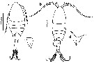
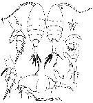 issued from : Mulyadi in Treubia, 2002, 32; [p.38, Fig.9]. Female (from Indonesian waters): a, habitus (dorsal); b, cephalon (lateral); c, metasomal somite 5 and urosome (lateral); d, rostrum (anterior view); e, P2; f, P3; g, P5. Male: h, habitus (dorsal); i, geniculate region of right A1; j, P5.
|
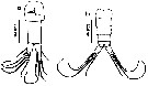
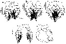
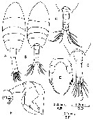 issued from : B.H.R. Othman & T. Toda in Coastal Mar. Sc., 2006, 30 (1). [p.307, Fig.2]. Female (from Sister's Island, Singapore): A, habitus (dorsal); D, posterior part of prosome and urosome (dorsal); E, P5. Male: B, habitus (dorsal); C, posterior part of prosome and urosome (dorsal); F, P5. Nota Female: Prosome to urosome length ratio 2.04 : 1. - Cephalon without lateral hook. - Prosome produced into posteriorly directed acute process. - Urosome 2-segmented. - Genital segment shorter than anal segment. - P5 symmetrical and uniramous; exopod 1-segmented apex terminates in 3 spines, inner being distinctly longer and plumose at its distal margin. Nota Male: Prosome to urosome length ratio 1.99 : 1. - Body similar to female. - Cephalon wiithout hook. - Right A1 geniculate. - Urosome 5-segmented, naked. - P5 asymmetrical and chelate; right leg 4-segmented, proximal inner margin of basis swollen, exopodal segment 1 with well developed thumb, claw spoo-shapede, slightly swollen at tip with 1 outer marginal seta, 1 terminal and 2 inner marginal; left leg basis swollen and gibbose, exopodal segment 1 with distolateral seta, terminal segment with 2 unequal apical spines.
|
 issued from : N. Phukham in Species diversity of calanoid copepods in Thai waters, Andaman Sea (Master of Science, Univ. Bangkok). 2008. [p.155, Fig.29]. Female (from W Malay Peninsula): a, habitus (dorsal); b, urosome; c, P5. Male: d, habitus (dorsal); e, P5. Body length after the drawings: F = 1.114 mm; M = 1.129 mm.
|
 ssued from : M.L.D.G. Lacuna, D.C. Sagrado, R.O. Mejorada, D.D. Simyunn & M.J.J. Pueblos in ABAH Bioflux, 2013, 5 (1). [p.65, Fig.13]. Female (from Iligan Bay, Mindanao): a, habitus (dorsal); c, rostrum. Male: b, habitus (dorsal); d, rostrum. Nota: The posterior corners of the last metasomal segment for both sexes are drawn out into spines.
|
 ssued from : M.L.D.G. Lacuna, D.C. Sagrado, R.O. Mejorada, D.D. Simyunn & M.J.J. Pueblos in ABAH Bioflux, 2013, 5 (1). [p.65, Fig.14]. Female: a, urosome (ventral). Male: b, urosome (dorsal).
|
 ssued from : M.L.D.G. Lacuna, D.C. Sagrado, R.O. Mejorada, D.D. Simyunn & M.J.J. Pueblos in ABAH Bioflux, 2013, 5 (1). [p.66, Figs.15, 16]. Female: a, left A1; b, right A1. Male: c, left A1; d, right A1. Nota: Both A1 female 17-segmented. For the male, left A1 17-segmented, while the right A1 is 12-segmented, geniculation at segments 7 and 8. The antennules of both sexes are terminated with 2 setae, each being distributed at the inner and outer margins.
|
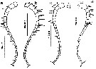
 ssued from : M.L.D.G. Lacuna, D.C. Sagrado, R.O. Mejorada, D.D. Simyunn & M.J.J. Pueblos in ABAH Bioflux, 2013, 5 (1). [p.67, Fig.17]. Female: a, P1; b, P2; c, P3; d, P4, e, P5.
|
 ssued from : M.L.D.G. Lacuna, D.C. Sagrado, R.O. Mejorada, D.D. Simyunn & M.J.J. Pueblos in ABAH Bioflux, 2013, 5 (1). [p.67, Fig.18]. Male: a, P1; b, P2; c, P3; d, P4; e, P5.
|
 Calanopia aurivilli Calanopia aurivilli. Female: 1 - P5 exopod 1-segmented. 2 - Exopod of P5 with 4 spines; 3 - Exopod of P5 with 3 small spines and 1 long spine; 4 - Exopodal segment of P5 with 3 subequal small lateral spines and 1 long medial spine (longer than segment itself). Male: 1 Left P5 longer than right one ; basis of left P5 swollen proximally. 2 2nd exopodal segment of right P5 nearly 2/5 length of 1st exopodal segment ; coxa of right P5 about or less than 1.4 times as long as basis. 3 Basis of left P5 swollen proximally without any spines or processes.
| | | | | Compl. Ref.: | | | Sewell, 1948 (p.323); C.B. Wilson, 1950 (p.174); Krishnaswamy, 1953 (p.138); Patel, 1975 (p.660); Carter, 1977 (1978) (p.36); Madhupratap & Haridas, 1986 (p.105, tab.1); Othman & al., 1990 (p.561, 564, Table 1); McKinnon, 1991 (p.471); Mauchline, 1998 (tab.8); Jitlang & al., 2008 (p.65, Table 1); Fernandes, 2008 (p.465, Tabl.2); McKinnon & al., 2008 (p.844: Tab.1); Cornils & al., 2010 (p.2076, Table 3); Shanthi & Ramanibai, 2011 (p.132, Table 1); Maiphae & Sa-ardrit, 2011 (p.641, Table 2, 3, Rem.); Jagadeesan & al., 2013 (p.27, Table 3, seasonal abundance); | | | | NZ: | 4 | | |
|
Distribution map of Calanopia aurivilli by geographical zones
|
| | | | | | | | | | Loc: | | | SE South Africa (off Durban, Natal), NW India (Saurashtra coast), Trivandrum coast, Sri Lanka, Madras, G. of Mannar, Bay of Bengal, Burman coast, Andaman Sea, W Malay Peninsula, Strait of Malacca (Singapore), G. of Thailand, Indonnesia (S Java, Semau Sound, SE Celebes Sea, Philippines, Mindanao (Iligan Bay), Viet-Nam (Cauda Bay), off Hong Kong, Australia (G. of Carpentaria, Noth West Cape, Great Barrier), off New Zealand (NW North Island)
Type locality: Semau Sound (southwestern Timor). | | | | N: | 23 | | | | Lg.: | | | (5) F: 1,34; M: 1,12; (34) F: 1,32-1,27; M: 1,18-1,17; (35) F: 1,45; M: 1,38; (334) F: 1,34; M: 1,12; (530) F: 1,2; M: 1,1; (795) F: 1,3; M: 1,1; (1086) F: 1,05-1,20; M: 0,98-1,08; (1087) F: 1,2-1,25; M: 1,05-1,1; (1130) F: 1,56; M: 1,50; {F: 1,05-1,56; M: 0,98-1,50} | | | | Rem.: | epiplagic. Inshore. Neritic.
For Mulyadi (2002, p.37)this species resembles C. minorand C. americana.
After Lacuna & al. (2013, p.77) the P5 of both sexes showed absence of setules on the inner and outer margins, however these attibutes were seen on the same organism from the Indian waters. Further, difference can also be observed between results from Mindanao and those in Indonesian waters. For instance, in Indonesian waters 18 segments for yje female A1 were observed, but our study described only 17, female swimming leg 5 reported as 3-segmented in Indonesian waters, however in Mindanao waters 4-segmented only. These variations exhibited between the same species from different places may be interpreted as an evidence of some kind of genetic switch mechanism. | | | Last update : 03/12/2020 | |
|
|
 Any use of this site for a publication will be mentioned with the following reference : Any use of this site for a publication will be mentioned with the following reference :
Razouls C., Desreumaux N., Kouwenberg J. and de Bovée F., 2005-2026. - Biodiversity of Marine Planktonic Copepods (morphology, geographical distribution and biological data). Sorbonne University, CNRS. Available at http://copepodes.obs-banyuls.fr/en [Accessed March 25, 2026] © copyright 2005-2026 Sorbonne University, CNRS
|
|
|
|


;)
;)
;)
;)
;)
;)
;)
;)
;)
;)

