|
|
 |
|
Calanoida ( Order ) |
|
|
|
Diaptomoidea ( Superfamily ) |
|
|
|
Pontellidae ( Family ) |
|
|
|
Calanopia ( Genus ) |
|
|
| |
Calanopia thompsoni A. Scott, 1909 (F,M) | |
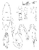
| | | | | | | Syn.: | Calanopia parathompsoni Gaudy, 1969 (p.45, figs.F,M); Silas & Pillai, 1973 (1976) (p.773); Conway & al., 2003 (p.123, figs.F,M, Rem.) | | | | Ref.: | | | A. Scott, 1909 (p.178, figs.F,M); Nicholls, 1944 (p.12); Sewell, 1932 (p.342, figs.F,M, juv., Rem.); Mori, 1937 (1964) (p.89, figs.M); C.B. Wilson, 1950 (p.176, fig.M); Shen & Lee, 1963 (p.582); Chen & Zhang, 1965 (p.95, figs.F,M); Saraswathy, 1966 (1967) (p.84); Kos, 1972 (Vol. I, figs.F, M, Rem.); Kasahara & al., 1974 (p.170, fig. egg); Li & Fang, 1984 (p.391, figs. N, Juv., Rem.); Silas & Pillai, 1976 (p.790, figs.F,M, Rem.); Zheng Zhong & al., 1984 (1989) (p.252, figs.F,M); Chihara & Murano, 1997 (p.865, Pl.146: F,M); Conway & al., 2003 (p.124, figs.F,M, Rem.); Othman & Toda, 2006 (p.308, F); Phukham, 2008 (p.75, figs.F,M); Al-Aidaroos & al., 2016 (p.19, Redescr. F,M, figs.F,M, Rem.) | 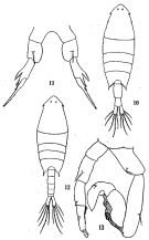 issued from: Q.-c Chen & S.-z. Zhang in Studia Marina Sinica, 1965, 7. [Pl.38, 10-13]. Female (from E China Sea): 10, habitus (dorsal); 11, P5 (posterior). Male: 12, habitus (dorsal); 13, P5 (posterior).
|
 Issued from : R.B.S. Sewell in Mem. Indian Mus., 1932, X (continued). [p.343, Fig.112]. Female (from N Indian): d, P4 (forma typica); e, P5 (variety from station 587). Male: a, right A1 (segments of the knee-joint); b, A2; c, Mxp; f, P5.
|
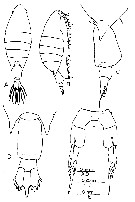
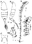
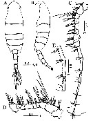
 issued from : A. Scott in Siboga-Expedition, 1909, XIX a. [Plate XLIX, Figs.1-8]. Female (from Indonesia-Malaysia): 1, habitus (dorsal); 2, last thoracic and genital segments (left side); 3, rostrum; 4, A1; 5, P5. Male: 6, urosome (dorsal); 7, right A1; 8, P5.
|
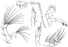
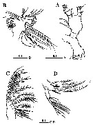
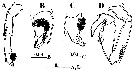
 issued from : E.G. Silas & P.P. Pillai in J. mar. biol. Ass. India, 1973 (1976), 15 (2). [p.790, Fig.6]. Female (from Indian Ocean): a, urosome (dorsal); b, rostrum (anterior view); c, P5; d, P5 (terminal part enlarged). Male: e, right A1 (geniculate part); f, P5. Nota: Ummerkutty (1969) collected an abnormal male from Gulf of Manaar. Scale as in C. minor.
|
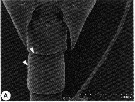
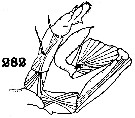 issued from : C.B. Wilson in U.S. National Museum. Bull.100, 14 (4), 1950. [Pl.20, Fig.282]. Male: 282, P5.
|
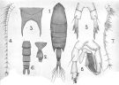
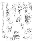
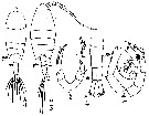
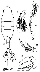 issued from : T. Mori in The pelagic Copepoda from the neighbouring waters of Japan, 1937 (2nd edit., 1964). [Pl.40, Figs.9-13]. Male (from Ki-Channel): 9, habitus (dorsal); 10, right A1; 11, A2; 12, Mxp; 13, P5. Nota: 2nd abdominal segment without spine on the right side. - Exopodite of left P5 moderately broad; The apex of the 2nd segment fusrnished with 2 spines, and a broad, flat, finely denticulated process. The terminal segment of the right exopodite is spoon-like. The shape of P5 is slightly different from the Scott's figure (Scott, 1909, Pl. XLIX, fig.8).
|
 issued from : B.H.R. Othman & T. Toda in Coastal Mar. Sc., 2006, 30 (1). [p.308, Fig.5]. Female (from Sister's Island, Singapore): A-B, habitus (dorsal and lateral, respectively); C-D, posterior part of prosome and urosome (lateral and dorsal, respectively); E, P5. Nota: Prosome to urosome length ratio 2.68 : 1. - Cephalon with minute lateral hook. - Thoracic segment produced posteriorly into strong process reaching almost half genital segment. - Urosome 2-segmented. - Genital segment much longer than urosomite 2. - P5 symmetrical and uniramous, exopod 2-segmented, exopodal segment 1 moderately long, distal portion of outer margin with 2 strong spiniform projections, exopodal segment 2 narrow and spiniform, terminating in a moderately long and stout serrated soine and 2 short spine on the outer margin.
|
 issued from : N. Phukham in Species diversity of calanoid copepods in Thai waters, Andaman Sea (Master of Science, Univ. Bangkok). 2008. [p.158, Fig.32]. Female (from W Malay Peninsula): a, habitus (dorsal); b, P5. Male: c, habitus (dorsal); d, P5. Body length after the drawings: F = 2.522 mm; M = 2.280 mm.
|
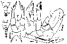
 issued from : R. Gaudy in Rec. Trav. Sta. mar. Endoume, hors série suppl. 9, 1969. [p.44, Pl. 1]. As Calanopia parathompsoni.
Female: 1, habitus (dorsal view); 2, urosome (right lateral view); 5, rostrum; 6, fifth feet; 7, distal part of fifth feet.
Male: 4, urosome (ventral view); 5, rostrum; 8, fifth feet (posterior view); 9, distal part of right foot (anterior view); 10, distal part of left foot (posterior view).
|
 issued from : R. Gaudy in Rec. Trav. Sta. mar. Endoume, hors série suppl. 9, 1969. [p.46, Pl. 2]. As Calanopia parathompsoni.
Female: A1; 3, A2; 4, Mxp; 5, Mx2; 6, Mx1; 7, Md; 8, gnathobase; 9, first foot; 10, second foot; 11, third foot; 12, fourth foot.
Male: 2, right A1.
|
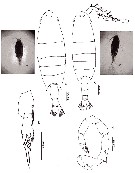
 Issued from : A.M. Al-Aidaroos, A.J. Salama & M.M. El-Sherbiny in ZooKeys, 2016, 552. [p.20, Fig.1]. Female (from 26°11.855'N, 36°25.58'E): A-B, habitus (dorsal and lateral, respectively); C, rostrum (frontal view); D, A1; E, enlarged proximal part of A1; F, A2. Scale bars in mm. Nota: Prosome about 2.5 times as long as urosome; - Cephalosome distinctly separated from 1st pediger, 4th and 5th pedigers somites fused. - One median eye. - Lateral hooks. - Posterolateral corners of prosome pointed in dorsal aspect reaching nearly 1/3 of way along genital compound somite. - Rostrum bearing pair of pointed processes with very small medial subterminal notch. - Urosome of 2 free somites. - 2nd urosomite symmetrical and slightly shorter than genital compound somite. - Caudal rami symmetrical and about 2.3 times as long as wide, each ramus with 5 plumose setae along distal margin and reduced seta (seta VVV) located on dorsal surface near medial distal angle. - A1 19-segmented, when extended reaxhing almost anterior border of 2nd urosomite. A2 biramous with short coxa bearing plumose seta at distomedial angle; basis with 2 subequal setae distomedially; exopod 5-segmented with setal formula 0, 4, 1, 2, 3; endopod 2-segmented, proximal segment with 2 unequal subterminal setae, distal segment bilobed , with medial (proximal) lobe bearing 8 setae, and with lateral (distal) lobe crowned with 6 setae and transverse row of fine setules.
|
 Issued from : A.M. Al-Aidaroos, A.J. Salama & M.M. El-Sherbiny in ZooKeys, 2016, 552. [p.22, Fig.3]. Female: A, Md; B, Mx1; C, Mx2; D, Mxp. Scale bars in mm. Nota: Md gnathobase with 8 teeth on cutting edge, 3rd to 7th teeth ornamented with row of short spinules anterodistally at base; palp biramous, basis with 4 unequal setae on medial margin; exopod 5-segmented with setal formula 1, 1, 1,1, 4; endopod 2-segmented, proximal segment with 2 setae at distomedial corner, distal segment with 7 long and 1 short setae. - Mx1 praecoxal arthrite bearing 9 marginal strong spines and 4 setae on posterior surface; coxal epipodite with 9 setae; coxal endite with 3 setae; basal exite with 1 seta; proximal and distal basal endites with 3 and 1 setae, respectively; exopod with 9 setae; endopod incorporated into basis with 3 setae laterally and 7 setae terminally.
|
 Issued from : A.M. Al-Aidaroos, A.J. Salama & M.M. El-Sherbiny in ZooKeys, 2016, 552. [p.24, Fig.4]. Female: A, P1; B, P2; C, P3; D, P4; E, P5. Scale bars in mm.
|
 Issued from : A.M. Al-Aidaroos, A.J. Salama & M.M. El-Sherbiny in ZooKeys, 2016, 552. [p.23]. Female: Armature of swimming legs P1 to P4.
|
 Issued from : A.M. Al-Aidaroos, A.J. Salama & M.M. El-Sherbiny in ZooKeys, 2016, 552. [p.21, Fig.2, A-B]. Female (SEM micrograph): A, abdomen (lateral view); B, rostrum (lateral view).
|
 Issued from : A.M. Al-Aidaroos, A.J. Salama & M.M. El-Sherbiny in ZooKeys, 2016, 552. [p.25, Fig.5]. Male: A-B, habitus (dorsal and lateral, respectively); C, right A1; D, enlarged proximal part of right A1; E, A1 segments 12-14. Scale bars in mm. Nota: Prosome approximately 2.2 times as long as urosome. - Cephalosome distinctly separated from 1st pediger, 4th and 5th pedigerous somites fused. - Rostrum bearing pair of pointed processes directed posteroventrally. - Urosome of 5 free symmetrical somites, 2nd somite longest. - Some male specimens from Red Sea show the presence of 1 and/or 2 fine spinules, ventrally on the right side in the 1st and 2nd urosomites respecyively. - Right A1 17-segmented, geniculate between segments XX (13) and XXI-XXIII (14). - Left A1, A2, mouthparts and swimming legs P1-P4 as in female.Left A1 - Anal somite shorter preceding somite. - Caudal rami symmetrical, 2.2 times longer than wide; caudal setae as in female.
|
 Issued from : A.M. Al-Aidaroos, A.J. Salama & M.M. El-Sherbiny in ZooKeys, 2016, 552. [p.26, Fig.7]. Male: A, left P5; B, distal segment of left P5 (ventral view); C, distal segment of left P5 (dorso-lateral view); D, right P5. Scale bars in mm. Nota: P5 uniramous, asymmetrical. Left P5 with short coxa; basis 1.8 times longer than coxa with plumose seta located posteriorly near proximal end. Exopod 2-segmented, 1st (proximal) segment slightly shorter than basis with pointed attenuation near distolateral corner, 2nd (distal) segment short, hirsute on posteromedial surface, with curved relatively long spine laterally, short spine with triangular base medially and 1 rounded and serrated process distally. Right leg, longer than left, coxa with 1 blunt process on posterior surface distally; basis with plumose seta laterally. Exopod 2-segmented, forming a stout subchela, 1st exopodal segment without thumb and nearly 4 times as long as wide, distal part of subchela with elongate depression medially and 1 seta on proximal border of the depression; 2nd exopodal segment (finger) elongate, curved at 1/3 its length, not acutely tapering with 1 medial seta proximally and 2 setae laterally nearly at midlength, distal part of finger with shallow depression medially.
|
 Issued from : A.M. Al-Aidaroos, A.J. Salama & M.M. El-Sherbiny in ZooKeys, 2016, 552. [p.26, Fig.6, A]. Male (SEM micrograph): 1st and 2nd urosomite. (spinules indicated by arrows) ventral view.
|
 Female: 1 - P5 exopod 2-segmented. 2 - Cephalic lateral hooks present. 3 - Genital compound somite nearly as long as 2nd urosomite. 4 - Caudal rami symmetrical; 2nd exopodal segment of P5 slightly shorter than first one. Male: 1 - Left P5 shorter than right one; basis of left P5 not swollen proximally. 2 -Prosomal posterolateral corner symmetrical. 3 - Cephalic lateral hooks present. 4 - Caudal rami symmetrical and not divergent posteriorly; 2nd exopodal segment of left P5 shorter than first one. 5 - 1st exopodal segment of right P5 with elongate, distomedial depression with 1 short seta on proximal border of depression.
|
 Issued from : M.C. Kos in Field guide for plankton. Zool Institute USSR Acad., Vol. I, 1972. After A. Scott, 1909. Female: 1, habitus (doesal); 2, P5. Male: 3, habitus (dorsal); 4, abdomen (dorsal); 5, P5.
| | | | | Compl. Ref.: | | | Sewell, 1912 (p.353, 368: juv.); 1914 a (p.232); 1948 (p.323, 324); Krishnaswamy, 1953 (p.139); Yamazi, 1958 (p.151, Rem.); Ganapati & Shanthakumari, 1962 (p.9, 15); Brodsky, 1972 (p.256); Patel, 1975 (p.660); Kasahara & al., 1975 (p.25, eggs, cycles); Chen Q-c, 1980 (p.794); Grice & Marcus, 1981 (p.125, Dormant eggs, Rem.: p.134, 135, 137); Madhupratap & Haridas, 1986 (p.105, tab.1); Yoo, 1991 (tab.1); Dai & al., 1991 (p.45, tab.1); Myung & al., 1994 (tab.1); Shih & Young, 1995 (p.71); Madhupratap & al., 1996 (p.77, Table 2: resting eggs); Marcus, 1996 (p.144); Park & Choi, 1997 (Appendix); Mauchline, 1998 (tab.40); Osore & al., 2003 (p.69); Rezai & al., 2004 (p.489, tab.2); Zuo & al., 2006 (p.163: tab.1); Ohtsuka & al., 2008 (p.115, Table 5); Jiang Z.-B. & al., 2009 (p.196, Table 1, 2); Maiphae & Sa-ardrit, 2011 (p.641, Table 2, 3, Rem.); Jagadeesan & al., 2013 (p.27, Table 3, seasonal abundance); Seo & al., 2013 (p.448, Table 1, occurrence); Nakajima & al., 2015 (p.19, Table 3: abundance); Trottet & al., 2017 (p. 7, Table 3: resting stage); Ohtsuka & Nishida, 2017 (p.565, Table 22.1) | | | | NZ: | 5 | | |
|
Distribution map of Calanopia thompsoni by geographical zones
|
| | | | | | | | | | Loc: | | | N Red Sea (Al Wajh), Indian, Madagascar, India (Saurashtra coast, Lawson's Bay, G. of Mannar, Palk Bay, Madras), W Malay Peninsula (Andaman Sea), Strait of Malacca, Singapore, G. of Thailand, Indonesia-Malaysia, Tioman Is., Philippines, Viet-Nam (Cauda Bay), G. of Tonkin, Hong Kong, China Seas ( Xiamen Harbour, Bohai Sea, Yellow Sea, East China Sea, South China Sea), S & W Korea, Japan Sea, Japan (Seto Inland Sea, Tanabe Bay), S Australia | | | | N: | 70 | | | | Lg.: | | | (5) F: 2,6; M: 2,4; (44) F: 2,303; M: 2,226; (91) M: ± 1,9; (256) F: 2,62-2,46; M: 2,31-2,19; (290) F: 1,8-2; M: 1,6-1,9; (351) F: 2,17; M: 2,08; (530) F: 2,2; (795) F: 2,4; M: 2; (991) F: 2,62; M: 2,19-2,31; (1086) F: 1,78-2,35; (1189) F: 1,92-1,98; M: 1,79-1,83; (Gaudy, 1969) F: 2,4; M: 1,9; {F: 1,78-2,62; M: 1,83-2,40} | | | | Rem.: | According to C.B. Wilson (1950, p.176) this is the largest species of the genus and is distinguished from any of the others by its size and by the lateral hooks on the sides of the head like those in the genus Pontella. The minimal dimensions (290) given by Chen & Zhang (1965) seem to correspond to local, particular conditions. Zuo & al. (2006) also give a minimum body length in the Eastern Chinese Sea (1.90 mm).
See in DVP Conway & al., 2003 (version 1)
Al-Aidaroos & al. (2016): doi: 103897/zookeys.552.6180.
For Al-Aidaroos & al. (2016, p.27) C. thompsoni from the Red Sea and C. parathompsoni Gaudy (1969) are conspecific. C. parathompsoni was distinguished from C. thompsoni mainly on : 1- asymmetr of female genital compound somite in outline (absence of Scott's protuberance); 2- presence of 2 fine spinules ventrally on the left side of male 2nd urosomite; 3- presence of a medial small spine swollen at base on the 1st segment of male right P5. Based on the examination of many specimens of C. thompsoni from the Red Sea, we consider these differences variability within one species since the structure of P5 in both sexes is very similar as are the two fine spines, detected on the ventral right side of the 2nd urosomite. Such variability is common in members of Pontellidae.
For Al-Aidaroos & al. (2016, p.27) the specimens collected from the Red Sea, closely resemble the original description by A. Scott (1909) from the Bay of Kankamaraan (south coast of Kangeang Island), although the Red Sea specimens varied in the absence of a rounded ventral protuberance on the female genital compound somite. Analyses of the shape of the urosome of C. thompsoni that have been reported in the literature between 1909 and 2008 reveal extensive variation in the shape of genital compound somite. The protuberance showed by A. Scott (1909) was absent in specimens collected from Andaman Sea by Sewell (1932), from the Yellow Sea by Chen & Zhang (1965) from Sister Island, Singapore waters by Othman & Toda (2006), and from Thailand waters by Phukham (2008). According to these previous descriptions and illustrations of C. thompsoni from different areas, there is some variation in the proportions of the genital compound somite, the 2nd urosomite, as wemll as the 1st and 2nd exopodal segments of the female P5. Another variation is noticed from Chen & Zhang (1965) from Yellow Sea specimens and Mori (1937) from Japanese waters: the 2nd (distal) exopodal segment of the male left P5 is longer than in other descriptions.
The authors note that the diversity of Red Sea pontellid copepods is remarkably low, given that the Indian Ocean is the origin of the Red Sea plankton.; Silas & Pillai (1973) recorded 71 species of pontellid copepods from the Indian Ocean compared to 15 species from the Red Sea. | | | Last update : 07/06/2020 | |
|
|
 Any use of this site for a publication will be mentioned with the following reference : Any use of this site for a publication will be mentioned with the following reference :
Razouls C., Desreumaux N., Kouwenberg J. and de Bovée F., 2005-2025. - Biodiversity of Marine Planktonic Copepods (morphology, geographical distribution and biological data). Sorbonne University, CNRS. Available at http://copepodes.obs-banyuls.fr/en [Accessed December 17, 2025] © copyright 2005-2025 Sorbonne University, CNRS
|
|
|
|


;)
;)
;)
;)
;)
;)
;)
;)
;)
;)
;)
;)
;)
;)
;)
;)
;)

