|
|
 |
|
Calanoida ( Order ) |
|
|
|
Epacteriscoidea ( Superfamily ) |
|
|
|
Ridgewayiidae ( Family ) |
|
|
|
Exumella ( Genus ) |
|
|
| |
Exumella mediterranea Jaume & Boxshall, 1995 (F,M) | |
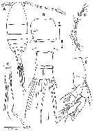
| | | | | | | Syn.: | Exumella polyarthra : Carola & al., 1995 (p.148, figs.F, st.5) | | | | Ref.: | | | Jaume & Boxshall, 1995 (p.93, figs.F,M); Barthélémy, 1999 a (p.10, fig.); Suarez-Morales & Iliffe, 2005 (p.420-421: Rem.); Vives & Shmeleva, 2007 (p.987, figs.F,M, Rem.); Laakmann & al., 2019 (p.330, fig. 2, 3, phylogenetic relationships) | 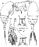 issued from : D. Jaume & G.A. Boxshall in Sarsia, 1995, 80. [p.95, Fig.1]. Female (from Mallorca, Balearic Islands): A-B, habitus (dorsal and lateral, respectively); C, rostrum (fro,tal view); D, anal somite and caudal rami (dorsal); E, idem (ventral); F, P5 (posterior). Nota: Rostral sensory complex composed of 2 raindrop shaped vesicles each opening via a small pore, 2 sensilla, and a pair of minute pores between sensilla. Head and 1st thoracic segment fused (incomplete transverse suture visible dorsally, Th4 and Th5 separated. Th5 segment asymmetrical, showing traces of a dorsal lobe on right side of posterior margin; 2 longitudinal rows of sensilla implanted along both sides of pedigerous somites 2-4. Urosome 3-segmented. Genital double-somite asymmetrical, produced on the right side, with single gonopore opening ventrolaterally on right side, operculum subcircular, 2 sensilla positioned anterodorsally and posteroventrally to operculum. Anal somite bearing striated distal hyaline frill forming 2 big triangular teeth on dorsal margin, and 2 smaller on ventral margin. Caudal rami symmetrical, about 1.7 times as long as wide, armed with 1 short subdistal dorsal seta and 5 distal setae; row of setules along medial margin of each ramus. A1 symmetrical, 27-segmented, not reaching distal end of prosome.
|
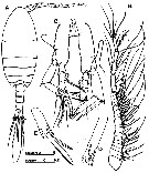
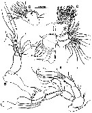 issued from : D. Jaume & G.A. Boxshall in Sarsia, 1995, 80. [p.96, Fig.2]. Female: A, A2; B, Md; C, Mx1; D, Mx1 (detail of basis, exopod and endopod). Nota: A2 with endopod 2-segmented; exopod 7-segmented. Md with coxal gnathobase stout, cutting blade well developed; mandibular palp with endopod reduced, 2-segmented; exopod 4-segmented. Mx1 with well defined praecoxa produced medially into arthrite bearing 10 stout, pectinate spines, submarginal row of 4 setae on ventral side, and isolated dorsal seta; coxa with epipodite armed with 9 setae, and endite with 4; basis fused with exopod and endopod; proximal basal endite discrete, armed with 4 setae; distal incorporated into segment, armed with 5 setae; endopod fully incorporated to basis, formed by 3 indistinctly fused lobes (perhaps remnants of 3 ancestral segments) with setal formula: 2, 4, 5; exopod bearing 10 marginal setae.
|
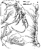 issued from : D. Jaume & G.A. Boxshall in Sarsia, 1995, 80. [p.97, Fig.3]. Female: A, right A1 (ventral); B, Mx2; C, Mx2 (detail of endopod); D, Mxp. Nota: Mx2 comprising completely fused praecoxa and coxa, each armed with 2 endites, allobasis, and 3-segmented endopod; proximal praecoxal endite with 4 long, stout setae, and 1 slender seta; distal with 3 stout setae, 1 longer than others; Both coxal endites armed with 3 setae (2 long and stout, 1 slender and reduced on proximal endite); 3 unequal, with 1 very stout, on distal endite; proximal allobasis endite well developed, swollen, armed with 4 stout unequal setae; distal bearing 3 unequal setae, 1 very reduced in size; free endopod setal formula: 3, 2, 2; 1 of setae on proximal segment very reduced, and another very thick and stout; 1 on distal segment shorter than other. Mxp 6-segmented; syncoxa armed with 7 setae distributed in 4 groups along medial margin; 1, 1, 2, 3; small lobe protruding on distal part of medial margin; row of short setules on distal margin of segment; basis plus fused 1st endopodal segment armed with 3 basal and 2 endopodal setae distally on medial margin; row of short setules along proximal portion of medial margin; free endopod 4-segmented; 1st and nd segments each with 4 setae on distal portion of medial margin; row of short setules along medial margin of 1st segment between 3rd and 4th setae; 4th seta on 2nd segment very long and stout, articulate, bearing specialized ornamentation; endopod segments 3 and 4 reduced; 3rd bearing 4 unequal setae; 4th formed by fusion of ancestral segments 5 and 6 (as indicated by presence of outer margin seta on this segment), with armature of 5 unequal setae
|
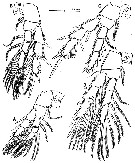 issued from : D. Jaume & G.A. Boxshall in Sarsia, 1995, 80. [p.99, Fig.4]. Female: A-D, P1 to P4 (anterior views).
|
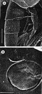 issued from : D. Jaume & G.A. Boxshall in Sarsia, 1995, 80. [p.101, Fig.6 A, B]. Female: A, genital double-somite (right lateral); B, detail of operculum and gonopore. Scale bar: 0.010 mm.
|
 issued from : D. Jaume & G.A. Boxshall in Sarsia, 1995, 80. [p.100, Fig.5]. Male: A, habitus (dorsal); B, right A1 (ventral); C, P5 (anterior); D, detail of left exopod segment 2 of P5; E, detail of right exopod segment 2 of P5. Nota: Prosome 3.6 times longer than urosome. 5th pedigerous somite symmetrical. Urosome 4-segmented. Genital somite asymmetrical, slightly produced and extended backwards on left side; single gonopore opening ventrolaterally on left side close to posterior margin of segment; operculum not so clearly defined as in female genital double-somite, forming narrow bulge dorsal to gonopore. Anal somite and caudal rami as in female. A1 asymmetrical, left as in female; right geniculate, 24-segmented, with segments II, III and IV, X and XI, and XII, XIII and XIV partially fused. P5 asymmetrical, both biramous; endopod 3-segmented; exopod 2-segmented.
|
 issued from : M. Carola, C. Razouls & J.L. Pretus in Vie Milieu, 1995, 45 (2). As Exumella polyarthra. Female (from Minorca, Balearic Islands): A, habitus (dorsal); B, urosome (ventral); C, urosome and spermatophore (lateral); D, A1; E, P5. Nota : no differences have been observed between the individuals of Minorca and those of Bahamas described by Fosshagen (1970).
|
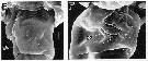 issued from : R.-M. Barthélémy in These Doc. Univ. Provence (Aix-Marseille I), 1999. [Fig.15, E, F]. Female (from La Ciotat, France): E, external ventral view of genital double-somite; F, right lateral position of genital area go = genital operculum; Scale bars: 0,010 mm.
| | | | | Compl. Ref.: | | | Giacomo & al., 2005 (p.98); Pansera & al., 2014 (p.221, Table 2, abundance); Belmonte, 2018 (p.273, Table I: Italian zones) | | | | NZ: | 1 | | |
|
Distribution map of Exumella mediterranea by geographical zones
|
| | | | | | 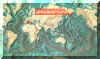 Chart of 1996 Chart of 1996 | |
| | | | Loc: | | | W Medit. (Baleares (Majorca, Minorca), La Ciotat (cave), Sicily (Lake Faro), Messina Strait | | | | N: | 5 | | | | Lg.: | | | (507) F: 1,06; M: 0,94; (511) F: 0,89-0,84; {F: 0,84-1,06; M: 0,94} | | | | Rem.: | For Jaume & Boxshall (1995, p.100) the differences between this species and E. polyarthra are subtle and lie in the minute details of the armature of the non geniculate A1 and A2, and in the distal portion of male P5; The proximal segment of endopod of the A2 bears 2 setae in the Mediterranean species, instead of only 1 in E. polyarthra.
The simultaneous discovery of this form, very near to E. polyarthra, at the Balearic Islands and near La Ciotat's cave, east of Marseille (R. Barthélemy, 1997, pers. comm.) in underwater caves, is an illustration of the isolation of a population at the time of the opening of the Atlantic. Its maintenance addresses the problem of the more-or-less important drying-out of the Mediterranean during the Miocene. Jaume & Boxshall (1995, p.104) do not find any major morphological differences between the Sardinian and the Balearic populations. The populations are isolated from each other by about 400 km of open sea with a depth in excess of 2000 m. However, similar wide distributions are known among other anchihaline Mediterranean crustaceans. | | | Last update : 06/02/2020 | |
|
|
 Any use of this site for a publication will be mentioned with the following reference : Any use of this site for a publication will be mentioned with the following reference :
Razouls C., Desreumaux N., Kouwenberg J. and de Bovée F., 2005-2025. - Biodiversity of Marine Planktonic Copepods (morphology, geographical distribution and biological data). Sorbonne University, CNRS. Available at http://copepodes.obs-banyuls.fr/en [Accessed August 23, 2025] © copyright 2005-2025 Sorbonne University, CNRS
|
|
|
|


;)
;)
;)
;)
;)
;)
;)

;)
