|
|
 |
|
Calanoida ( Order ) |
|
|
|
Clausocalanoidea ( Superfamily ) |
|
|
|
Aetideidae ( Family ) |
|
|
|
Chiridiella ( Genus ) |
|
|
| |
Chiridiella pacifica Brodsky, 1950 (F,M) | |
| | | | | | | Syn.: | ? Chiridiella macrodactyla : Tanaka, 1957 a (p.57, figs.F); Tanaka & Omori, 1970 a (p.153) | | | | Ref.: | | | Brodsky, 1950 (1967) (p.193, Descr.F, figs.F); Bradford, 1971 b (p.21, Rem.); Deevey, 1974 (p.458, Descr.F,M, figs.F,M); Markhaseva, 1996 (p.99, figs.F,M) | 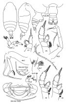 issued from : E.L. Markhaseva in Proc. Zool. Inst. RAN, St. Petersburg, 1996, 268. [p.102, Fig.77]. Female. P.md: mandibular palp; Gntb: gnathobase of Md (partial). Specimen from Kuril Trench. Nota: Mxp protopodite with 2 setae in distal group; endopodal segment 1 with 3 setae in medial part, 1.5 times longer than protopodite.
|
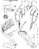
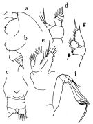 issued from : O. Tanaka in Publ. Seto Mar. Biol. Lab., 1957, VI (1). [p.57, Fig.35]. As Chiridiella macrodactyla. With doubt. Female: a, forehead (lateral); b, last thoracic segment and urosome (lateral left side); c, urosome (ventral); d, Md (mandibular palp); e, Mx1; f, Mx2; g, P1. Nota: Head and 1st thoracic segment fused, 4th and 5th fused. Rostrum absent. Proportional lengths of urosomites and furca 40:12:12:12:20:16 = 100. Furcal rami wider than long, carries short setae. A2 has 7-segmented exopod which is 1.23-times as long as endopod. Md: endopod with 3 setae on the distal segment, endopod with 5 setaeMx1: exopod wuith 5 setae, endopod with 4 setae, 3rd inner lobe with 3 setae, 1st inner lobe with 10 setae.
|
 Issued from : K.A. Brodskii in Calanoida of the Far Eastern Seas and Polar Basin of the USSR. Opred. Fauna SSSR, 1950, 35 (Israel Program for Scientific Translations, Jerusalem, 1967) [p.194, Fig.109]. Female (from NW Pacif.): habitus (dorsal and lateral left side); Mp2, Mx2, S1, P1; S2, P2; S3, P3; S4, P4.
|
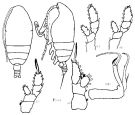
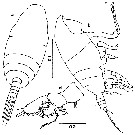
 issued from : G.A. Deevey in Bull. Mar. Sc., 1974, 24 (2). [p.460, Fig.12]. Female (from 32°10'N, 6°30'W): a-b, habitus (dorsal and lateral, respectively); c, Mx1; d, Md; e, P1; f, P2. Scale at left for parts a and b; at lower right for parts c-f. Scales in mm. Nota : Head and 1st thoracic segment separate. Cephalothorax 82-83 % of total length. Urosome about 21 % of cephalothorax length. Genital segment very protuberant ventrally. A1 24-segmented, not longer than cephalothorax. Endopod of A2 ¾ length of exopod.
|
 issued from : G.A. Deevey in Bull. Mar. Sc., 1974, 24 (2). [p.461, Fig.13]. Female: a, A2; b, P3; c, P4; d, Mxp; e, Mx2. Scale in mm.
|
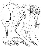
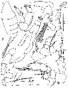 issued from : G.A. Deevey in Bull. Mar. Sc., 1974, 24 (2). [p.462, Fig.14]. Male: a, habitus (lateral); b, A2; c, Md; d, left A1; e, Mx1; f, left P5; g, right P5; h, P3 (exopod segments 2 and 3 missing); i, P2; j, Mx2; k, Mxp (endopod setae not shown). Scale on left margin for parts a; on right margin for parts b-k. Scales in mm. Nota : Md and Mx2 greatly reduced (suggesting that the male does not eat after his final molt. A1 longer than in female, reaching the urosome, both similar, neither geniculate. Endopod of A2 larger and slightly longer than exopod. Endopodal segment 2 of Md with 8 setae. Mxp and P1 similar to that in females. Endopod of P2 2-segmented. Endopod and exopod of P3-P4 3-segmented. Coxopodite of P4 without hairs. P5 biramous, endopod 1-segmented.
|
 issued from : G.A. Deevey in Bull. Mar. Sc., 1974, 24 (2). [p.459, Fig.11, a-b, d]. Male: d, P1. Immature Male: a-b, habitus (dorsal and lateral, respectively). Scales in mm.
|
 Chiridiella pacifica Chiridiella pacifica female; 1 - Mx2 with 3 endites (1st and 2nd endites reduced, 3rd present without setae, 4th with spines of pincer-form, 5th transformed into single curved spine; distal part of endopod represented by 2 setae).
|
 Chiridiella pacifica Chiridiella pacifica female ; 1 - Penultimate lobe of Mx2 with 1 long and 1 short spine arranged as pincers. 2 - Mx2 with only 2 highly modified lobes
| | | | | Compl. Ref.: | | | Deevey & Brooks, 1977 (p.256, tab.2, Station "S"); Ikeda & al., 2006 (p.1791, Table 2); Galbraith, 2009 (pers. comm.); Homma & al., 2011 (p.29, Table 3, abundance, feeding pattern: suspension feeders) | | | | NZ: | 5 + 1 doubtful | | |
|
Distribution map of Chiridiella pacifica by geographical zones
|
| | | | | | 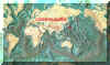 Chart of 1996 Chart of 1996 | |
| | | | Loc: | | | NW Pacif., Kuril-Kamchatka, Aleutian, Is. Japan (NE), ? Japan (Suruga), Mariana Trench, S Aleutian Is., Vancouver Is., Sargasso Sea: off Bermuda: Station "S" (32°10'N, 64°30'W), W Indian.
Type locality: NW Pacific. | | | | N: | 8 | | | | Lg.: | | | (22) F: 3,1; (37) F: 3,1-2,5; M: 2,85; ? (39) F: 2,13; (224) F: 2,75-2,5; M: 2,85; {F: 2,50-3,10; M: 2,85} | | | | Rem.: | bathy & abyssopelagic. Sargasso Sea: 1000-2000 m (Deevey & Brooks, 1977, station "S");
For Deevey (1974, p.463), since the diagnostic characteristics of the female appendages are all altered in the male, and only P1 is similar in the two sexes, it is difficult to be certain of identification. However, only C. pacifica was found fairly often over the 1500- to 2000- m depth) level. Also, endopod of P1 is oval shaped, as is characteristic of females of C. pacifica and of no other species found by Deevey, and an almost spine-shaped point on the outer surface of the endopods of the female P2-P4 is clearly visible in the 1st endopod segment of P2-P4 in the male. Brodsky (1950) did not say on what grounds he identified his male as C. reducta, but in this specimen and also in Tanakas (1957) male the mouthparts were much reduced and leg segmentation increased. | | | Last update : 26/07/2018 | |
|
|
 Any use of this site for a publication will be mentioned with the following reference : Any use of this site for a publication will be mentioned with the following reference :
Razouls C., Desreumaux N., Kouwenberg J. and de Bovée F., 2005-2026. - Biodiversity of Marine Planktonic Copepods (morphology, geographical distribution and biological data). Sorbonne University, CNRS. Available at http://copepodes.obs-banyuls.fr/en [Accessed March 01, 2026] © copyright 2005-2026 Sorbonne University, CNRS
|
|
|
|


;)
;)
;)
;)
;)
;)
;)

;)
