|
|
 |
|
Calanoida ( Order ) |
|
|
|
Epacteriscoidea ( Superfamily ) |
|
|
|
Ridgewayiidae ( Family ) |
|
|
|
Ridgewayia ( Genus ) |
|
|
| |
Ridgewayia marki (Esterly, 1911) (F,M) | |
| | | | | | | Syn.: | Lampoidopus marki Esterly, 1911 b (p.219, figs.F,M, Rem) | | | | Ref.: | | | M.S. Wilson, 1958 (p.138, 146, Rem.F,M); Yeatman, 1969 (p.7, figs.F,M, Rem.); Humes & Smith, 1974 (p.130, Rem.); Ferrari, 1995 (p.196, figs.F,M, Rem.); Figueroa & Hoefel, 2008 (p.145, Table 2, 3, Rem.); Vives & Shmeleva, 2007 (p.991, figs.F,M, Rem.) | 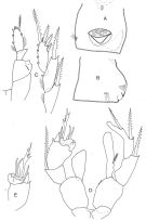 issued from : F.D. Ferrari in Proc. Biol. Soc. Washington, 1995, 108 (2). [p.187, Fig.12]. Female: A, genital complx (ventral); B, idem (latera lright side); C, P5 (posterior, exopod detached). Male: D, P5 (posterior); E, left exopod 1 and 3 of P5 (anterior). bar: 0.1 mm.
|
 issued from : H.C. Yeatman in J. Tenessessee Acad. Sci., 1969, 44 (1). [p.9, Pl.I]. Female (from Bermuda): 1, habitus (lateral); 3, caudal rami (left ramus with normal setation, right with abnormally forked setae; dorsal view); 5, A1; 7, A2; 8, Md; 9, Mx1; 10, Mx2; (showing variations in number of setae); 12, P1; 13, proximal segments of exopod of P1; 15, P3; 16, P4. Nota: A1 26-segmented (Esterly reported 25 segments by error); these antennae reach at least to the distal ends of the caudal rami. A prominent rostrum, bent ventrally. Urosome 4-segmented, the 4th very short (Esterly failed to note the joint separating the 4th segment. Caudal rami generally divergent, about 3 times longer than broad, and with hairs on the inner borders. Male: 2, habitus (lateral); 6, right A1. Nota: Left A1 26-segmented; right 24-segmented, geniculate.
|
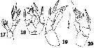 issued from : H.C. Yeatman in J. Tenessessee Acad. Sci., 1969, 44 (1). [p.10, Pl.II, figs.17-20]. Female: 17, P5. Male: 18, P5 (anterior view); 19, right leg of P5 (posterior view); 20, left leg of P5 (posterior view).
|
 issued from : M.S. Wilson in Proc. U.S. Nat. Mus., 1958, 108 (No 3398). [p.170]. Female: P5 of five species Ridgewayia. Ratio of certain characters of endopod segment 2, expressed as percentage of total length of inner margin of segment. A: distance between base of inner margin and placement of 1st inner seta; B, distance between base of inner margin and placement of 1st outer seta; C: length of outer spinous process; D: length of basal joint of outer apical seta.
|
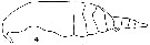 issued from : C.O. Esterly in Proc. Am. Acad. Arts Sci., 1911, 47 (7). [Pl.I, Fig.4]. As Lampoidopus marki. Female (from Bermuda Is.): 4, habitus (lateral). Nota: A1 25-segmented, reach to the end of the caudal rami
|
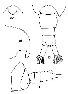 issued from : C.O. Esterly in Proc. Am. Acad. Arts Sci., 1911, 47 (7). [Pl.2, Figs.13, 14, 20, 21]. As Lampoidopus marki. Female: 13, part of last thoracic segment and urosome (dorsal); 21, forehead (lateral). Male: 14, last two thoracic segments and urosome (lateral); 20, forehead, shopping rostrum (ventral).
|
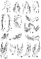
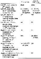
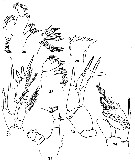 issued from : C.O. Esterly in Proc. Am. Acad. Arts Sci., 1911, 47 (7). [Pl.3, Figs.25, 26, 28, 29, 30, 31, 34]. As Lampoidopus marki. Female: 25, Mx1; 26, Md (cutting edge); 28, Mx2; 29, P5; 31, Md (mandibular palp). Nota: Right and left P5 symmetrical, endopodite 2-segmented, exopodite 3-segmented, a peculiarity is the attachment of the terminal segment of the exopodite at the middle of the inner margin of the 2nd segment. Male: 30, right P5; 34, left P5. Nota: Each endopodite is 1-segmented, that of right leg being club-shaped, that of the left shorter and broader; Exopodite of the right leg is 2-segmented, that of the left 3-segmented with the terminal segment peculiarly modified
|
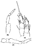 issued from : C.O. Esterly in Proc. Am. Acad. Arts Sci., 1911, 47 (7). [Pl.4, Figs.35, 38, 42]. As Lampoidopus marki. Female: 38, Mxp (bristles not shown); 42, P1. Male: geniculating part of right A1. Nota: Right A1 23-segmented, with a grasping organ, the terminal portion of the grasping is 4-segmented; left A1 25-segmented
|
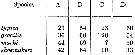
 issued from : D.F. Figueroa & K.L. Hoefel in J. Crustacean Biol., 2008, 28 (1). [p.145, Table 2]. Characters female of the R. marki species-group.
|
 issued from : D.F. Figueroa & K.L. Hoefel in J. Crustacean Biol., 2008, 28 (1). [p.145, Table 3]. Characters male of the marki species-group.
| | | | | Compl. Ref.: | | | Giacomo & al., 2005 (p.98) | | | | NZ: | 2 | | |
|
Distribution map of Ridgewayia marki by geographical zones
|
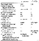
| | | | | | | Loc: | | | Bermuda Is. (Agar Is., Walsingham Cave), W Medit. (Sicily: Lake Faro) | | | | N: | 3 | | | | Lg.: | | | (146) F: ± 1; M: ± 1,05; (519) F: 1,05-0,98; M: 1,03-0,9; {F: 0,98-1,05; M: 0,90-1,05} | | | | Rem.: | The only place where Esterly (1911 b, p.222) found this species was in a cave in the small ledge-like island at Agar's Island. The coloration of the copepod is strikingly lyke that of the coral (Agaricia fragilis), the same brownish tint characterizing both. After Steuer (1910 c, p.282) and copepods' color, it seems to regard the color of this species as protective and that their colors should be so similar to that of the corals.
This species resembles R. klausruetzleri, but can be separated by different characters: position of the copulatory pore on the genital segment, the shape of the distomedial corner of the 2nd (proximal) endopodal segment of the female P5, and so on (see Ferrari, 1995, p.196).
See in Ridgewayia marki minorcaensis and remarks.
For Giacomo & al. (2005) these hyperbenthic forms would be relics of the Tethys Sea. | | | Last update : 19/07/2021 | |
|
|
 Any use of this site for a publication will be mentioned with the following reference : Any use of this site for a publication will be mentioned with the following reference :
Razouls C., Desreumaux N., Kouwenberg J. and de Bovée F., 2005-2025. - Biodiversity of Marine Planktonic Copepods (morphology, geographical distribution and biological data). Sorbonne University, CNRS. Available at http://copepodes.obs-banyuls.fr/en [Accessed August 23, 2025] © copyright 2005-2025 Sorbonne University, CNRS
|
|
|
|


;)
;)
;)
;)
;)
;)
;)
;)

