|
|
 |
|
Calanoida ( Order ) |
|
|
|
Clausocalanoidea ( Superfamily ) |
|
|
|
Scolecitrichidae ( Family ) |
|
|
|
Scolecithricella ( Genus ) |
|
|
| |
Scolecithricella minor (Brady, 1883) (F,M) | |
| | | | | | | Syn.: | Scolecithrix minor Brady, 1883 (p.58, figs.F, non Pl.XVI, fig.16; M); Giesbrecht, 1892 (p.266); Giesbrecht & Schmeil, 1898 (p.46); Fernandes, 2008 (p.465, Tabl.2);
Scolecithrix glacialis Giesbrecht, 1902 (p.25, Descr.F, figs.F);
no Scolecithrix glacialis : Wolfenden, 1911 (p.251, figs.M); Sewell, 1948 (p.574); Vervoort, 1957 (p.101);
Scolecithrix römeri Mrazek, 1902 (p.513, 522, figs.F,M); Brady, 1918 (p.23); Sewell, 1948 (p.496, Rem.);
Scolecithricella glacialis : Farran, 1929 (p.209, 247, Rem.); Hardy & Gunther, 1935 (1936) (p.165, Rem.); Sewell, 1948 (p.570, 573); Vervoort, 1951 (p.95, figs.M, Rem.); 1957 (p.101, Rem.); Tanaka, 1960 (p.40, figs.F, juv.M); 1964 (p.8); Bradford, 1971 b (p.23, fig.F); 1972 (p.44, figs.F,M); Björnberg, 1973 (p.332); Séret, 1979 (p.125, figs.F,M); De Decker, 1984 (p.316, 363: carte); Prado Por, 1986 (p.517); Zmijewska, 1987 (tab.2a);
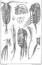
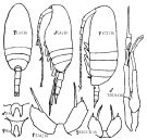
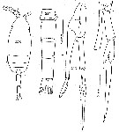
S. minor : Ward & al., 1995 (p.195, Table 2); | | | | Ref.: | | | Sars, 1902 (1903) (p.55, figs.F,M); Farran, 1908 b (p.51); Lysholm, 1913 (p.6); With, 1915 (p.204, figs.F, juv.); Sars, 1925 (p.188); Farran, 1929 (p.209, 247); Wilson, 1932 a (p.83, figs.F,M); Rose, 1933 a (p.157, figs.F,M); Jespersen, 1934 (p.91); Mori, 1937 (1964) (p.51, figs.F,M); Jespersen, 1940 (p.38); Wilson, 1942 a (p.208, figs.M, juv.); Lysholm & al., 1945 (p.28); Brodsky, 1950 (1967) (p.269, figs.F,M, Rem: var. orientalis and occidentalis); C.B. Wilson, 1950 (p.334, fig.M); Chiba, 1956 (p.55, figs.F,M); Chiba & al., 1957 (p.308); 1957 a (p.12); Vervoort, 1965 (p.82, Rem.); Park, 1968 (p.554, figs.F); Shih & al., 1971 (p.46, 152, 207); Minoda, 1971 (p.29); Vidal, 1971 a (p.13, 25, figs.F,M); Bradford, 1973 (p.142); Sullivan & al., 1975 (p.176, figs.Md); Arcos, 1976 (p.85, Rem.: p.91, Table II, figs.F,M); Park, 1980 (p.31, 35, Rem.); Björnberg & al., 1981 (p.639, figs.F); Gardner & Szabo, 1982 (p.302, figs.F,M); Bradford & al., 1983 (p.103, 107, figs.F,M, Rem.); Zheng Zhong & al.,1984 (1989) (p.240, figs.F,M); Kim & al., 1993 (p.270); Razouls, 1994 (p.129, figs.F,M, Rem.); Mazzocchi & al., 1995 (p.204, figs.F,M, Rem); Chihara & Murano, 1997 (p.901, Pl.173,176: F,M); Bradford-Grieve & al., 1999 (p.881, 933, figs.F,M); Vyshkvartzeva, 1999 (2000) (p.234); Conway & al., 2003 (p.203, figs.F,M, Rem.); G. Harding, 2004 (p.44, figs.F,M); Vives & Shmeleva, 2007 (p.783, figs.F,M, Rem.); Soh & al., 2013 (p.98, figs.F,M) | 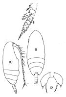 Issued from : T.S. Park in Fishery Bull. Fish Wild. Serv. U.S., 1968, 66 (3). [p.553, Pl.8, Figs.9-12]. Female: 9, habitus (dorsal); 10, idem (lateral right side); 11, P2; 12, P5.
|
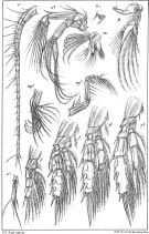
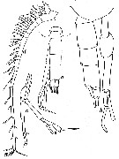
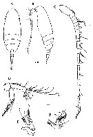
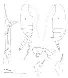 Issued from : J.M. Bradford, L. Haakonssen & J.B. Jillett in Mem. N.Z. oceanogr. Inst., 1983, 90. [p.108, Fig.65]. Female (Antarctic): A, habitus (lateral left side); B, posterior metasome (dorsal and lateral of another specimen); C, P5; D, P5 (other specimen). Male (Antarctic): E, habitus (lateral right side); F, P5.
|
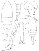
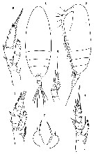
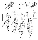
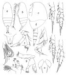 issued from : T. Park in Antarct. Res. Ser. Washington, 1980, 31 (2). [p.32, Fig.2]. Female: a, habitus (dorsal); b, idem (lateral left side); c, forehead (lateral); d, last thoracic segment and urosome (lateral left side); e, rostrum (anterior); f, A2; g, Md; h, Mx1; i, distal part of Mx2; j, Mxp; k, P1 (anterior); l, P2 (posterior); m, P3 (posterior); n, P4 (posterior); o, P5 (posterior). Nota: Cepphalosome and 1st metasomal segment fused, 4th and 5th fused.
|
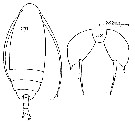
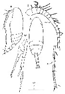
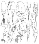 issued from : T. Park in Antarct. Res. Ser. Washington, 1980, 31 (2). [p.33, Fig.3]. Male: a, habitus (dorsal); b, idem (lateral left side); c, forehead (lateral); d, rostrum (anterior); e, A2; f, Md; g, Mx1; h, distal part of Mx2; i, Mxp; j, P1 (anterior); k, P2 (posterior); l, P3 (posterior); m, P4 (posterior); n, P5 (anterior).
|
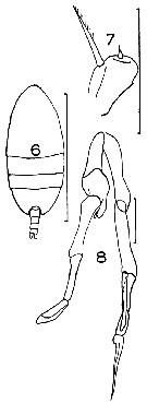
 issued from : G.O. Sars in An Account of the Crustacea of Norway. Vol. IV. Copepoda Calanoida. Published by the Bergen Museum, 1903. [Pl. XXXVII]. Female & Male.
|
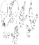
 issued from : G.O. Sars in An Account of the Crustacea of Norway. Vol. IV. Copepoda Calanoida. Published by the Bergen Museum, 1903. [Pl. XXXVIII]. Female. Nota: M = Md (Mx = cutting edge); m, Mx1; mp1 = Mx2; mp2 = Mxp.
|
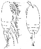
 Issued from: M.G. Mazzocchi, G. Zagami, A. Ianora, L. Guglielmo & J. Hure in Atlas of Marine Zooplankton Straits of Magellan. Copepods. L. Guglielmo & A. Ianora (Eds.), 1995. [p.205, Fig.3.37.1]. Female: A, habitus (dorsal); B, idem (lateral left side); C, P5. Nota: Head and 1st thoracic somite fused, 4th and 5th fused. A1 reaching posterior margin of genital somite. Proportional lengths of urosomites and furca 39:18:18:8:17 = 100. Male: D, habitus (dorsal); E, urosome (dorsal); F, P5. Nota: Proportional lengths of urosomites and furca 16:26:20:25:4:9 = 100. Right exopod of P5 with spatula-like distal segment; left exopod ending in \\\"bayonet-shaped\\\" segment.
|
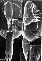 Issued from: M.G. Mazzocchi, G. Zagami, A. Ianora, L. Guglielmo & J. Hure in Atlas of Marine Zooplankton Straits of Magellan. Copepods. L. Guglielmo & A. Ianora (Eds.), 1995. [p.206, Fig.3.37.2]. Female (SEM preparation): A, habitus (dorsal); B, idem (lateral right side); C, rostrum (frontal view); D, urosome ( (ventral) showing genital aperture); E, urosome (lateral right side); F, P5. Bars: A, B 0.100 mm; C, D, E 0.050 mm; F 0.010 mm.
|
 Issued from : K.A. Brodskii in Calanoida of the Far Eastern Seas and Polar Basin of the USSR. Opred. Fauna SSSR, 1950, 35 (Israel Program for Scientific Translations, Jerusalem, 1967) [p.269, Fig.178]. Female: Arb, habitus (dorsal); CHb, habitus (lateral left side); Rb, rostrum; S5CH, P5; S5Ja, P5. Male: Arb, habitus (lateral left side); Rb, rostrum; S5Ar, P5. Ar = from Arctic; CH = from Chukchi Sea; J, = Sea of Japan; a = var. orientalis; b = var. occidentalis.
|
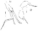
 issued from : W. Vervoort in Verh. K. ned. Akad. Wet., Afd. Natuurk., 1951, (Sect. 2) 47 (2). [p.97, Fig.51]. As Scolecithricella glacialis. Male (from 64°31'S, 10050'W): a-b, habitus (lateral and dorsal, respectively; rostrum on ventral aspect). According to Vervoort (1951, p.96) Scolecithricella glacialis has originally been described by Giesbrecht (1902) from female specimens. The species, as is apparent from its description and figures, differs only slightly from Scolecithricella minor (Brady, 1883), the most obvious charcters are the shape of the postero-lateral thoracic border (broadly rounded in minor, slightly produced and angular in and slight differences in the shape of P5. The present males attributed to glacialis, measure 1.34-1.35 mm, agree better with the length of the female.
Nota: Proportional lengths of cephalothorax and abdomen as 65:26. Head and 1st thoracic segment, 4th and 5th completely fused ( the back shows a weakly indicated line, which may represent a rudimentary line of separation between the cephalon and the 1st thoracic somite). Two fine filifom rostral appendages present. Abdomen 5-segmented, anal segment completely telescoped into 4th urosomal somite; 1st urosomal somite produced on the left side; proportional lengths of urosomal somites and caudal rami 11:30:19:23:4:13 = 100. Caudal rami twice as long as wide, each ramus with 4 marginal setae, allsetae plumose,; the outer edge of each ramus haired.
|
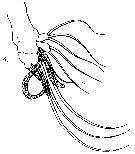
 issued from : W. Vervoort in Verh. K. ned. Akad. Wet., Afd. Natuurk., 1951, (Sect. 2) 47 (2). [p.98, Fig.52]. As Scolecithricella glacialis. Male: a, urosome and P5 (dorsal); b, idem (lateral); c, P5 (rt = right foot, lt = left foot); d, right A1. Nota: A1 19-segmented (8th-13th segments completely fused, 14th segment imperfectly separated from the preceding, as also the 15th from the 16th and the 24th from the 25th). In structure the P5 resemble those of S. minor (cf. Sars, 1902 [1903], Pl.37); there are, however, small differences in the structure of the various segments; the right 1st basal segment is short, stubby and partly fused with the left 1st basal segment; 2nd basal segment on the right side slightly swollen; right endopod short, triangular; 1st and 2nd exopodal segments of right side fused, slightly notched at the place of fusion; right 3rd exopodal segment elongate ovoid in outline, slightly curved and with some elevated ridges on the interior surface; left 1st and 2nd basal segments long and slender; left endopod cylindrical, elogate, cut off at the apex; left exopod 3-segmented, 1st segment cylindrical, 2nd almost cylindrical, slightly swollen near the apex and there with a small spiniform hair along the outer edge; 3rd exopodal segment of left side elongate, dagger-like, with swollen basal portion, tapering towards the end but not acutely pointed.
|
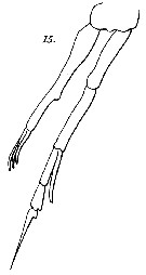
 issued from : T. Mori in The pelagic Copepoda from the neighbouring waters of Japan, 1937 (2nd edit., 1964). [Pl.25, Figs.1-7]. Female: 1, habitus (lateral); 6, P5. Male: 3-4, habitus (dorsal and lateral, respectively); 5, P4 (posterior); 7, P5.
|
 issued from : J.M. Bradford in Mem. N. Z. Oceonogr. Inst., 1972, 54. [p.43, Fig.10, (6-8)]. As Scolecithricella glacialis. Female (from Kaikoura, New Zealand): 6, habitus (dorsal); 7, P5. Male: 8, P5. Scale bars: 1 mm (6); 0.1 mm (7, 8).
|
 issued from : W. Giesbrecht in Copepoden. Res. voyage du S. Y. Belgica. Rapports scientifiques, Zoologie, 1902. [Taf. IV, Figs.1-7]. As Scolecithrix glacialis. Female (from S Peter Ist Island, Bellingshausen Sea): 1-2, habitus (dorsal and lateral, respectively); 3-6, P1 to P4; 7, P5.
|
 Issued from : G.S. Brady in Rep. Scient. Results Voy. Challenger, Zool., 1883, 8 (23). [Pl.XVIII, Figs.1-3, 5]. As Scolecithrix minor. Female: 1, habitus (latera; a = P5); 2, A1; 3, Mx1; 5, Mx2.
|
 Issued from : G.S. Brady in Rep. Scient. Results Voy. Challenger, Zool., 1883, 8 (23). [Pl.XVIII, Fig.4]. As Scolecithrix minor. Male: 4, Mx2.
|
 Issued from : G.S. Brady in Rep. Scient. Results Voy. Challenger, Zool., 1883, 8 (23). [Pl.XVI, Fig.15]. As Scolecithrix minor. Male: P5.
|
 issued from : G. Harding in Key to the adullt pelagic calanoid copepods found over the continental shelf of the Canadian Atlantic coast. Bedford Inst. Oceanogr., Dartmouth, Nova Scotia, 2004. [p.44]. Female & Male.
|
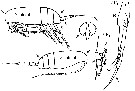
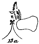 issued from : C. With in The Danish Ingolf-Expedition, 1915, III (4). [Pl. VII, Fig.13, a]. Female (from 62°00N, 21°36'W): a, serrula 6-dentata.
|
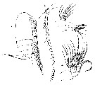
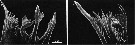 issued from : B.K. Sullivan, C.B. Miller, W.T. Peterson & A.H. Soeldner in Mar. Biol., 1975, 30. [p.181, Fig.6, A-B]. Scolecithricella minor (from 50°N, 145°W) female: A, SEM of left Md (posterior surface); B, detail of dorsal end.
|
 issued from : D.F. Arcos in Rev. Com. Perm. Pacifico Sur, 1976, 5. [Figs. 12, 13]. Female (from ± 54°22.3, 64°34.3): 12, P5. Male: 13, P5. Scale bars: 50 µ (12); 100 µ (13). Nota: environmental conditions: temperature5.40 °C to 6.09 °C and salinity 29.59 to 30.69 p.1000.
|
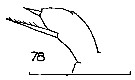 Issued from : J.M. Bradford in N.Z. Oceanogr. Inst., 1971, 206, Part 8, No 59. [p.22, Fig.78]. As Scolecithricella glacialis. Female (from 75°09'S, 171°00'W): 78, P5. Scale bar: 100 µm.
|
 Issued from : O. Tanaka in Spec. Publs. Seto mar. biol. Lab., 10, 1960 [Pl. XVI, 1-7]. As Scolecithricella glacialis. Female (from 66°59'S-67°03'S, 41°19'E-10°44'E): 1-2, habitus (dorsal and lateral, respectively); 3, P1; 4, P2; 5, P3; 6, P4; 7, P5. Nota: Cephalothorax and abdomen in the proportional lengths 78 to 22. Abdominal segments and caudal rami in the proportional lengths 36 : 19 : 19 : 7 : 19 = 100. A1 20-segmented, extends about to the posterior thoracic margin; segments 3, 4, 5; 8, 9, 10 and 24, 25 fused.
|
 Issued from : C. Séret in Thesis 3ème Cycle, UPMC, Paris 6. [Pl. XXXIV, Figs.211, 212]. As Scolecithricella glacialis. Female (from off Kerguelen Is.): 211, habitus (dorsal); 212, P5.
|
 Issued from : C. Séret in Thesis 3ème Cycle, UPMC, Paris 6. [Pl. XXXIV, Figs.213-216]. As Scolecithricella glacialis. Male: 213, habitus (dorsal); 214, urosome (ventral view); 215, P5; 216, left P5.
|
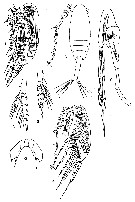
 Issued from : H.Y. Soh, S.Y. Moon & J.H. Wi in Invertebrate Fauna of Korea (eds) Incheon: NIBR, 2013, 21 (28). [p.99, Fig.56]. Female (from Korean waters): A-B, habitus (dorsal and lateral, respectively); C, A1; D, A2; E, Md; F, Mx1. Scale bars: A, B = 300 µm; C-F = 20 µm.
|
 Issued from : H.Y. Soh, S.Y. Moon & J.H. Wi in Invertebrate Fauna of Korea (eds) Incheon: NIBR, 2013, 21 (28). [p.100, Fig.57]. Female: A, Mx2; B, Mxp; C, P1; D, P2; E, P3; F, P4; G, P5. Scale bars: A-F = 20 µm; G = 10 µm.
|
 Issued from : H.Y. Soh, S.Y. Moon & J.H. Wi in Invertebrate Fauna of Korea (eds) Incheon: NIBR, 2013, 21 (28). [p.101, Fig.58]. Male: A-B, habitus (dorsal and lateral, respectively); C, A1; D, P5. Scale bars: A, B = 300 µm; C, D = 20 µm.
|
 Issued from : C. Razouls in Ann. Inst. océanogr., Paris, 1994, 70 (1). [p.129]. Caractéristiques morphologiques de Scolecithricella minor femelle et mâle adultes. Terminologie et abbréviations: voir à Calanus propinquus. The station ''Kerfix'' est située à l'entrée de la baie du Morbihan aux Ïles Kerguelen.
| | | | | Compl. Ref.: | | | Cleve, 1904 a (p.197); Pearson, 1906 (p.18, Rem.); Damas & Koefoed, 1907 (p.396, tab.II); Lysholm & Nordgaard, 1921 (p.19); Hardy & Gunther, 1935 (1936) (p.165, distribution charts); Jespersen, 1939 (p.59, Rem., Table 26); Sewell, 1948 (p.391, 406, 502, 514); Gundersen, 1953 (p.1, 26, seasonal abundance); Østvedt, 1955 (p.15: Table 3, p.69); Minoda, 1958 (p.253, Table 1, 2, abundance); Fagetti, 1962 (p.25); Grice & Hart, 1962 (p.287, table 3); Grice, 1963 a (p.495); Gaudy, 1963 (p.24, Rem.); M.W. Johnson, 1963 (p.89, Table 1, 2); Unterüberbacher, 1964 (p.25); Grice & Hulsemann, 1965 (p.224); Mazza, 1966 (p.71); Harding, 1966 (p.17, 65, 66); Furuhashi, 1966 a (p.295, vertical distribution vs mixing Oyashio/Kuroshio region, Table 10); Matthews, 1967 (p.159, Table 1, Rem.); Fleminger, 1967 a (tabl.1); Maclellan D.C., 1967 (p.101, 102: occurrence); Vinogradov, 1968 (1970) (p.256); Dunbar & Harding, 1968 (p.319); Delalo, 1968 (p.137); Björnberg, 1973 (p.332, 389); Peterson & Miller, 1975 (p.642, 650, Table 3, interannual abundance); 1976 (p.14, Table 1, 2, 3, abundance vs interannual variations); 1977 (p.717, Table 1, seasonal occurrence); Pipe & Coombs, 1980 (p.223, figs.1, 2, 4, table 1); Kovalev & Shmeleva, 1982 (p.84); Mackas & Sefton, 1982 (p.1173, Table 1); Buchanan & Sekerak, 1982 (p.41, vertical distribution); Vives, 1982 (p.292); Huntley & al., 1983 (p.143, Table 2, 3); Sameoto, 1984 (p.213, Table 1); 1984 a (p.767, vertical migration); Tremblay & Anderson, 1984 (p.6); Roe, 1984 (p.357); Longhurst, 1985 (tab.2); Hopkins, 1985 (p.197, Table 1, gut contents); Mikhailovsky, 1986 (p.83, Table 1, ecological modelling); Mackas & Anderson, 1986 (p.115, Table 2); Ward, 1989 (tab.2); Kosobokova, 1989 (p.26); Anderson J.T., 1990 (p.127, Rem.: p.131); Hirakawa & al., 1990 (tab.3); Coyle & al., 1990 (p.764); Atkinson & al., 1990 (p.1213, tab.1); Fransz & al., 1991 (p.9); Shih & Marhue, 1991 (tab.2, 3); Hattori, 1991 (tab.1, Appendix); Hirakawa, 1991 (p.376: fig.2); Santos & Ramirez, 1991 (p.79, 80); Mumm, 1993 (tab.1); Richter, 1994 (tab.4.1a); Shih & Young, 1995 (p.73); Krause & al., 1995 (p.81, Fig.15, abundance, Rem.: p.131); Padmavati & Goswami, 1996 a (p.85, fig.3); Kotani & al., 1996 (tab.2); Falkenhaug & al., 1997 (p.449, spatio-temporal pattern); Errhif & al., 1997 (p.423); Park & Choi, 1997 (Appendix); Yamaguchi & al., 1999 (p.54, vertical migration, life cycle, Rem.); Dolganova & al., 1999 (p.13, tab.1); Nicholas & Nash R, 1999 (p.367); Halvorsen & Tande, 1999 (p.279, tab.2, 3, Rem.: p.282); Voronina & Kolosova, 1999 (p.71); Bragina, 1999 (p.195); Goldblatt & al., 1999 (p.2619, tabl. 2); Ansorge & al., 1999 (p.135, Table 2, abundance v.s. TS diagram); Razouls & al., 2000 (p.343, tab. 5, Appendix); Atkinson & Sinclair, 2000 (p.46, 50, 51, 54, 55, zonal distribution); Pinchuk & Paul, 2000 (p.4, table 1, % occurrence); ; Chiba & al., 2001 (p.95, tab.4, 7); Holmes, 2001 (p.59); Hunt & al., 2001 (p.374, tab.1); Auel & Hagen, 2002 (p.1013, tab.2); Yamaguchi & al., 2002 (p.1007, tab.1); Ringuette & al., 2002 (p.5081, Table 1); Cabal & al., 2002 (p.869, Table 1, abundance); Ward & al., 2002 (p.2183, tab.2); Sameoto & al., 2002 (p.13); Ward & al., 2003 (p.121, tab.4); Hsiao & al., 2004 (p.326, tab.1); Lan & al., 2004 (p.332, tab.1); Gislason & Astthorsson, 2004 (p.472, tab.1); Lo & al., 2004 (p.89, tab.1); Fernandez & al., 2004 (p.501, tab.5); Hunt, 2004 (p.1, 47, 74, Table 3.2); Hopcroft & al., 2005 (p.198, table 2); Mackas & al., 2005 (p.1011, tab.2, 3); Blachowiak-Samolyk & al., 2006 (p.101, tab.1); Ward & al., 2006 (p.83: tab.4); Hop & al., 2006 (p.182, Table 4); Tsujimoto & al., 2006 (p.140, Table1); Hooff & Peterson, 2006 (p.2610); Deibel & Daly; 2007 (p.271, Table 1, 2, 4, Rem.: Arctic polynyas); Dur & al., 2007 (p.197, Table IV); Blachowiak-Samolyk & al., 2007 (p.2716, Table 2); Ward & al., 2007 (p.1871, Table 2, abundance); Blachowiak-Samolyk & al., 2008 (p.2210, Table 3, 5, biomass, composition vs climatic regimes); Schnack-Schiel & al., 2008 (p.1046: Tab.2); Ward & al., 2008 (p.241, Tab.2, Appendix II ); Humphrey, 2008 (p.84: Appendix A); Gaard & al., 2008 (p.59, Table1, N Atlantic Mid-Ridge); Ayon & al., 2008 (p.238, Table 4: Peruvian samples); Darnis & al., 2008 (p.994, Table 1); Lan Y.-C. & al., 2008 (p.61, Table 1, % vs stations); C.-Y. Lee & al., 2009 (p.151, Tab.2); Galbraith, 2009 (pers. comm.); Chiba & al., 2009 (p.1846, Table 1, occurrence vs temperature change); Lan Y.-C. & al., 2009 (p.1, Table 2, % vs hydrogaphic conditions); Park & Ferrari, 2009 (p.143, Table 4, 7: common deep water species, Appendix 1, biogeography); Eloire & al., 2010 (p.657, Table II, temporal variability); Kosobokova & Hopcroft, 2010 (p.96, Table 1, fig.7); Homma & Yamaguchi, 2010 (p.965, Table 2); Bucklin & al., 2010 (p.40, Table 1, Biol mol.); Swadling & al., 2010 (p.887, Table 2, 3, A1, fig.6, abundance, indicator species); Dvoretsky & Dvoretsky, 2010 (p.991, Table 2); 2011 a (p.1231, Table 2: abundance, biomass); Kosobokova & al., 2011 (p.29, Table 2, figs.4, 6, as S. minor var. occidentalis, Rem.: Arctic Basins); Yang & al., 2011 a (p.921, Table 2, inter-annual variation 1999-2006); Hsiao S.H. & al., 2011 (p.475, Appendix I); Hsiao & al., 2011 (p.317, Table 2, indicator of seasonal change); Marrari & al., 2011 (p.1599, abundance, composition, ineterannual variation); 2011 a (p.1614, Table 2, Fig.2A, 5, 6); Homma & al., 2011 (p.29, Table 2, 3, 5, abundance, feeding pattern: detritivores); Pepin & al., 2011 (p.273, Table 2, seasonal abundance); Matsuno & al., 2011 (p.1349, Table 1, abundance vs years); Guglielmo & al., 2012 (p.1301, Table 3); Matsuno & al., 2012 (Table 2); Ward & al., 2012 (p.78, Table A1); Michels & al., 2012 (p.369, Table 1, occurrence frequency); Uysal & Shmeleva, 2012 (p.909, Table I) ; DiBacco & al., 2012 (p.483, Table S1, ballast water transport); Sigurdardottir, 2012 (p.1, Table 2.3); Takahashi M. & al., 2012 (p.393, Table 2, water type index); in CalCOFI regional list (MDO, Nov. 2013; M. Ohman, pers. comm.); Ohashi & al., 2013 (p.44, Table 1, Rem.); Questel & al., 2013 (p.23, Rem. p.31); Ojima & al., 2013 (p.1293, Table 2, 3, 4, abundance); Lee D.B. & al., 2013 (p.1215, Table 1, 2: abundance, composition); Lidvanov & al., 2013 (p.290, Table 2, % composition); Arendt & al., 2013 (p.105, fig.3, abundance); Bonecker & a., 2014 (p.445, Table II: frequency, horizontal & vertical distributions); Coyle & al., 2014 (p.97, table 3); Dias & al., 2015 (p.483, Table 2, abundance, biomass, production); Smoot & Hopcroft, 2016 (p.1, fig.7, vertical distribution); El Arraj & al., 2017 (p.272, table 2); Belmonte, 2018 (p.273, Table I: Italian zones); Dias & al., 2018 (p.1, Table 5: % vs. season); Dias & al., 2019 (p.1, Table II, IV, fig.5, occurrrence, vertical distribution, Rem.: p.12, 14); Acha & al., 2020 (p.1, Table 3: occurrence % vs ecoregions). | | | | NZ: | 22 | | |
|
Distribution map of Scolecithricella minor by geographical zones
|
| | | | | | | | | | | | | | | | | | 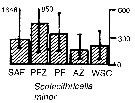 Issued from : A. Atkinson & J.D. Sinclair in Polar Biol., 2000, 23. [p.50, Fig.3] Issued from : A. Atkinson & J.D. Sinclair in Polar Biol., 2000, 23. [p.50, Fig.3]
Scolecithricella minor from Scotia Sea.
Median and interquartile ranges of copepods (nos /m2) in the five water zones; from north to south these are SAF Subantractic Front area, PFZ Polar frontal Zone, PF Polar Front area, AZ Antarctic Zone, WSC Weddell-Scotia Confluence area/ East Wind Drift.
Numbers on the plots are upper interquartiles where these could not be scaled. |
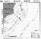
 Issued from : K.M. Swadling, So. Kawaguchi & G.W. Hosie in Deep-Sea Research II, 2010, 57. [p.898, Fig.6 (continued)]. Issued from : K.M. Swadling, So. Kawaguchi & G.W. Hosie in Deep-Sea Research II, 2010, 57. [p.898, Fig.6 (continued)].
Distribution of indicator species Scolecithricella minor from the BROKE-West survey (southwest Indian Ocean) during January-February 2006.
Sampling with a RMT1 net (mesh aperture: 315 µm), oblque tow from the surface to 200 m.
The survey area was located predominantly within the seasonal ice zone, and in the month prior to the survey there was considerable ice coverage over the western section but none over the east.
See map showing sampling sites in Calanus propinquus. |
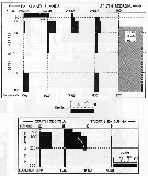
 Issued from : M. Krause, J.W. Dippner & J. Beil in Prog. Oceanog., 1995, 35. [p.102, Fig.15]. Issued from : M. Krause, J.W. Dippner & J. Beil in Prog. Oceanog., 1995, 35. [p.102, Fig.15].
Horizontal distribution pattern of Scolecithricella minor (individuals per m2) in the winter North Sea.
Collected by WP2-net. Numbers (ind/m3) depth-integrated, extrapoled to the bottom (but at a maximum to a depth of 500 m) and expressed as ind per m2 of water surface. |
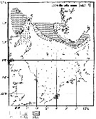
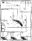 Issued from : A.C. Hardy & E.R. Gunther in Discovery Reports, 1935 (1936), 11. [p.166, Fig.76]. Issued from : A.C. Hardy & E.R. Gunther in Discovery Reports, 1935 (1936), 11. [p.166, Fig.76].
Charts showing the distribution of Scolecihricella minor in the upper layers of waters at stations in the 1926-7 surveys around South Georgia.
The squares represent the average numbers per 50 m vertical haul from 250 m (or less at shallow-water stations) to the surface with N 70 V nets. |
 Issued from : C. Séret in Thesis 3ème Cycle, UPMC, Paris 6. 1979, Annexe. [p.42]. Issued from : C. Séret in Thesis 3ème Cycle, UPMC, Paris 6. 1979, Annexe. [p.42].
Geographical occurrences of Scolecithricella glacialis (= S. minor) in the Antarctic indian zone. [after publications from: Brady, 1883, 1918; Thompson, 1900; Wolfenden, 1908, 1911; With , 1915; Rosendorn, 1917; Farran, 1929; Sewell, 1929, 1947; Brady & Gunther, 1935; Steuer, 1929, 1392, 1933; Ommaney, 1936; Vervoort, 1957; Tanaka, 1960; Brodsky, 1964; Seno, 1966; Andrews, 1966; Grice & Hulsemann, 1967; Seno, 1966; Frost & Fleminger, 1968; Voronina, 1970; Zverva, 1972].
Nota: C. Séret notes the occurrence at stations 50°.5S, 71°E; 51°S, 65°E and 56°S, 70°E. |
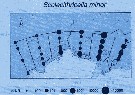
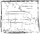
 Issued from : A.C. Hardy & E.R. Gunther in Discovery Reports, 1935 (1936), 11. [p.170, Fig.78]. Issued from : A.C. Hardy & E.R. Gunther in Discovery Reports, 1935 (1936), 11. [p.170, Fig.78].
Vertical distribution of Scolecithricella minor at stations between the Falkland Islands and South Georgia February 1927 and between South Georgia and Tristan da Cunha February 1926.
The scale represents the numbers per 50 m vertical haul taken by a series of closing N 70 V nets.
Horizontal broken lines show the ranges of these vertical hauls. |
 Issued from : C.O. Dias, A.V. de Araujo & S.L.C. Bonecker in An. Acad. Bras. Ciênc., 2019, 91 (1). [p.11, Fig.5, b] Issued from : C.O. Dias, A.V. de Araujo & S.L.C. Bonecker in An. Acad. Bras. Ciênc., 2019, 91 (1). [p.11, Fig.5, b]
Study area in The Campos basin (Brazil) showing the seasonal variation of Scolecithricella minor (ind.m3) at 1 m depth, during the dry season.
Nota: This species is considered a detritus feeder. The species was found in the Campos Basin with greater abundance recorded in the 1 m and 250 m layers, mainly during the dry season. |
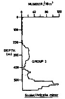 Issued from : R.K. Pipe & S.H. Coombs in J. Plankton Res., 1980, 2 (3). [p.230, Fig.4]. Issued from : R.K. Pipe & S.H. Coombs in J. Plankton Res., 1980, 2 (3). [p.230, Fig.4].
Scolecithricella minor from Wyville Thomson Ridge (60°03.4' N, 07°03.9' W to 60°08' N, 07°02.9' W) on 24 April 1978: Vertical distribution.
Members of this group (Group 3) contained six of the most abundant taxa and would not be associated with a single water mass. This species was found in and just above Norwegian Sea Deep water with a secondary peak in mid-depths; that is, in North Atlantic Oceanic water.
This group 3 includes Metridia lucens, Calanus finmarchicus, Euchaeta (= Paraeuchaeta) norvegica, Oithona spp., and Micromesistius poutassou.
A computed coefficient of dissimilarity was used in association with a 'group-average' clustering strategy (see Clifford & Stephenson, 1975) to classify the plankton taxa on the basis of their vertical distribution after the indices of similarity and the results arranged diagramatically in the form of a dendogram (Fig.2). Classification was carried out using the Bray-Curtis measure which gives a coefficient of dissimilarity. |
| | | | Loc: | | | Cosmopolite : Antarct. (Amundsen Sea, Croker Passage, Peninsula, Scotia Sea, Weddell Sea, S South Georgia, SW & SE Atlant., Indian, Lützow-Holm Bay, SW & SE Pacif., Prydz Bay), sub-Antarct. (N South Georgia, Atlant. (SW & S), off Prince Edward Is., Indian, Kerguelen Is., Pacif. (SW, SE), Magallanes region, Straits of Magellan, Atlant., Brazil (Campos Basin), Azores, off SE Nova Scotia, Newfoundland (Flemish Cape), W Greenland (Nuuk), Godthaab Fjord, SE Greenland, Fram Strait, Spitsbergen, Wyville Thomson Ridge, Kongsfjorden, Iceland, Norwegian Sea, Malangen fjord, Raunefjorden (all the year), Barents Sea, Franz Josef Land, Porcupine Bank, North Sea, Morocco-Mauritania, Mediterranean Sea (W-E), Red Sea, Madagascar, Indian, Goa, Bay of Bengal, Pacif., China Seas (East China Sea, South China Sea), Taiwan Strait, Taiwan (SW, E, NW, N: Mienhua Canyon), Korea, Japan Sea, Japan (Onagawa, Toyama Bay, off Sanriku), Station Knot, Station "P", off British Columbia, Fjord System (Alice Arm & Hastings Arm), Portland Inlet, off Washington coast, Oregon (off Newport), Arct. (Barrow Strait, Nansen Basin, Amundsen Basin, Makarov Basin, Chukchi Sea, Fletcher's Ice Is., SE Beaufort Sea, Canadian abyssal plain, Canada Basin, Barents Sea), Bering Sea, Aleutian Basin, S Aleutian Is., Okhotsk Sea, St. Lawrence Island, W Baffin Bay, N Baffin Sea, Greenland Sea, Fram Strait, Norwegian Sea (Nordvestbanken), S Iceland, Peru (in Abanto, 2001) | | | | N: | 251 | | | | Lg.: | | | (7) F: 1,52; M: 1,34; (9) F: 1,46-1,08; M: 1,46-1,2; (16) F: 1,32; (25) F: 1,46-1,32; M: 1,45-1,37; (31) F: 1,38-1,28; M: 1,35-1,34; (33) F: 1,3-1,4; (35) F: 1,41-1,2; (36) F: 1,27-1,37; M: 1,27-1,31; (45) F: 1,5-1,25; M: 1,4-1,2; (47) F: 1,6; (65) F: 1,4; M: ± 1,4; (66) F: 1,41-1,28; (72) F: 1,42; (114) F: 1,28; (116) F: 1,2; M: 1,24; (208) F: 1,7-1,3; M: 1,4; (246) F: 1,26-1,4; M: 1,3; (432) F: 1,55-1,43; (523) F: 1,46-1,08; M: 1,46-1,34; (991) F: 1,08-1,46; M: 1,2-1,46; (1174) F: 1,02-1,04; M: 1,07-1,09; {F: 1,02-1,70; M: 1,07-1,46} | | | | Rem.: | epi- bathypelagic. 0-580 m (Pipe & Coombs, 1980, Arctic: 60°N).
Scolecithricella minor var. orientalis Brodsky, 1948 (F).
Ref.: Brodsky, 1948 (p.53, Pl.XII, figs.3-6); 1950 (1967) (p.270, fig.F)
Loc.: N pacific, Sea of Japan, Okhotsk Sea, Bering Sea, Chukchi Sea.
Nota: P5 female with slightly curved and elongate distal segment; outer lateral spine shifted far toward the apical spine.
Scolecithricella minor var. occidentalis Brodsky, 1950
Ref.: Sars, 1903 (p.55, Pl.XXXVII & Pl. XXXVIII); Brodsky, 1950 (1967) (p.270, figs.F,M); Kosobokova & al., 2011 (p.29, Table 2, figs.4, 6, Rem.: Arctic Basins).
Loc.: N Atlantic, Arctic, S Chukchi Sea.
Nota: P5 female with short rounded distal segment; distance from apical to outer lateral spine equal to distance from latter spine to proximal margin of segment.
See in DVP Conway & al., 2003 (version 1)
For Vervoort (1957, p.102) the adult females as Scolecithricella glacialis) in the Banzare collection show a considerable amount of variation in the development of P5. The P5 always consists of a pallet shaped gree segment, attached to a communal basal portion. This segment is 2.5-2 times as long as wide. The internal margin has a rather long spiniform seta, about as long as the sgment or slightly shorter, which is haired along its outer edge. The apex of the segment has a short spine, variable in length (1/2-1/8 the total length of the free segment) and always completely nude. At the external corner of the spine a small denticle is occasionally to be observed. The external margin of the segment is usually denticulated opposite the insertion of the internal seta ; in many specimens, however, such denticules appear to be absent. For Tanaka (1960, p.41) S. glacialis is only closely allied to S. minor.
For Park (1980, p.35) this species is the most common of the genus in Antarctic waters; It seemed to be the only species that inhabits mainly the epipelagic parts of the antarctic seas.
For Mazzocchi & al. (1995) this form is similar to the occidentalis variety, distinguished mainly for the shape of the P5 in females.
After Dias & al. (2019) this species is considered as a detritus feeder. | | | Last update : 25/10/2022 | |
|
|
 Any use of this site for a publication will be mentioned with the following reference : Any use of this site for a publication will be mentioned with the following reference :
Razouls C., Desreumaux N., Kouwenberg J. and de Bovée F., 2005-2026. - Biodiversity of Marine Planktonic Copepods (morphology, geographical distribution and biological data). Sorbonne University, CNRS. Available at http://copepodes.obs-banyuls.fr/en [Accessed March 02, 2026] © copyright 2005-2026 Sorbonne University, CNRS
|
|
|
|


;)
;)
;)
;)
;)
;)
;)
;)
;)
;)
;)
;)
;)
;)
;)
;)
;)
;)
;)
;)
;)
;)
;)
;)
;)
;)
;)
;)

;)
;)
;)
;)
;)
;)
;)
;)
