|
|
 |
|
Calanoida ( Order ) |
|
|
|
Clausocalanoidea ( Superfamily ) |
|
|
|
Scolecitrichidae ( Family ) |
|
|
|
Scolecithricella ( Genus ) |
|
|
| |
Scolecithricella tropica Grice, 1962 (F) | |
| | | | | | | Syn.: | Scolecithricella beata Tanaka, 1962 (p.50, Descr.F, figs.F) | | | | Ref.: | | | Grice, 1962 (p.211, Descr.F, figs.F); Bradford, 1973 (p.142); Gopalakrishnan, 1974 a (p.97, figs.F, Rem.); Bradford & al., 1983 (p.103); Vyshkvartzeva, 1999 (2000) (p.236: Rem.) | 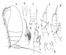 issued from : O. Tanaka in Publ. Seto Mar. Biol. Lab., 1962, X (1). [p.51, Fig.134]. As Scolecithricella beata; Female: a, habitus (left lateral side); b, rostrum; c, urosome (dorsal); d, Mx2; e, P1; f, P2; g, P3; h, P5. Nota Female: - Head and 1st pediger segment fused, 4th and 5th thoracic segments fused. - Cephalothorax about 4.95 times the length of abdomen. - Posterolateral corners of the last thoracic segment rounded, and notched at apex. - Rostrum gradually attenuates into 2 long filaments, rather thick. - Abdomen 4-segmented; proportional lengths with caudal rami 36 : 16 : 16 : 12 : 20 = 100. - Genital segment not swollen ventrally. - Caudal rami longer than wide. - A1 22-segmented (segments 8, 9, 10 and 24, 25 fused); extends to the distal margin of cephalothorax. - A2 exopod 1.3 times as long as the endopod. - Mx1 with 7 setae on the outer lobe, 6 setae on exopod, 5+3 setae on endopod, 4 setae on the 2nd basal, 1 seta on the 3rd inner lobe, 2 setae on the 2nd inner lobe, 11 setae on the 1st inner lobe (praecoxal arthrite). - Mx2 with only worm-like filaments on the endopod. - P1 without outer edge spine on the exopodal segment 1. - P2 with a fairly long and curved spine on the 1st exopodal segment, reaching the middle of the next segment; 2nd exopodal segment with the outer edge spine on the 1st exopodal segment. - P3 with a group of small spines on the distal border where the endopod articulates with the basis. - P4 with several spines near the inner margin on the posterior surface; the 2nd and 3rd segments both exopod and endopod are more finely covered with groups of minute spinules on the posterior surface than those on P2 or P3. - P5 2-segmented; segments cylindrical in shape; terminal segment with 2 spines, subequal in lengths, on the distal margin. Remarks: For Tanaka, the species is closely related to Scolecithrix auropecten Giesbrecht (1892) [= Archeoscolecithrix auropecten], in the structure of the P5, but can be easily distinguished from it in its slmall size and in the length of the terminal spines on the P5.
|
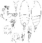 issued from : G.D. Grice in Fish. Bull. Fish Wildl. Serv., 1962, 61. [p.210, Pl.18, Figs.10-18]. Female (from equatorial Pacific): 10-11, habitus (dorsal and lateral, respectively); 12, rostrum; 13, Mx1; 14, terminal part of Mx2; 15, P1; 17, P5; 18, P5 (abnormal). Paratype. Nota: The posterior thoracic border has a well-defined indentation at a point just anterior to the apex. Genital segment longer than the combined lengths of the next two segments. Mx1 with 2 setae on the 2nd inner lobe, 3 on the 3rd inner lobe, 5 on the 2nd basal segment, 8 on the endopod, 6 on the exopod. Terminal portion of Mx2 with 5 sensory and 3 worm-like appendages. P5 variable, most of the specimens have 2, approximately equal, terminal spines, but at least 2 specimens were observed to have 3 on one side and 2 on the other.
|
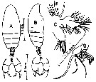 Issued from : Gopalakrishnan in Mahasagar, 1974, 7 (1 & 2). [p.98, Fig.1]. Female (from 04°57'S, 77°59'E & 21°04'S, 112°50'E): A-B, habitus (dorsal and lateral, respectively); C, Mx1; D, terminal part of Mx2; E, P5; F, P5 (abnormal). Noya: As described by Grice, these specimens can easily be identified from the structure of the posterior thoracic margin and P5. - There is a notch on the posterior thoracic margin just anterior to the apex. - P5 show variations in a few specimens. In some the legs are symmetrical by having 2 approximately equal terminal spines; whereas in a few they are asymmetrical with 3 terminal spines on one side and 2 on the other. - In Mx1 Grice has shown 8 setae on the 1st inner lobe (arthrite), Tanaka has mentionede 11 setae, and here also have 11 setae. The 3rd lobe (basal endite in the specimen from the equatorial Pacific has 3 setae, so also the Indian Ocean specimens, whereas Tanaka has mentioned only 1 seta. - On the endopod of Mx2 the Indian specimens have 5 sensory and 3 worm-like elements (as described by Grice); Tanaka has mentioned only about worm-like elements.
|
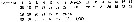 issued from : O. Tanaka in Publ. Seto Mar. Biol. Lab., 1962, X (1). [p.50]. As Scolecithricella beata; Female: Proportional lengths of segments of A1.
| | | | | Compl. Ref.: | | | Delalo, 1968 (p.138); Carter, 1977 (1978) (p.35); Guangshan & Honglin, 1984 (p.118, tab.); Madhupratap & Haridas, 1986 (p.105, tab.1); Gopalakrishnan & Balachandran, 1992 (p.167, figs.1, 7, Table 1, 2); Shih & Young, 1995 (p.73); Padmavati & al., 1998 (p.347); Cornils & al., 2010 (p.2076, Table 3) | | | | NZ: | 7 | | |
|
Distribution map of Scolecithricella tropica by geographical zones
|
| | | | | | | | | | Loc: | | | South Africa (E) (Natal), Red Sea, Arabian Sea, S India, Indonesia (SW Celebes), Pacif. (W equatorial), W Australia, Pacif. (equatorial), China Seas (East China Sea, South China Sea), Japan (Sagami Bay), Galapagos-Ecuador | | | | N: | 13 | | | | Lg.: | | | (101) F: 1,3-1,13; (117) F: 1,25; {F: 1,13-1,30} | | | | Rem.: | Probable in the Genus Amallothrix.
For Grice & Hulsemann (1965, p.242) this species is similar to Scolecithricella unispinosa from which it differs in P5 and the form of the posterior lateral corner of the 5th thoracic segment.
After Gopalakrishnan (1974, p.98) the similarities between S. tropica and S. beata Tanaka, 1962 are obvious enough to consider them synonymous. | | | Last update : 09/12/2020 | |
|
|
 Any use of this site for a publication will be mentioned with the following reference : Any use of this site for a publication will be mentioned with the following reference :
Razouls C., Desreumaux N., Kouwenberg J. and de Bovée F., 2005-2025. - Biodiversity of Marine Planktonic Copepods (morphology, geographical distribution and biological data). Sorbonne University, CNRS. Available at http://copepodes.obs-banyuls.fr/en [Accessed August 27, 2025] © copyright 2005-2025 Sorbonne University, CNRS
|
|
|
|


;)
;)
;)

