|
|
 |
|
Calanoida ( Order ) |
|
|
|
Spinocalanoidea ( Superfamily ) |
|
|
|
Spinocalanidae ( Family ) |
|
|
|
Monacilla ( Genus ) |
|
|
| |
Monacilla typica Sars, 1905 (F,M) | |
| | | | | | | Syn.: | Oxycalanus spinifer Farran, 1908 b (p.25, figs.F);
Oxycalanus semispinus A. Scott, 1909 (p.33, Descr.F, figs.F);
Monacilla dubia A. Scott, 1909 (p.35, Descr.M, figs.M, Rem.); Sewell, 1948 (p.553);
M. semispina C.B. Wilson,1950 (p.266, fig.M);
M. typica asymmetrica Tanaka, 1956 c (p.398);
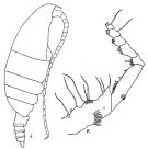
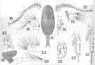
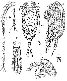
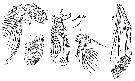
no M. typica : Wheeler, 1970 (p. 9, fig.M) | | | | Ref.: | | | Sars, 1905 b (p.9, Descr.F); 1925 (p.38, figs.F,M); Farran, 1926 (p.245, Rem.); Rose, 1933 a (p.87, figs.F,M); Lysholm & al., 1945 (p.11); Vervoort, 1946 (p.158, Rem.); C.B. Wilson, 1950 (p.267, fig.M); Farran & Vervoort, 1951 h (n°40, p.3, figs.F,M); Tanaka, 1956 c (p.396, figs.F); Vervoort, 1963 b (p.120, Rem.); Owre & Foyo, 1967 (p.42, figs.F,M); Mazza, 1967 (p.99, 100, 381, figs.F,M, juv.); Minoda, 1971 (p.23, Rem.); Grice, 1971 (p.275, 279, figs.F,M); Damkaer, 1975 (p.64, Rem.F,M); Brodsky & al.,1983 (p.325, figs.F,M); Schulz, 1989 (p.187, figs.F); Lapernat, 1999 (p.27, 55, Rem.); Bradford-Grieve & al., 1999 (p.878, 914, figs.F,M); Vives & Shmeleva, 2007 (p.829, figs.F,M, Rem.); Bode & al., 2017 (p.600, Table I, III, fig. 2, 3, 4, morphology vs genetic). | 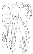 issued from : O. Tanaka in Publ. Seto Mar. Biol. Lab.,1956, V (3). [p.397, Fig.18]. Female: a, habitus (dorsal); b, forehead (left lateral ); c, genital somite (ventral); d, last thoracic segment and genital somite (left); e, Mxp; f, P1; g, P2; h, P4.
|
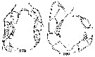
 issued from : Brodsky K.A., Vyshkvartseva N.V., Kos M.S. & Markhaseva E.L. in Opred. Faune SSSR, 1983, 135. [p.326, Fig.161] Female. Abd = urosome.
|
 issued from : Brodsky K.A., Vyshkvartseva N.V., Kos M.S. & Markhaseva E.L. in Opred. Faune SSSR, 1983, 135. [p.327, Fig.161]. Female. Gnb = gnathobase of Mx1. lb = left, np = right; Bap = variant.
|
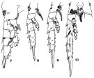
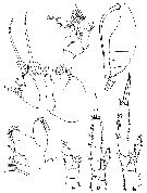
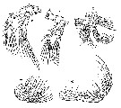
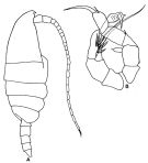 issued from : G.D. Grice in Cah. Biol. Mar., 1971, XII. [p.278, Fig.3 A-B]. Male (from Medit.): A, habitus (lateral right side); B, P5. Nota: Rostrum present.
|
 issued from : G.D. Grice in Cah. Biol. Mar., 1971, XII. [p.277, Fig.2 J-K]. Female (from Medit.): J, habitus (lateral right side); K, Mxp. Nota: Rostrum present.
|
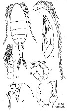
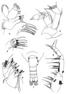 issued from : K. Schulz in Mitt. hamb. zool. Mus. Inst., 1989, 86. [p.200, Figs.1-6]. Female (off Mauritania): 1, urosome (dorsal); 2, A2; 3, Mx1 (anterior); 4, Mx1 (inner lobe 1, posterior); 5, Mx2; 6, Mxp. Nota: Genital segment in dorsal view asymmetrical, protruding on right, bearing some chitinous ridges dorsolaterally. Caudal rami length about 1.6 times width, with 1 short outer seta (sometimes overlooked) and 4 terminal setae; inner seta located ventrally, symmetrical on both rami. Inner lobe1 of Mx1 with a total of 15 setae (1 anterior, and 4 posterior, 1 marginal anteriorly and 9 ordinary strong); inner lobe 2 and 3 with 5 and 4 setae, respectively; basis with 5 and endopod with 4, 5, 7 setae; exopod carrying 11 setae; outer lobe 2 with 1 minute seta.
|
 issued from : K. Schulz in Mitt. hamb. zool. Mus. Inst., 1989, 86. [p.201, Figs.7-10]. Female: 7, P1 (anterior); 8, P2 (posterior); 9, P3 (posterior); 10, P4 (posterior).
|
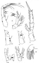
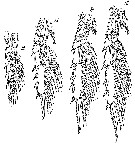
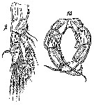
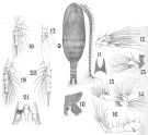 issued frm : A. Scott in Siboga-Expedition, 1909, XIX a. [Plate II, Figs.9-21]. As Oxycalanus semispinus. Female (from Indo-Malaysia): 9, habitus (dorsal); 10, last thoracic and genital segments (left side); 11, rostrum; 12, A2; 13, Md; 14, Mx1; 15, Mx2; 16, Mxp; 17, P1; 18, P2; 19, P3; 20, P4 (right side); 21, basipod 1 of P4. Nota: Head and 1st thoracic segment fused, 4th and 5th fused. A1 24-segmented, the terminal segment, small, is furnished with one sensory filament. Caudal rami slightly longer than broad, about as long as the anal segment. Genital segment asymmetrical. 1st basipodite 1 of P3with 2 rows of fine spines on its surface, Surface of the 2nd exopodal segment of P3 with 1 row and the 3rd with 2 rows of fine spines; 2nd and 3rd endopodal segments of P3 with each 2 rows of fine spines. 2nd exopodal segments of P4 with 2 rows of fine spines, 3rd covered with fine spines; Basipod 1 of P4 with about 6 spines on the inner margin; basipodite 1 of left P4 only traversed with a single row of long acicular spines on the posterior surface.
|
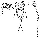
 issued frm : A. Scott in Siboga-Expedition, 1909, XIX a. [Plate III, Figs.17-29]. As Monacilla dubia. Male (from Banda Sea): 17, habitus (dorsal); 18, last thoracic and genital segments (left side); 19, right A1; 20, left A1; 21, A2; 22, Md; 23, Mx1; 24, Mx2; 25, Mxp; 26, P1; 27, P2; 28, P4; 29, P5. Nota: Head and 1st thoracic segment separate, 4th and 5th fused. Front with a small knob. A1 20-segmented, reaching the end of the 4th urosomal segment, 8th segment of right A1 represents 4 fused segments, and the same segment of the left side represents 5 segments.
|
 issued from :C.B. Wilson in Bull. U.S. natn Mus., 100, 14 (4). [Pl.26, Figs.379, 380]. Male: P5. 379, as Monacilla semispina (from 6°29'15''N, 126°18'45''E). 380, as Monacilla typica (from the same station and 13°45'30''N, 120°30'15''E; 15°45'54''N, 119°42'45''E)..
|
 issued from : G.P. Farran in Fish. Ire. Sci. Invest., 1906, II [1908]. [Pl. I, Figs.11-17]. As Oxycalanus spinifer. Female (from 50°0'N, 10°48'W): 11, habitus (lateral); 12, P3; 13, P2; 14, P1; 15, A2; 16, Mx2; 17, Mx1.
|
 issued from : K.A. Brodsky, N. V. Vyshkvartzeva, M.S. Kos & E.L. Markhaseva in Opred. Faune SSSR, 1983, 135 (1). [p.328, Fig.162]. Male. Ce = cephalon; L = left leg of P5; R = right leg of P5. P5 variant from Wilson (1950); other figures from Sars (1924).
|
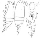
 Issued from : G.O. Sars in Résult. Camp. Scient. Prince Albert I, 69, pls.1-127 (1924). [Pl.XI, figs. 1-6, 6a, ]. Female: 1, habitus (dorsal); 2, idem (lateral); 3, forehead (lateral); 4, rostrum; 5, distal part of urosome; 6, A1; 6a, distal segments of A1 (enlarged).
|
 Issued from : G.O. Sars in Résult. Camp. Scient. Prince Albert I, 69, pls.1-127 (1924). [Pl.XI, figs. 7-11]. Female: 7, A2; 8, Md; 9, Mx1; 10, Mx2; 11, Mxp.
|
 Issued from : G.O. Sars in Résult. Camp. Scient. Prince Albert I, 69, pls.1-127 (1924). [Pl.XI, figs. 12-15]. Female: 12, P1; 13, P2; 14, P3; 15, P4.
|
 Issued from : G.O. Sars in Résult. Camp. Scient. Prince Albert I, 69, pls.1-127 (1924). [Pl.XII, figs. 1-3]. Male: 1, habitus (dorsal); 2, idem (lateral); 3, forehead (lateral).
|
 Issued from : G.O. Sars in Résult. Camp. Scient. Prince Albert I, 69, pls.1-127 (1924). [Pl.XII, figs. 4-8]. Male: 4, A2; 5, Md; 5a, Md (masticatory edge; enlarged); 6, Mx1; 7, Mx2; 8, Mxp.
|
 Issued from : G.O. Sars in Résult. Camp. Scient. Prince Albert I, 69, pls.1-127 (1924). [Pl.XII, figs. 9-10]. Male: 9, P1; 10, P5.
|
 issued from : P.E. Lapernat & C. Razouls in Vie Milieu, 2002, 52 (1). [p.21, Pl. II, fig.2]. Masticatory edge of Md gnathobase female (from off Malta, Mediterranean Sea).
|
 Issued from : V.N. Andronov in Russian Acad. Sci. P.P. Shirshov Inst. Oceanol. Atlantic Branch, Kaliningrad, 2014. [p.85, Fig.22: 7]. Monacilla typica after C.B. Wiulson, 1950. Male P5.
| | | | | Compl. Ref.: | | | Massuti Alzamora, 1942 (p.110); Sewell, 1948 (p.499, 508, 545, 550); Grice, 1963 a (p.495); Unterüberbacher, 1964 (p.20); De Decker & Mombeck, 1964 (p.13); Mazza, 1966 (p.70); Grice & Hulsemann, 1965 (p.223); Ehrhardt, 1967 (p.738, geographic distribution, Rem.); Hulsemann, 1967 (p.14); Park, 1970 (p.475); Roe, 1972 (p.277, tabl.1, tabl.2); Vives & al., 1975 (p.41, tab.II); Deevey & Brooks, 1977 (p.256, tab.2, Station "S"); Kovalev & Schmeleva, 1982 (p.83); Scotto di Carlo & al., 1984 (1041); Lozano Soldevilla & al., 1988 (p.58); Pancucci-Papadopoulou & al., 1990 (p.199); Scotto di Carlo & al., 1991 (p.270); Kouwenberg, 1994 (tab.1); Shih & Young, 1995 (p.74); Hure & Krsinic, 1998 (p.44, 100, 112); Lapernat & Razouls, 2001 (p.123, tab.1); Holmes, 2001 (p.41); Vukanic, 2003 (139, tab.1); Hsiao & al., 2004 (p.326, tab.1); Vukanic & Vukanic, 2004 (p.9, tab. 2); Fernandes, 2008 (p.465, Tabl.2); Licandro & Icardi, 2009 (p.17, Table 4); Mazzocchi & Di Capua, 2010 (p.427); Medellin-Mora & Navas S., 2010 (p.265, Tab. 2); Brugnano & al., 2012 (p.207, Table 2); Siokou & al., 2013 (p.1313, fig.4, 8, biomass, vertical distribution); Zaafa & al., 2014 (p.67, Table I, occurrence); Benedetti & al., 2016 (p.159, Table I, fig.1, functional characters); El Arraj & al., 2017 (p.272, table 2, spatial distribution); Chaouadi & Hafferssas, 2018 (p.913, Table II: occurrence); Belmonte, 2018 (p.273, Table I: Italian zones); Hure M. & al. (2018, p.12: Rem.) | | | | NZ: | 12 | | |
|
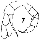
Distribution map of Monacilla typica by geographical zones
|
| | | | | | | | | | | | | Loc: | | | South Africa (E), Namibia, off Cape Verde Is., Canary Is., E Atlant. (11°N-13°S), Madeira, Portugal, Azores, Barbados Is., Caribbean Sea, Caribbean Colombia, G. of Mexico, Florida, Sargasso Sea, Station "S" (32°10'N, 64°30'W), Bay of Biscay, Ireland, Maroccan coast, Ibero-moroccan Bay, off W Tangier, Medit. (Alboran Sea, Sidi Fredj coast, Marseille, Ligurian Sea, Tyrrhenian Sea, Strait of Messina, off Malta, Adriatic Sea, Ionian Sea, Aegean Sea, Lebanon Basin), Indian, Bay of Bengal, Indonesia-Malaysia, Philippines, China Seas (South China Sea), Japan, off SE Hokkaido, NW Pacif., off S Aleutian Is., off SW California. | | | | N: | 57 | | | | Lg.: | | | (1) F: 2,3; M: 1,9; (5) F: 2,4; M: 2,3; (13) F: 2,3; M: 1,75; 1,72; (14) F: 2,25-2,16; (24) F: 2,32; (28) F: 2,34-1,95; M: 1,62-1,01; (38) F: 2,28-2,24; (55) F: 2,25; (131) F: 2,5-1,95; M: 2,3-1,59; (199) F: 2,43-2,28; (207) F: 2,1-1,96; M: 1,96-1,9; (208) F: 2,43; (340) F: 2; (432) M: 1,72; (1252) F: 2,05-2,58; {F:1,95-2,58; M: 1,01-2,30}
The mean female size is 2.234 mm (n = 19; SD = 0.1703), and the mean male size is 1.797 mm (n = 11; SD = 0.3552). The size ratio (male : female) is 0.80.
(1252) F: Pr/Ur = 2,5-3,7. | | | | Rem.: | bathypelagic. Sargasso Sea: 500-2000 m. 500-1000 m in the Aegean Sea (Siokou & al., 2013).
This species differs M. tenera by absecence of crest.
R. Stephen, 2007 : Data sheets of NIO, Kochi, India (on line).
After Lapernat & Razouls (2002, p.19) the Itoh's index value = 730.2 (number of teeth: 9) on the right Md, and 751.2 on the left Md (number of teeth: 9).
Depth: 400-2000 m (in Bode & al., 2017) | | | Last update : 25/10/2022 | |
|
|
 Any use of this site for a publication will be mentioned with the following reference : Any use of this site for a publication will be mentioned with the following reference :
Razouls C., Desreumaux N., Kouwenberg J. and de Bovée F., 2005-2025. - Biodiversity of Marine Planktonic Copepods (morphology, geographical distribution and biological data). Sorbonne University, CNRS. Available at http://copepodes.obs-banyuls.fr/en [Accessed November 02, 2025] © copyright 2005-2025 Sorbonne University, CNRS
|
|
|
|


;)
;)
;)
;)
;)
;)
;)
;)
;)
;)
;)
;)
;)
;)
;)
;)
;)
;)
;)
;)

