|
|
 |
|
Misophrioida ( Order ) |
|
|
|
Misophriidae ( Family ) |
|
|
|
Misophriella ( Genus ) |
|
|
| |
Misophriella schminkei Martinez Arbizu & Jaume, 1999 (F) | |
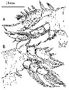
| | | | | | | Ref.: | | | Martinez Arbizu & Jaume,1999 (p.110, Descr.F, figs.F) | 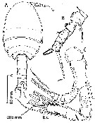 issued from : P. Martinez Arbizu & D. Jaume in Helgol. Mar. Res., 1999, 53. [p.110, Fig.8]. Female (from 72°28'S, 17°23.5'W): A, habitus (dorsal); B, P5 (anterior); C, A2 (arrow pointing to insertion of accidentally lost seta). Nota: Naupliar eye absent. Prosome 5-segmented, having 1st pedigerous somite not incorporated into cephalothorax but completely covered by posterior, carapace-like extension of dorsal cephalic shield. Rostrum completely fused to cephalosome with one marginal sensilla at each side and central pore on anterior surface. A2 exopod shows some suture lines on the 1st, 2nd and apical segments; these suture lines indicate that the 1st segment is a triple segment, while the 2nd and apical ones are double segments; the antennary exopod could therefore be derived from an ancestral 10-segmented exopod (which represents the groundpattern in Copepoda) displaying the following fusion pattern: (I-III), (IV-V), VI, VII, VIII, (Ix-X).P5 uniramous, jointed by very narrow intercoxal sclerite; legs 4-segmented (coxa, basis and 2-segmented exopod).
|
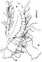
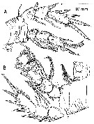
 issued from : P. Martinez Arbizu & D. Jaume in Helgol. Mar. Res., 1999, 53. [p.111, Fig.9]. Female: A, urosome with 5th pedigerous somite omitted; ventral); B, idem (dorsal). Roman numerals identify caudal setae. Nota: Urosme 5-segmented, with surface of both genital double-somite and abdominal somites covered with epicuticular hyaline ridges. Genital and 1st abdominal segment partially fused to form genital double-somite retaining articulation dorsally about midway of somite; articulation fringed with hyaline frill belonging to posterodorsal margin of genital somite; patch of short spinules dorsolaterally at each side of genital somite; pair of gonopores located ventrally at about 2/5 of distance along double-somite, each closed off by operculum derived from P6 (6th legs), armature of latter consisting of 2 long setae implanted on discrete pedestals; single copulatory pore located midventrally. Anal operculum ornamented with finely serrated frill on posterior margin. Caudal rai about as long as wwide, armed with 7 setae, tube pore located mid-ventrally.
|
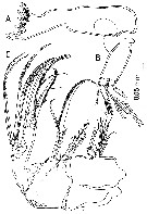
 issued from : P. Martinez Arbizu & D. Jaume in Helgol. Mar. Res., 1999, 53. [p.112, Fig.10]. Female: A, right A1 (medial; arrows pointing to insertion of accidentally lost setae on segment 3 and 9; B, rostrum (anterior). Nota: A1 19-segmented. Misophriella tetraspina differs from M. schminkei in the spiniform condition of some setae on the 1st and 2nd antennulary segments, the homologous armature elements being normal setae in the Antarctic species
|
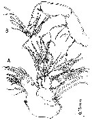
 issued from : P. Martinez Arbizu & D. Jaume in Helgol. Mar. Res., 1999, 53. [p.113, Fig.11]. Female: A, right Md (mandibular coxal gnathobase); B, right Md (mandibular palp (dashed seta on exopod present only on left mandibular palp); C, Mx2. Nota: Mx2 6-segmented; praecoxa and coxa separate, endite formula: 7, 2, 3, 3; 1 seta on distal coxal endite hypertrophied; basal endite drawn out into stout, curved medial claw basally incorporated into segment, 3 setae (1 hypertrophied) subdistally on endite; endopod 3-segmented, setal formula 1, 2, 3.
|
 issued from : P. Martinez Arbizu & D. Jaume in Helgol. Mar. Res., 1999, 53. [p.114, Fig.12]. Female: A, Mx1; B, Mxp. Nota: Mx1 with discrete praecoxa, praecoxal arthrite with 8 stout spines plus 1 seta distally, 2 setae subdistally on medial margin and other 2 on anterior surface; coxal epipodite with 6 setae, 3 of them reduced; coxam endite with 6 setae distally; basal endites each with 4 setae; disal endite largely incorporated into segment; endopod unsegmented , elongate, with armature distributed in 3 groups of 2, 1, and 5 setae, respectively; exopod unsegmented (about twice as long as wide) with 6 distal setae; surface of maxillulary segments ornamented with spinules, denticles and setules. Mxp comprising syncoxa, basis and 5-segmented endopod.
|
 issued from : P. Martinez Arbizu & D. Jaume in Helgol. Mar. Res., 1999, 53. [p.115, Fig.13]. Female: A, P1; B, P2.
|
 issued from : P. Martinez Arbizu & D. Jaume in Helgol. Mar. Res., 1999, 53. [p.116, Fig.14]. Female: A, P4; B, P3 (with disarticulated exopod; arrow pointing to insertion of accidentally lost outer basal seta.
| | | | | NZ: | 1 | | |
|
Distribution map of Misophriella schminkei by geographical zones
|
| | | | | | | Loc: | | | Antarct. (E Weddell Sea) | | | | N: | 1 | | | | Lg.: | | | (844) F: 0,61; { F: 0,61} | | | | Rem.: | hyperbenthic (300 m).
According to P. Martinez Arbizu & Jaume (1999, p.116) the gut contents consisted of parts of copepod limbs (cyclopinids). This indicates that the species is carnovorous; it may be a predator or a scavenger. | | | Last update : 19/01/2015 | |
|
|
 Any use of this site for a publication will be mentioned with the following reference : Any use of this site for a publication will be mentioned with the following reference :
Razouls C., Desreumaux N., Kouwenberg J. and de Bovée F., 2005-2026. - Biodiversity of Marine Planktonic Copepods (morphology, geographical distribution and biological data). Sorbonne University, CNRS. Available at http://copepodes.obs-banyuls.fr/en [Accessed March 02, 2026] © copyright 2005-2026 Sorbonne University, CNRS
|
|
|
|


;)
;)
;)
;)
;)
;)
;)

