|
|
 |
|
Misophrioida ( Order ) |
|
|
|
Misophriidae ( Family ) |
|
|
|
Misophriopsis ( Genus ) |
|
|
| |
Misophriopsis okinawensis Ohtsuka, Huys, Boxshall & Itô, 1992 (F,M) | |
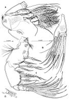
| | | | | | | Ref.: | | | Ohtsuka & al., 1992 a (p.860, 872, figs.F,M); Chihara & Omori, 1997 (p.932, Pl.190: F,M); Martinez Arbizu & Jaume, 1999 (p.109); Humes, 1999 (p.976) | 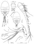 issued from : S. Ohtsuka, R. Huys, G.A. Boxshall & T. Itô in Zool. Sc., 1992, 9. [p.861, Fig.1]. Female (from Kume island, Okinawa): A, habitus (dorsal); B, urosome (dorsal); C, A1; D, A2; E, basal two exopod segments of A2; F, distal exopod segment of A2. Nota: A1 18-segmented. A2: coxa bearing row of fine setules near anterior margin; basis with 2 apical setae of unequal lengths and patch of fine spinules on posterior surface; endopod 3-segmented; segment 1 with 2 small, subfdistal inner setae of unequal lengths and patch of minute spinules on posterior surface; segment 2 with 2 lateral and 3 distal setae along inner margin; segment 3 with 6 long setae and short seta apically; exopod 6-segmented ( setal formula: 0, 2, 1, 1, 1, 3, respectively). 1st pedigerous somite entirely concealed beneath carapace-like expansion from posterior end of maxilliped-bearing somite. 3rd and 4th pedigerous somites produced posteriorly. Urosome 5-segmented. 5th pedigerous somite posterolaterally produced into acute process on both sdes. Genital and 1st abdominal somites fused ( to form double-somite). Pair of slit-lake genital pores located ventrolaterally on double-somite and covered by operculum (derived from P6). 4th abdominal somite completely concealed beneath 3rd. Anal somite fringed with minute prominences dorsoposteriorly. Caudal rami wider than long. Male: G, habitus (dorsal). Nota: Both A1 12-segmented, geniculate with geniculation between 10th (XIX-XX) and 11th (XXI-XXIII) segments Urosome 6-segmented. 5th pedigerous somite with acute posterolateral angles. Genital somite large. 3rd abdominal somite small, largely concealed beneath 2nd.
|
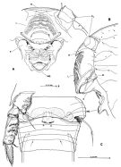 issued from : S. Ohtsuka, R. Huys, G.A. Boxshall & T. Itô in Zool. Sc., 1992, 9. [p.862, Fig.2]. Female: A, oral area and rostrum (ventral view); B, idem (lateral left side); C, P5 and P6 and genital double-somite (dotted area indicates seminal receptacle). A1: antennule; R: rostrum; L: labrum; P: paragnath; M: mandible; IMS: intermaxillary swelling. Nota: Rostrum partly fused with labrum, posteroventrally produced, and furnished with a pair of sensilla near its pointed tip. Naupliar eye absent. Labrum covered with numerous minute spinules, bearing row of fine hairs near its posterior margin and pair of shotrt prominences on both sides of posterior end. Pair of paragnaths located posterior to end of labrum, ornamented with fine spinules on tip. Intermaxillary swelling arising from mid-ventral surface, with row of minute spinules along posterior margin.
|
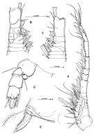
 issued from : S. Ohtsuka, R. Huys, G.A. Boxshall & T. Itô in Zool. Sc., 1992, 9. [p.863, Fig.3]. Female: A, Md (mandibular gnathobase); B, Md (mandibular palp); C, P6 and genital and copulatory pores on genital double-somite (lateral left side); D, caudal ramus (ventral). Nota: mandibular gnathobase with 5 multicusped teeth, 3 serrate blades and 1 spiniform seta; ornamentation of fine spinules and relatively long setules present on anterior surface near palp; mandibular palp biramous; basis bearing patches of fine spinules and inner medial seta; endopod 2-segmented, proximal segment with inner suterminal seta, distal segment with 1 medium length, 1 short and 6 long setae; exopod 5-segmented (setal formula: 1, 1, 1, 1, 2, respectively)
|
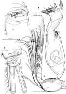
 issued from : S. Ohtsuka, R. Huys, G.A. Boxshall & T. Itô in Zool. Sc., 1992, 9. [p.864, Fig.4]. Female: A, caudal ramus (lateral view); B, Mx1. Nota: Mxi with praecoxal arthrite with 7 strong spines and 8 setae (2 of which arising from anterior surface); coxal endite with 1 thick serrate seta and 5 pinnate setae, distal basal endite with 4 setae; epipodite of coxa with 7 setae of unequal lengths; endopod comprising single compound segment (representing fused 1st to 3rd segments), armature divided into groups of 3 inner medial, 3 inner subterminal and 6 terminal setae representing original segmental elements; exopod 1-segmented with 3 inner lateral and 5 terminal setae and relatively long setules along inner and outer margins.
|
 issued from : S. Ohtsuka, R. Huys, G.A. Boxshall & T. Itô in Zool. Sc., 1992, 9. [p.865, Fig.5]. Female: A, Mx2 (setae of allobasis and endopod omitted); B, B, Mx2 (allobasis and endopod); C, Mxp; D, Mxp (distal segment of endopod). Nota: with praecoxa partly fused with coxa, with 2 endites (proximal with 6 setae, distal with 3 setae); coxa with 2 endites and outer patch of fine spinules, each endite with 3 distal setae; allobasis derived from fusion of basis and 1st endopodal segment, produced into strong curved claw and bearing 6 setae (3 of which derived from 1st endopodal segment); free endopod indistinctly 3-segmented (setal formula: 1, 2, 4, respectively). Mxp with 4 endites on syncoxa (setal formula: 0, 1, 3, 2, respectively), row of long spinules along outer medial margin, proximal patch of minute spinules and irregular rows of small spinules near bases of setae on middle and subdistal endites; basis with 3 spinulose inner setae medially and row of long spinules proximally, fringed with short spinules along posterior half of inner margin; free endopod 5-segmented (setal formula: 2, 2, 2, 2, 5, respectively)
|
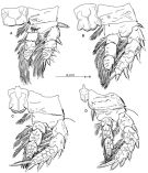 issued from : S. Ohtsuka, R. Huys, G.A. Boxshall & T. Itô in Zool. Sc., 1992, 9. [p.866, Fig.6]. Female: A, P1 (anterior); B, P2 (anterior); C, P3 (anterior); D, P4 (anterior).
|
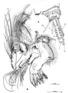
 issued from : S. Ohtsuka, R. Huys, G.A. Boxshall & T. Itô in Zool. Sc., 1992, 9. [p.867, Fig.7]. Male: A, antennule; B, A1 (segments 3 to 7, dorsal view); C, A1 (segment 3 to 7, ventral view); D, P5 (anterior); E, P6 (anterior). Nota: P5 biramous; compound protopodal segment fringed by minute spinules along inner margin with 1 setaderived from basis; endopod represented by small subcircular segment, with small seta distally; exopod 3-segmented, proximal segment unarmed, middle segment with thick, plumose seta at distal inner angle, apical segment with 1 inner seta and 1 seta on each side of apical serrate spine. P6 forming opercular plate overlying genital opening, bearing large serrate inner spine and 2 plumose setae.
|
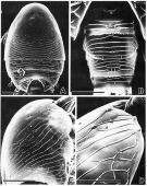 issued from : S. Ohtsuka, R. Huys, G.A. Boxshall & T. Itô in Zool. Sc., 1992, 9. [p.868, Fig.8]. Female (SEM micrographs): A, prosome (dorsal); B, urosome (dorsal); C, cephalosome (lateral right side); D, epicuticular ornamentation on cephalosome. Scale bars = 0.100 mm (A, C); 0.050 mm (B, D). Nota: The body surface is almost entirely covered with shallow, epicuticular lamellae except for the anterior half of the dorsal cephalic shield which has a smooth surface with a number of hair-like sensilla and small pores but no lamellae. The lamellae are perpendicular to the surface on the prosome, whereas they incline posteriorly on the urosome. Cone organs lacking on the lateral side of cephalosome as in Benthomisophria palliata Sars,1909 and Misophriopsis dichotoma Boxshall,1983. The 5th pedigerous somite has a relatively large pore on the dorsomedial surface; the genital somite, anterior and posterior dorsomedial pores; 1st abdominal somite, posterior medial pore dorsally; 4th somite with anterior pore dorsomedially.
|
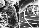 issued from : S. Ohtsuka, R. Huys , G.A. Boxshall & T. Itô in Zool. Sc., 1992, 9. [p.869, Fig.9]. Female (SEM micrographs): A, labrum (pore indicated by an arrow); B, P6. Scale bars = 0.050 mm (A); 0.010 mm (B). Nota: The labrum is densely covered with ornamentation consisting of dentate scales separed by open smooth areas. The rostrum and the labrum are partly separate from each other (Fig.9A); a pore located medially on the anterior part of labrum is arrowed (Fig.9A). P6 is armed with a plumose seta on a low cylindrical process, a small spine, and smooth and serrate processes (Fig.9B)
|
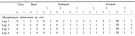
 issued from : S. Ohtsuka, R. Huys, G.A. Boxshall & T. Itô in Zool. Sc., 1992, 9. [p.870, Table 1]. Armatures of A1. Number of antennulary segment of ancestral copepod represented by Roman numeral; number of setae on the segment represented by Arabic numeral: a+b = (number of anterior setae) + (number of posterior setae). ae: aesthetasc; proc: spinous process derived from modification of setal element.
|
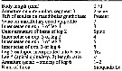
 issued from : S. Ohtsuka, R. Huys, G.A. Boxshall & T. Itô in Zool. Sc., 1992, 9. [p.871, Table 2]. Seta and spine formula of legs 1 to 4. Seta represented by Arabic numeral, and spine by Roman numeral. o = outer border of segment; t = terminal border of segment; i = inner border of segment.
|
 issued from : P. Martinez Arbizu & D. Jaume in Helgol. Mar. Res., 1999, 53. [p.109, Table 2]. Diagnostic features for identification. exp. 2 = exopod segment 2; exp. 3 = exopod segment 3.
| | | | | NZ: | 1 | | |
|
Distribution map of Misophriopsis okinawensis by geographical zones
|
| | | | Loc: | | | Japan (Kume Is.) | | | | N: | 1 | | | | Lg.: | | | (615) F: 0,79-0,66; M: 0,55-0,52; {F: 0,66-0,79; M: 0,52-0,55} | | | | Rem.: | hyperbenthic (neritic).
Ohtsuka & al, 1992 (p.871, Table 2) indicate 3 setae on the inner border of exopodal segment 3 of P1 and 4 setae on the inner border of exopodal segment 3 of P2 (for 4 and 5, respectively in M. dichotoma and M. sinensis. | | | Last update : 20/01/2015 | |
|
|
 Any use of this site for a publication will be mentioned with the following reference : Any use of this site for a publication will be mentioned with the following reference :
Razouls C., Desreumaux N., Kouwenberg J. and de Bovée F., 2005-2025. - Biodiversity of Marine Planktonic Copepods (morphology, geographical distribution and biological data). Sorbonne University, CNRS. Available at http://copepodes.obs-banyuls.fr/en [Accessed November 20, 2025] © copyright 2005-2025 Sorbonne University, CNRS
|
|
|
|


;)
;)
;)
;)
;)
;)
;)
;)
;)
;)
;)
