|
|
 |
|
Calanoida ( Order ) |
|
|
|
Clausocalanoidea ( Superfamily ) |
|
|
|
Aetideidae ( Family ) |
|
|
|
Euchirella ( Genus ) |
|
|
| |
Euchirella venusta Giesbrecht, 1888 (F,M) | |
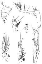
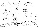
| | | | | | | Syn.: | Euchirella tanseii Omori, 1965 a (p.60, figs.F); Tanaka & Omori, 1969 (p.61, Rem.); 1969 a (p.160, Rem.) | | | | Ref.: | | | Giesbrecht, 1892 (p.233, 244, figs.F); Giesbrecht & Schmeil, 1898 (p.35); Wolfenden, 1905 a (p.18, Rem.F); A. Scott, 1909 (p.57, Rem.); Farran, 1929 (p.208, 237, Rem.); Sciacchitano, 1930 (p.14); Sewell, 1947 (p.85, figs.F,M); Vervoort, 1949 (p.20, figs.F); C.B. Wilson, 1950 (p.226, figs.F, Rem.); Vervoort, 1952 f (n°47, p.4: Rem., fig.F); Tanaka, 1957 b (p.182, figs.F,M); Grice, 1962 (p.194, figs.F,M, Rem.); Omori, 1965 a (p.64, figs.F, Rem.) ; Owre & Foyo, 1967 (p.48, figs.F,M); Tanaka & Omori, 1969 (p.59, figs.F,M); 1969 a (p.159, Rem.F); Vaupel Klein, 1972 (p.502, 505, fig.F); Bradford, 1972 (p.38, figs.F); Bradford & Jillett, 1980 (p.43, figs.F,M, fig. 73, distribution chart); Vaupel Klein, 1980 (p.152); 1984 a (p.47, Table II: characters, Rem.); Markhaseva, 1996 (p.175, figs.F,M); Chihara & Murano, 1997 (p.686, Pl.37,41: F,M) | 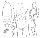 issued from : Tanaka O. in Publ. Seto Mar. Biol. Lab., 1957, 6 (2). [Fig.47, p.182]. Female (from Suruga): a, habitus (dorsal aspect); b, head (lateral aspect); c, last thoracic segment and genital segment (lateral aspect); d, P1; e, basal joints of P4. Male (from Suruga): f, P5; g, a part of distal joint of exopodite of right P5. Nota Female: - Cephalothorax about 3.88 times the abdomen length. - Abdominal segments and caudal rami in the proportional lengths 54 : 13 : 11 : 5 : 18 = 100. - Genital segment asymmetrical, a remarkable protuberance on the left side of the segment; proximal dorsal surface hollowed; ventral protuberance situated anterior to the genital opening very prominent. - Caudal rami about as long as wide. - A1 23-segmented, extends to distal end of genital segment. - A2 exopod about 3.4 times as long as endopod. - Md blade robust with a long inner edge seta. - Mx1 with 11 setae on exopod; 4 setae on endopod; 6 setae on outer lobe; 3 setae on 2nd basal segment. - Mxp as that of E. messinensis; outer margin of 2nd basal segment with 2 swellings on the distal half of segment. - P1 with 2 outer edge spines on outer margin of 1st exopodal segment (as in other members of the genus). - Terminal spine of exopod of P2 to P4 with 20, 21, and 20 teeth respectively. - P4 with 2 spines reaching the proximal margin of basis on the inner margin of coxa at the base of inner marginal seta. Nota Male: - Cephalothorax about 3.93 times the abdomen length (2.95 : 0.75). - Head without crest. - A1 reaches back to distal margin of 1st abdominal segment. - P5 closely resemble those of E. messinensis. Nota male after Markhaseva (1996, p.175): Crest absent. Rostrum faintly developed. Endopodla segment 2 of A2 with 6 setae on external and 7 setae on internal lobes. 1st to 3rd inner lobes of Mx1 highly reduced, external lobe with 5 setae, exopod with 11 setae, exopodal segment 1 with 2 very small spines. Basipodite of left P5 reaching the distal border of right coxopodite; exopod of left P5 faint, its distal end exceeding the third projection of the exopod of right P5.
|
 issued from : E.L. Markhaseva in Proc. Zool. Inst. RAN, St. Petersburg, 1996, 268. [p.174, Fig.136]. Female (specimen from NW Indian). CP4: coxopod of P4. Nota: Rostrum well developed. Genital segment asymmetrical, its shape varying. A1 reaching the midlength of urosome.
|
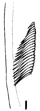
 issued from : J.M. Bradford & J.B. Jillett in Mem. N.Z. Oceanogr. Inst., 86, 1980. [p.47, Fig.30]. Male: E, habitus (lateral right side); F, Mx1; G, P5; H, terminal part of left exopod of P5. Nota: The two males from the south-west Pacific are identical to those described by Sewell (1947) except that the terminal exopod joint of the right P5 has 31 semicircular teeth. Since female E. speciosa are very similar to E. venusta it is possible that teir males are also very alike, in which case the present two males may be attribued to E. speciosa if compared more closely with E. venusta.
|
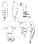
 Issued from : W. Vervoort in Zool. Verh., Leiden, 1949, 5. [p. 21, Fig.10]. Female (from Arafura Sea): a, c, distal part of thoracic segment and urosome (dorsal, lateral left side, respectively); b, forehead (lateral); d, basal portion of P4, showing the spines on the 1st basal segment. Nota: A1 (24 free-segments) reaches the middle of the genital segment. Head and 1st thoracic segment, 4th and 5th completely fused. The mouth parts agree closely with those of E. messinensis. The abdominal segments and furca in the proportional lengths 55:12:11:7:15 = 100.
|
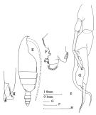
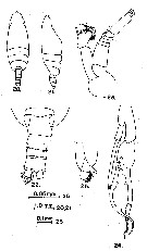
 issued from : R.B.S. Sewell in The John Murray Expedition, 1933-34, Scientific Reports, VIII (1), 1947. [p.87, Fig.18]. Female (from S Arabian Sea): A, urosome (lateral right side); B, Mxp; C, P1; D, P4. Nota: Proportional lengths of cephalothorax and abdomen as 80 to 20. The proportional lengths of the various segments of the body (cephalon to caudal rami) as 386:141:91:84:81:91:34:34:24:34=1000. Head and 1st pediger segment separate, 4th and 5th fused. A rounded prominence on the left side of the genital segment, and a high ridge runs across the ventral part of this segment in front of the genital aperture. A1 23-segmented (8-9, 24-25, respectively fused) Male: E, P1; F, P5; G, terminal claw of left P5. Nota: Proportional lengths of cephalothorax and abdomen as 79 to 21. The proportional lengths of the various segments of the body (cephalon to caudal rami) as 484:84:80:96:68:56:48:40:12:32=1000. Forehead without crest. Head and 1st pediger segment fused, 4th and 5th fused; the 5 th segment merely forming a low crest along the lateral part of the region. Anal segment with a tuft of hairs ventrally, entirely telescoped into the preceding one. Left A1 22-segmented (segments 8-9, 12-13, 24-25 respectively fused), right 21-segmented (segments 8-9, 12-13, 20-21, 24-25 respectively fused). Exopod of A2 7-segmented; endopod bears on its two lobes 6 setae on the inner, and 5 well developed and 1 small and delicate setae on the outer. Md devoided of any biting blade. Mx1 and Mx2 reduced. In Mx2 a fleshy lobe bears at its extremity a single segment with 5 delicate setae.
|
 issued from : J.C. von Vaupel Klein in Crustaceana, Supplt 9, Studies on Copepoda, III, 1984. [p.75, Fig.13, l]. Female: arrangementof spinules on the proximal part of the medial face of basosipodite 2 of Mxp (right appendage in medial view). Nota: 20 long and slender spinules in a single, contiguous, distally decreasing row.
|
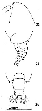
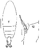 issued from : J.M. Bradford in Mem. N. Z. Oceonogr. Inst., 1972, 54. [p.39, Fig.7, (11-12) Female (from Kaikoura, New Zealand): 11, habitus (dorsal); 12, basipod segment 1 of P4. Scale bars: 1 mm (1); 0.1 mm (12). Nota: Characterised by the genital segment and by the two spines on the P4, 1st basipod segment which reach its hind margin.
|
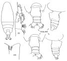
 issued from : M. Omori in J. Oceanol. Soc. Japan, 1965, 21 (2). [p.61, Figs.1-5]. As Euchirella tanseii. Female (from 38°58.9'N, 139°35.4'E): 1-2, habitus (dorsal and lateral, respectively); 3, forehead (lateral); 4-5, posterior prosome and urosome (dorsal and left lateral, respectively). Nota : Cephalothorax is a little more than 4 times the length of urosome. Rostrum distinct. Head and 1st thoracic segment fused, 4th and 5th fused. Last thoracic segment with scattered bristles on distal surface. Proportional of the urosomal segments and caudal rami 49 :14 :12 :9 :16 = 100. Genital segment as long as broad, asymmetrical ; two remarkable protuberances on the left side, the right side is very smooth ; in lateral aspect a high ridge runs across the ventral part ; the ventral protuberance situated anterior to the genital opening is very prominent ; the left side shows a transversal groove, close to the distal margin. 1st and 2nd urosomal segments armed with a row of small, triangular spinules along the dorsal distal margin. Caudal rami about as long as wide, with 4 strong plumose subequal setae, plus 1 outer short and 1 curved ventral seta on each ramus ; the inner margins are densely haired.
|
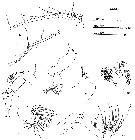 issued from : M. Omori in J. Oceanol. Soc. Japan, 1965, 21 (2). [p.62, Figs.6-12]. As Euchirella tanseii. Female: 6, A1; 7, A2; 8, Md; 9, Md (masticatory edge of gnathobase); 10, Mx1; 11, Mx2; 12, Mxp. Nota : A1 23-segmented (segments 8-9 completely fused, articulation between 24th and 25th segments very incomplete). A2 with exopodite about 3.6 times the length of the endopodite ; outer endopodal lobe with 6 setae, inner lobe with 4 minute setae. Mouth parts agree closely with those of E. venusta. Exopodite of Md with 6 setae and 9 on the endopodite ; gnathobase robust with a long inner marginal seta. Mx1 with 11 setae on exopodite, 4 setae on endopodite, 4 setae on 2nd inner lobe, 3 setae on 3rd inner lobe, and 3 setae on 2nd basal segment. Mxp has 4 setae on 1st, 3 setae on 2nd, 3 setae on 3rd, 3 setae and 1 external seta on 4th, 3 setae and 1 external seta on 5th segment, respectively ; 2 swellings on the distal half of the outer margin of 2nd basal segment are not distinct.
|
 issued from : M. Omori in J. Oceanol. Soc. Japan, 1965, 21 (2). [p.63, Figs.13-18]. As Euchirella tanseii. Female: 13, P1; 14, P2; 15, P3; 16, distal portion of the exopodite of P3 (left side); 17, P4; 18, basal portion of P4. Nota : The structure of P3 is different from those in other members of the genus that it is doubtful whether thisis the normal type ; the legs are asymmetrical, the endopodite of the right leg is 3-segmented, the left one is 2-segmented ; both the exopodite are 3-segmented, but the 2nd segment of the left leg has 3 small spines at the base of the outer spine, the 3rd segment has 2 outer spines, there is a notch on the outer margin between those 2 spines , in place of the terminal spine, there is a plumose seta.
|
 issued from : M. Omori in J. Oceanol. Soc. Japan, 1965, 21 (2). [p.64, FTable 1]. Comparison of characters among three species: E. tanseii, E. venusta and E. pulchra. Nota: E. tanseii closely resembles E. venusta and E. pulchra, but it may be distinguished from these two species.
|
 issued from : M. Omori in J. Oceanol. Soc. Japan, 1965, 21 (2). [p.64, Figs.22-24]. Female: 22, forehead (lateral); 23, posterior prosome (left lateral); 24, idem (dorsal). Nota : Head without trace of crest. Genital segment with a small but clear indent in the middle ofthe segment (C.B. Wilson, 1950, figured E. venusta without indent on the identical portion, but is a posible mistake).
|
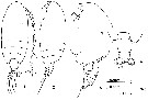
 issued from : O. Tanaka & M. Omori in Publ. Seto mar. Biol. Lab., 1969, XVII, 1. [p.60, Fig.10]. Female (from W Pacific): a, forehead (lateral); b-c, last thoracic segment and urosome (dorsal and lateral, rsepectively); d, idem (another specimen). Male: e, forehead (lateral); f, P5; g, left P5 (distal segment of exopod); h, idem (another vuew); i, idem (another specimen).
|
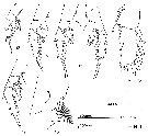
 issued from : G.D. Grice in Fish. Bull. Fish and Wildl. Ser., 1962, 61. [p.193, Pl.9, Figs.14-19]. Female (from 01°10'S, 134°57'W): 14-15, habitus (dorsal and lateral, respectively); 16-17, urosome (dorsal and lateral, respecticely); 18, endopod of A2; 19, P4. Nota: A knob-like protrusion from the left posterior margin of the genital segment. Presence of 4 setae on the inner lobe and 5 setae on the external lobe of the endopod of A2. 2 long spines on the 1st basipodal segments of the P4.
|
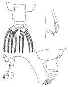
 issued from : G.D. Grice in Fish. Bull. Fish and Wildl. Ser., 1962, 61. [p.193, Pl.9, Figs.20-25]. Male: 20-21, habitus (dorsal and lateral, respectively); 22, urosome (dorsal); 23, A2; 24, P5; 25, terminal part of left P5. Nota: A2 with 6 setae on the outer lobe and 7 setae on the inner lobe, 2 of which are small (Sewell reported 6 setae on each lobe).
|
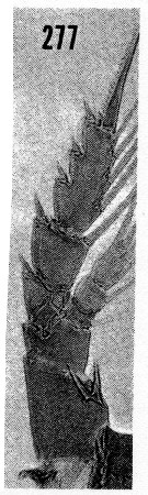 issued from : H.B. Owre & M. Foyo in Fauna Caribaea, 1967, 1, Crustacea, 1: Copepoda. [p.48, Fig.277]. Female (from Florida Current): 277, P4. Nota: Apparently the first record of this species in the western North Atlantic (Owre & Foyo, 1964) from 292 m in the Caribbean Sea.
|
 Euchirella venusta Euchirella venusta female: 1 - Genital segment asymmetrical. 2 - Crest absent. 3 - Genital segment with left lateral side swelled (dorsal view). 4 - Coxopodite of P4 with 2 spines. 5 - Endopodal segment 1 of Md without seta. 6 - Endopodal segment 2 of A2 with 5 setae on external lobe and 4 setae on internal lobe. 7 - Genital segment in dorsal view left side with projection not bilobated (dorsal view).
|
 issued from : Tanaka O. in Publ. Seto Mar. Biol. Lab., 1957, 6 (2). [p.182]. Female A1: proportional lengths of segments. - A1 23-segmented, extends to distal end of genital segment.
|
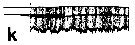 issued from : J.C. von Vaupel Klein in Crustaceana, (Supplt 9), 1984 a. [p.61, Fig.4, k]. caudal fringe of the urosomite 3 in sector of the dorsal ridge (hair-fringe only). Nota: Types of caudal hair- fringes on the urosomites in dorsal ridges vs. additional fringe of spinules adjacent to caudal margin.
|
 issued from : J.C. von Vaupel Klein in Crustaceana, Supplt 9, Studies on Copepoda, III, 1984. [p.66, Fig.7, k, l]. Euchirella venusta: Structural feature of exopodite 1 and exopodite 2 of A2 (lateral aspects of left appendages and medial views of right antennal parts; compare with Fig.7n which represents left A2 medial face drawn from a lateral view). k, lateral; l, medial. Nota: Degree of fusion exopodites 1 and 2: only faint indications of a suture present both laterally and medially.
|
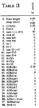 Issued from : J.C. von Vaupel Klein in Crustaceana, (Supplement) 9, 1984. [p.93, Table II]. Euchirella venusta Female: Datamatrix stating observed states of characters from Table I (p.87-90) presently examined; nos. refer to the input nos. used in Table I (see to the family Aetideidae).
|
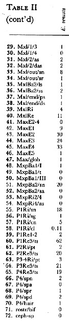 Issued from : J.C. von Vaupel Klein in Crustaceana, (Supplement) 9, 1984. [p.94, Table II (cont' d)]. Datamatrix stating observed states of characters from Table I (p.87-90) presently examined; nos. refer to the input nos. used in Table I (see to the family Aetideidae).
|
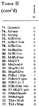 Issued from : J.C. von Vaupel Klein in Crustaceana, (Supplement) 9, 1984. [p.95, Table II (copnt' d)]. Datamatrix stating observed states of characters from Table I (p.87-90) presently examined; nos. refer to the input nos. used in Table I (see to the family Aetideidae).
| | | | | Compl. Ref.: | | | Cleve, 1904 a (p.190); Sewell, 1948 (p.331, 519, 531, 540, 552); Fagetti, 1962 (p.20); De Decker & Mombeck, 1964 (p.12); Grice & Hulsemann, 1967 (p.15); Carter, 1977 (1978) (p.35); Cahoon, 1982 (p.202); Arashkevich & al., 1982 (p.477, Table 2, diet); Guangshan & Honglin, 1984 (p.118, tab.); Brinton & al., 1986 (p.228, Table 1); Suarez & al., 1990 (tab.2); Suarez, 1992 (App.1); Shih & Young, 1995 (p.67); Suarez-Morales & Gasca, 1998 a (p108); Padmavati & al., 1998 (p.349); Hsiao & al., 2004 (p.325, tab.1); Dur & al., 2007 (p.197, Table IV); Tutasi & al., 2011 (p.791, Table 2, abundance distribution vs La Niña event); Sano & al., 2013 (p.11, Table 7, food habits) | | | | NZ: | 12 | | |
|
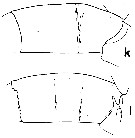
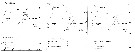
Distribution map of Euchirella venusta by geographical zones
|
| | | | | | | | |  Chart of 1996 Chart of 1996 | |
 Issued from : J.M. Bradford & J.B. Jillett in New Zealand Ocean. Inst. Memoir, 86, 1980. [p.88-89, Figs.65-67]. Issued from : J.M. Bradford & J.B. Jillett in New Zealand Ocean. Inst. Memoir, 86, 1980. [p.88-89, Figs.65-67].
Distribution of several species of Euchirella in the Tasman Sea and around New Zealand.
Nota: Euchirella indica (= Euchirella massinensis indica). |
| | | | Loc: | | | South Africa (E), off S Cape Verde Is., Brazil, Barbada Is., Caribbean Sea, G. of Mexico, Florida, Arabian Sea, Natal, Indian, Indonesia-Malaysia, China Seas (East China Sea, South China Sea) , Taiwan (SW), Japan (Izu), Pacif. (W equatorial), New Zealand (Kaikoura), Pacif. (central), Pacif. (equatorial), Gulf of California, off SW Mexico, off Peruvian coast, Galapagos-Ecuador, Chile.
Type locality: Pacific (tropical). | | | | N: | 45 | | | | Lg.: | | | (11) F: 4,383; M: 3,8; (35) F: 5-4,62; (37) F: 4,88-4,25; M: 4,16-3,57; (41) F: 4,6-4,25; (56) F: 4,88; M: 3,7; (101) F: 4,84-4,37; M: 3,8; (110) F: 4,9-4,2; M: 4,16-3,57; (116) F: 5,06; (201) M: 3,9; (242) F: 5-4,05; (1125)* F: 3,4-3,5; (1257) F: 4,05-4,8; {F: 4,05-5,06; M: 3,57-4,16}
* in Arashkevich & al. (1982) from the upwelling Peruvian coast in March 1978.
The mean female size is 4.619 mm (n = 15; SD = 0.3421), and the mean male size is 3.833 mm (n = 8; SD = 0.2319). The size ratio (male : female) is 0.83. [Arashkevich's forms not included in the computation]. | | | | Rem.: | epi & mesopelagic.
For Omori (1965, p.60 and 64) E. venusta resembles very closely E. tanseii, but may be distinguished by: the two species have two protuberances on left side of the genital segment, but the protrusion of the proximal one of E. tanseii is more distinct than that of E. venusta; the right side in E. tanseii is very smooth whereas in E. venusta there is a small but clear indent in the middle of the segment (C.B. Wilson, 1950, figured E. venusta without indent on the identical portion, but this might have been a mistake). The number of setae on the 2nd segment of endopodite of A2 in E. tanseii seems to slightly differ from that of E. venusta; the internal lobe has 6 and the external lobe has 6 setae in the former species, while they are 4 and 5 in the latter one as described by Vervoort (1949). The 1st outer spine of the 1st segment of exopodite of P1 of E. tanseii curves to the inside. The strucure of P3 of E. tanseii seems so abnormal that it might be safe to not consider the characteristic feature of the species. A doubt subsists concerning the synonimy between the two species, the single specimen found is perhaps abnormal in relation to the habitat on a sandy and gravely substrate at a depth of 100 m in Sagami Bay.
R. Stephen: Data sheets of NIO, Kochi, India (on line).
| | | Last update : 22/02/2021 | |
|
|
 Any use of this site for a publication will be mentioned with the following reference : Any use of this site for a publication will be mentioned with the following reference :
Razouls C., Desreumaux N., Kouwenberg J. and de Bovée F., 2005-2025. - Biodiversity of Marine Planktonic Copepods (morphology, geographical distribution and biological data). Sorbonne University, CNRS. Available at http://copepodes.obs-banyuls.fr/en [Accessed January 04, 2026] © copyright 2005-2025 Sorbonne University, CNRS
|
|
|
|


;)
;)
;)
;)
;)
;)
;)
;)
;)
;)
;)
;)
;)
;)
;)
;)
;)
;)
;)

;)
