|
|
 |
|
Monstrilloida ( Order ) |
|
|
|
Monstrillidae ( Family ) |
|
|
|
Cymbasoma ( Genus ) |
|
|
| |
Cymbasoma javensis (Isaac, 1974) (M) | |
| | | | | | | Syn.: | Monstrilla javensis Isaac, 1974 a (p.132, Descr.M, figs.M); Grygier, 1995 a (p.43, 68, Rem.) | | | | Ref.: | | | Suarez-Morales, 2000 d (p.144, figs.M, Rem.); 2011 (p.8, 10, as C. javense); Grygier & Suarez-Morales, 2018 (p.504: Rem.. | 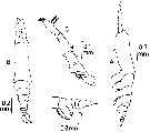 issued from : M.J. Isaac in Mitt. Zool. Mus., Berlin, 1974, 50 (1). [p.133, Fig.2]. Male (from Bankga Strait,Java Sea): A-B, habitus (lateral and dorsal, respectively); C, left A1 (dorsal); D, 5th thoracic segment and abdomen (lateral). A1 approximately 2/3 of the length of the cephalothorax, and the first three segments appear fused; the tip bears 2 curved spines and there is the usual articulating joint between the two end segments. cephalothorax is just under half the body length. the \"mouth\" is about half-way along the ventral surface. P5 partly obscured by the swimming legs, but appear from the side as small knobs, each tipped with 2 curved, parallel setae. The genital segment bears a long protuberance ventrally, with a pointed tip and a pair of lappets on the posterior side. The 3 other abdominal segments have approximate length ratios of 6:4:3.
|
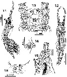 issued from : E. Suarez-Morales in Beaufortia, 2000, 50 (7). [p.145, Figs.11-16]. Male (holotype): 11, habitus (lateral); 12, idem (dorsal); 13, head (ventral); 14, oral papilla (lateral); 15, anterior part of cephalothorax (lateral); 16, four paired structures on ventral surface between antennule bases. Nota: A dorsal hump present on anterior 1/3 of cephalothorax, visible in lateral view (arrowed in figs.11, 15). Nota: Cephalothorax representing 55 % of total body length. Oral papilla protruding from ventral surface, located at almost 40 % of way back along surface of cephalothorax. Forehead with central depression. Dorsalocelli present, close to anteriormost end of head, pigment cups small and widely separated by a distance equal to 1.5 times an eye diameter, ovoid in dorsal view.
|
 issued from : E. Suarez-Morales in Beaufortia, 2000, 50 (7). [p.146, Figs.17-20]. Male (holotype): 17, right A1 (dorsal); 18, detail of seta 3 showing ornamented base area; 19, left A1 (dorsal); 20, detail of seta IVv area showing ornamentation of basal area. Nota: A1 5-segmented, length ratio of antennular segments: 10:25.6:16.4:23.6:24.4 = 100.
|
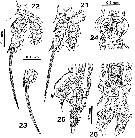 issued from : E. Suarez-Morales in Beaufortia, 2000, 50 (7). [p.147, Figs.21-26]. Male (holotype): 21, P1; 22, P3; 23, terminal exopodal segment of P4; 24, genital complex (ventral); 25, distal urosomites and genital complex (lateral); 26, idem (dorsal). Nota: Two subtriangiular processes present on tip of genital structure (arrowed in Fig.25). Nota: Urosome consisting of 4 somites: 5th pedigerous (with no appendages), genital somite (with genital complex), and 2 free somites, the last of which is the anal somite; from dorsal view, genital somite slightly longer than anal and preanal urosomites. Anal somite being the shortest of the urosome. Caudal rami subtriangular, about twice as long as wide, specimen with terminal seta only, sockets of 2 additional setae were observed.
|
 issued from : E. Suarez-Morales in Beaufortia, 2000, 50 (7). [p.148]. Male (holotype): Armature of swimming legs P1 to P4. Roman number = spine; arabic number = seta.
| | | | | NZ: | 1 | | |
|
Distribution map of Cymbasoma javensis by geographical zones
|
| | | | Loc: | | | Indonesia (Bangka Strait, W Java Sea) | | | | N: | 3 | | | | Lg.: | | | (739) M: 1,39; (842) M: 1,18; {M: 1,18-1,39} | | | | Rem.: | For Isaac (1974 a, p.132) the specimen is in rather a poor condition, with many setae missing.
Suarez-Morales (2000 d, p.148) : in the original description Isaac (1974 a, p.133) depicted 3 segments after the genital complex, but this probably resulted from misinterpretation of the urosomite segmentation. The specimen is relocated in the genus Cymbasoma mainly by having only 2 free somites after the genital complex, and secondarily by showing probably only 3 caudal setae; furthermore, the absence of P5 is another secondary character supporting its inclusion within Cymbasoma. | | | Last update : 17/02/2020 | |
|
|
 Any use of this site for a publication will be mentioned with the following reference : Any use of this site for a publication will be mentioned with the following reference :
Razouls C., Desreumaux N., Kouwenberg J. and de Bovée F., 2005-2026. - Biodiversity of Marine Planktonic Copepods (morphology, geographical distribution and biological data). Sorbonne University, CNRS. Available at http://copepodes.obs-banyuls.fr/en [Accessed January 07, 2026] © copyright 2005-2026 Sorbonne University, CNRS
|
|
|
|


;)
;)
;)
;)
