|
|
 |
|
Monstrilloida ( Order ) |
|
|
|
Monstrillidae ( Family ) |
|
|
|
Monstrillopsis ( Genus ) |
|
|
| |
Monstrillopsis dubia (T. Scott, 1904) (F,M) | |
| | | | | | | Syn.: | Monstrilla dubia T. Scott, 1904 (p.247, Descr.F, figs.F, Rem.); van Breemen, 1908 a (p.217, figs.F); ? Fontaine, 1955 (p.890); ? Lacroix & Filteau, 1971 (p.718); ? Davis, 1949 a (p.248); ? Tremblay & Anderson, 1984 (p.8); ? Dias de O., 1996 (p.253, Rem.);
no M. dubia : Sars, 1921 (p.26, figs.F,M); Rose, 1933 a (p.352, figs.F,M); Vilela, 1968 (p.45, fig.F); | | | | Ref.: | | | Isaac, 1974 (p.136); 1975 (p.3, 6, 9, figs.F,M); McAlice & Jaeger, 1982 (p.46, Rem.); Huys & Boxshall, 1991 (p.155, 168, 440, 465, no figs.M); Grygier, 1994 a (p.28); 1995 a (p.63); Suarez-Morales & Ivanenko, 2004 (p.42, Rem.: p.43 & suiv.); Suarez-Morales & al., 2006 (p.101, 105, Tab.1); Vives & Shmeleva, 2010 (p.187, figs.F,M, Rem.); Suarez-Morales, 2011 (p.6, 10, 12, Rem.) |  issued from : E. Suarez-Morales & V.V. Ivanenko in Arctic, 2004, 57 (1). [p.42, Table 1]. Comparison of taxonomic features and morphometry in females of selected species of Monstrillopsis. A1 = antennule; AN = anal somite; CT = cephalothorax; FP = 1st pediger; GS = genital double-somite; LA = last antennular segment; TL = total length; UR = urosome. All ratios are presented in percentage form.
|
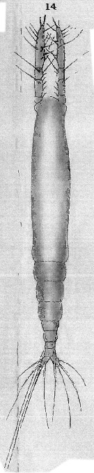 Issued from : T. Scott in Rep. Fishery Bd Scotl., 1904, 12 (3). [Pl. XIII, fig.14]. As Monstrilla dubia. Female (from Scotland): 14, habitus (dorsal). Nota: Cephalothorax about one and a half times the entire length of the ramaining thoracic segments and abdomen. Abdomen 3-segmented; the 1st segment about equal in size to the last thoracic segment, the 2nd is smaller rhan the next, while the 2nd and 3rd are together scarcely as long as the 1st segment.
|
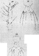 Issued from : T. Scott in Rep. Fishery Bd Scotl., 1904, 12 (3). [Pl. XIV, figs.16-18]. As Monstrilla dubia. Female: 16, A1; 17, P5; 18, Abdomen and caudal rami. Nota: A1 4-segmented, moderately stout, and about half as long as the cephalothoracic segment; the 1st and 3rd segments small, the 2nd is about half as long again as the 3rd, while the 4th is equal to the entire length of the 3 segments. P5 narrow and sub-cylindrical at the proximal end, but becomes wider distally and terminates in 2 lobes; the outer lobe is larger than the inner and is furnished with 3 moderately long setae, the inner lobe is narrow and appears to be devoid of setae. Caudal rami with 4 elongated hairs, one of them springs from near the base of the outer margin, 2 spring from the apex, while the 4th is attached on the inner aspect and near the middle of the segment.
|
 Issued from : E. Suarez-Morales, A. Bello-Smith & S. Palma in Zool. Anz., 2006, 245. [p.103, Table 1]. Structural characteristics. Morphological characters for comparative morphology of the species assigned to the genus Monstrillopsis by different authorities. See : M. dubioides, M. fosshageni, M. reticulata, M. sarsi, Cymbasoma gracilis, M. ferrari, M. zernovi, M. angustipes, Monstrillopsis sp. Key for the identification of the currently known species of Monstrillopsis (female only) after Suarez-Morales & al. (2006, p.105): 1 - Outer (exopodal) lobe of P5 with distal elongation; last antennular segment relatively long, more than 50% of A1 length; large species (more than 3 mm) ..... 2. 2 - Genital double-somite relatively short, representing 40% of urosomal length; A1 length over 30% of total body length, element 1 (on 1st segment) developed normally; caudal rami 1.6 times longer than wide.
|
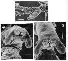 Issued from : R. Huys & G.A. Boxshall in The Ray Society, 1991, 159. [p.168, Fig. 2.5.11, C, D, E]. Monstrillopsis dubia male (from Raunefjorden, Norway): C, lateral view of urosome; D, detail of genital prominence showing gonopores (posterior view); E, same (ventral view). Scale bars: 100 µm (C); 30 µm (D, E). Nota: - Urosome 5-segmented. - Genital somite possesses a massive, posteroventrally directed genital prominence which is bifid distally. - two distal lobes unarmed. - Genital openings positioned at the tip of the genital prominence between the distal lobes. The paired openings are adjacent at the ventral midline, but each is closed off by a separate opercular flap (possibly representing the P6. - The paired vasa deferentia open close together on the genital prominence and elongate spermatophores are extruded. In Monstrilla the genital prominence is smaller than in Monstrillopsis species.
| | | | | Compl. Ref.: | | | ? Bernier & al., 2002 (p.651, tab.1); Dias & Bonecker, 2007 (p.270, 272, fig.3, tab.II); Dvoretsky & Dvoretsky, 2010 (p.991, Table 2); El Arraj & al., 2017 (p.272, table 2); | | | | NZ: | 2 + 4 doubtful | | |
|
Distribution map of Monstrillopsis dubia by geographical zones
|
| | | | Loc: | | | ? [ G. of Maine, Bay of Fundy, Baie des Chaleurs, Ungava Bay, Cambois Bay & St. Mary's lighthouse], Maroccan coast ?, Scotland (Firth of Forth, Loch Fyne, Firth of Clyde), ? Banyuls, Norway (Raunfjorden), Barents Sea, North Sea, Brazil (Vitoria, Rio de Janeiro) | | | | N: | 10 ? | | | | Lg.: | | | (727) F: 3,3; ? (748) F: 2,8-2,5; ? (466) F: 1,19-2,48; {F: 3,30} | | | | Rem.: | The north-American locality records are to be confirmed.
See remarks in Suarez-Morales & al. (2006, p.101) concerning comparison with M. chilensis.
After Dias (1996), the females differ from the description by Sars (1921) in body length only.
For Suarez-Morales (2011, p.6, 12) the once purportedly cosmopolitan M. dubia (T. Scott, 1904) is now known to contain at least three different species. After the author the species is resticted to Scotland (60°N). For the author, the records of M. dubia from the Southern hemisphere could refer to M. chilensis, M. igniterra, or undescribed taxa.
After Lee J. & al; (2016, p.421), genitalia male with a well developed, long median shaft and relatively short, ovoidal or globose lateral lappets that are more or less separated from the median shaft belongs subgroup ''Type I'' (Suarez-Morales & McKinnon, 2014). | | | Last update : 19/06/2023 | |
|
|
 Any use of this site for a publication will be mentioned with the following reference : Any use of this site for a publication will be mentioned with the following reference :
Razouls C., Desreumaux N., Kouwenberg J. and de Bovée F., 2005-2026. - Biodiversity of Marine Planktonic Copepods (morphology, geographical distribution and biological data). Sorbonne University, CNRS. Available at http://copepodes.obs-banyuls.fr/en [Accessed March 24, 2026] © copyright 2005-2026 Sorbonne University, CNRS
|
|
|
|


;)
;)
;)
;)
