|
|
 |
|
Monstrilloida ( Order ) |
|
|
|
Monstrillidae ( Family ) |
|
|
|
Monstrillopsis ( Genus ) |
|
|
| |
Monstrillopsis reticulata (Davis, 1949) (F,M) | |
| | | | | | | Syn.: | Monstrilla reticulata Davis, 1949 a (p.251, figs.F,M); Björnberg, 1971 (p.348); Grygier, 1995 a (p.46,73); Dias de O., 1996 (p.254, Rem.); Suarez-Morales, 2011 (p.9) | | | | Ref.: | | | Isaac, 1975 (n°144/145, p.2, 6, 9, figs.F,M); Suarez-Morales & al., 2006 (p.102, Rem., Tab.1); Grygier & Ohtsuka, 2008 (p.501, Rem.); Grygier & Suarez-Morales, 2018 (p.506: Rem.). | 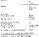 Issued from : E. Suarez-Morales, A. Bello-Smith & S. Palma in Zool. Anz., 2006, 245. [p.103, Table 1]. Structural characteristics. Morphological characters for comparative morphology of the species assigned to the genus Monstrillopsis by different authorities. See : M. chilensis, M. dubioides, M. fosshageni, dubia, sarsi, Cymbasoma gracilis, M. ferrari, M. zernovi, M; angustipes, Monstrillopsis sp.
|
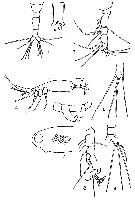 Issued from : C.C. Davis in Trans. am. microsc. Soc., 1949, 68 (3). [p.253, Fig. 1-9]. As Monstrilla reticulata. Female (from Biscayne Bay, Florida): 1, urosome (dorsal view); 3, left A1; 4, habitus (lateral view); 6, P5; 7, portion of the cephalic segment showing reticulation (lateral view). Male (from Biscayne Bay): 2, genital segment showing genital protuberance and spermatophore (ventral view); 5, urosome (lateral view); 8, A1 (ventral view); 9, urosome, posterior portion with furcal ramus. Nota: 1 female found off Coconut Grove (March 10, 1948); all others individuals obtained off Chicken Key in Biscayne Bay: 1 ripe female (December 13, 1947; 1 female (January 16, 1948); 2 females and 1 male (March 30, 1948); 1 female, 1 ripe, 2 females carrying eggs and 1 male (April 6, 1948); 1 female (April 13, 1948); 1 female (April 27, 1948); 1 female (May 4, 1948; 2 females and 1 male (June 1, 1948); 12 females and 1 male 'June 8, 1948). Female: - Colorless, transparent or translucent for the most paert, however, the distal 3/4 of A1, the area around the eyes, that between the segments of the metasome, ventral surface of urosome posterior to ovigerous spines, furcal rami, were blackish-brown, while eyes themselves reddish-black. - Cephalic segment approximately same length as the remainder body, and its surface is finely reticulate (reticulation sometimes difficult to ascertain). - Oral papilla very prominent, located about 1/5 of the distance from the anterior margin. - 2 dorsal eyes and 1 ventral, prominent. - On the anterior margin of the cephalic segment, between bases of A1, frontal organ prominent. - Cephalic segment very clear and transparent in all except 2 specimens, in these two the entire cephalic segment filled with a large number of eggs. - The four thoracic segments (2nd, 3rd, 4th and 5th) posterior to the cephalic segment subequal in length, but progressively narrower posteriorly. - Genital segment about twice as long as the segments that precede and follow it, and it is relatively slightly swollen ventrally, on the anterior half of the segment. It bears the ventral ovigerous spines, closely appressed or slightly fused together near their bases. - Ovigerous spines extend only slightly beyond the furcal rami. Anal segment very short, and fused with the furcal rami dorsally and ventrally, being clearly set off only laterally. - A1 consist of 4 clear segments. Antennal seta not well developed, and none of them dichotomously branched. - P5 short and thick, with an inner lobe bearing a single terminal seta, and an outer lobe bearing 3 terminal setae; the two inner of these arise very close together. - Furcal rami somewhat longer than broad. Setae very fine, the largest is borne on the outer margin, while terminally there is a group of 3, borne close together near the inner corner, and a 4th on the outer corner. Male: -Cephalic segment opaque and without obvious reticulation - Genital segment with genital protuberance, very prominent, wkith relatively small, unornamented genital lappets. - There are 3 segments between the genital segment and the furcal rami, with as in the female, the anal segment very short and fused dorsally and ventrally with the furcal rami. - Furcal rami bear 1 strong seta on outer margin and 3 terminal setae, outermost of which is weaker and shorter than the rest. A1: 5-segmented, geniculate, with a rather strong hooked spine and 3 dichotomously branced setae on terminal segment. - P5 lacking.
| | | | | NZ: | 3 | | |
|
Distribution map of Monstrillopsis reticulata by geographical zones
|
| | | | | | | Loc: | | | Brazil, Caribbean, SE Florida (Chicken Key, Biscayne Bay), Burma (Tenasserim coast).
Type locality: 25°33'57'' N, 80°12'50'' W. | | | | N: | 3 | | | | Lg.: | | | (466) F: 3,01-2,39; (743) F: 0,74-0,67; M: 0,56; {F: 0,67-3,01; M: 0,56} | | | | Rem.: | In a tank with Perna perna (Bivalve).
For Davis (1949, p.252), this species resemble M. longipes A. Scott and M. wandelii Stephensen in possessing a reticulated cephalic segment in the female, but differs these in the structure of P5. Also, oral papilla more strongly developed than in the other species. The male differs from all other species in size and in the presence and arrangement of the 4 furcal setae.
For Dias (1996), the species show two kinds of differences from the description given by Davis (1949): they are longer and have the caudal rami with 6 setae (4 terminal, 1 of them shorter) and 2 lateral setae. Four collected in aquarium water with Perna perna (Bivalvia).
For Suarez-Morales & al. (206, p.102), this species from Florida, was included in Monstrillopsis by Isaac (1975); type female of this species should be retained in Monstrilla owing both to its possession of an inner lobe of the P5 armed with 1 seta and the presence of 5 caudal setae. The same two features, inconsistent with the definition of Monstrillopsis, are shared by another species that was incorrectly assignated to Monstrillopsis by Suarez-Morales (1993), Monstrillopsis ciqroi which is also transferred to the genus Monstrilla.
After Lee J. & al; (2016, p.421), genitalia male with a well developed, long median shaft and relatively short, ovoidal or globose lateral lappets that are more or less separated from the median shaft belongs subgroup ''Type I'' (Suarez-Morales & McKinnon, 2014).
For Grygier & Suarez-Morales (2018, p.506), resolution of the problem posed by assignment of the specific name reticulata to supposedly non-conspecitic males and females in the genus Monstrillopsis requires the designation of a neotype by the CZN.
For Suarez-Morales & al. (2006, p.104), it is very difficult to reliably link both sexes of monstrilloids unless they were collected from the same host or they share a clearly distinctive set of characters; specimens of different sexes obtained in plankton samples, and even in the same sample, as in Davis (1949), do not necessarily belong to the same species. | | | Last update : 28/07/2020 | |
|
|
 Any use of this site for a publication will be mentioned with the following reference : Any use of this site for a publication will be mentioned with the following reference :
Razouls C., Desreumaux N., Kouwenberg J. and de Bovée F., 2005-2025. - Biodiversity of Marine Planktonic Copepods (morphology, geographical distribution and biological data). Sorbonne University, CNRS. Available at http://copepodes.obs-banyuls.fr/en [Accessed January 05, 2026] © copyright 2005-2025 Sorbonne University, CNRS
|
|
|
|


;)
;)

