|
|
 |
|
Cyclopoida ( Order ) |
|
|
|
Lubbockiidae ( Family ) |
|
|
|
Atrophia ( Genus ) |
|
|
| |
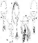
Atrophia minuta (Wolfenden, 1905) (F,M) | |
| | | | | | | Syn.: | Lubbockia glacialis : Olson, 1949; Heron & Damkaer, 1969 (p.16, figs.M); Boxshall, 1977 a (p.118, figs.M, Rem.); Deevey & Brooks, 1977 (p.287);
Lubbockia minuta Wolfenden, 1905 a (p.15, Descr.F); van Breemen, 1908 a (p.193); Heron & Damkaer, 1978 (p.19, figs.F,M); Gardner & Szabo,1982 (p.126, figs.F,M); Mazzocchi & al., 1995 (p.226, figs.F,M, Rem.); Böttger-Schnack, 1997 (p.409); Razouls & al., 2000 (p.343, Appendix); Galbraith, 2009 (pers. comm.); Hsiao S.H. & al., 2011 (p.475, Appendix I) | | | | Ref.: | | | Huys & Böttger-Schnack, 1996/97 (p.259); Boxshall & Halsey, 2004 (p.577); Vives & Shmeleva, 2010 (p.254, figs.F,M, Rem.) | 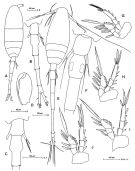 Issued from: M.G. Mazzocchi, G. Zagami, A. Ianora, L. Guglielmo & J. Hure in Atlas of Marine Zooplankton Straits of Magellan. Copepods. L. Guglielmo & A. Ianora (Eds.), 1995. [p.227, Fig.3.42.1]. As Lubbockia minuta. Female: A, habitus (dorsal); B, urosome (dorsal); C, last thoracic somite and genital somite (dorsal); D, Mxp. Nota: Prosome 1.2 times longer than urosome. Proportional lengths of urosomites and furca 15:25:17:20:10:13 = 100. Male: E, habitus (dorsal); F, last thoracic somite and 1st and 2nd urosomal somites (dorsal); G, P1; H, P2; I, P3; J, P4. Nota: Urosome 1.5 longer than Prosome. Proportional lengths of urosomites and furca 9:12:16:19:29:5:10 = 100.
|
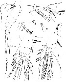 issued from : G.A. Boxshall in Brit. Mus. nat. Hist., Zool., 1977, 31 (3). [p.119, Fig.8]. As Lubbockia glacialis. Male (from 18°N, 25°W): a, habitus (dorsal); b, A1 (ventral); c, A2 (anterior); d, Mx1 (posterior); e, Mx2 (posterior); f, Mxp (anterior); g, P1 (posterior); h, P4 (posterior). Nota: Relative lengths of urosome somites 8:12:16:23:4:9.
|
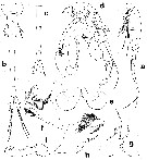 issued from : G.A. Heron & D.M. Damkaer in Smiths. Contr. Zool., 1978, 267. [p.20, Fig.13]. As Lubbockia minuta. Female: a, habitus (lateral); b, urosome (dorsal); c, postgenital segments and caudal rami (ventral); d, cephalosome (ventral); e, labrum (ventral); f, right Md; g, left Mx1; h, left Mx2; i, right Mxp. Scale bars: A (fig.a); C (figs.b, c); E (fig.d); G (figs.e-i).
|
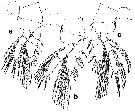 issued from : G.A. Heron & D.M. Damkaer in Smiths. Contr. Zool., 1978, 267. [p.21, Fig.14]. As Lubbockia minuta. Female: a-c, P1 to P3. Scale bar: E (figs.a-c).
|
 issued from : G.A. Heron & D.M. Damkaer in Smiths. Contr. Zool., 1978, 267. [p.22, Fig.151]. As Lubbockia minuta. Female: a, P4; b, last prosomal segment and first two urosomal segments (lateral). male: c, habitus (ventral). Stage V: d, habitus (dorsal). Stage IV: e, habitus (dorsal). Scale bars: A (fig.c); B (fig.d, e); E (figs.a, b).
|
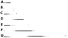 issued from : G.A. Heron & D.M. Damkaer in Smiths. Contr. Zool., 1978, 267. [p.2, Fig.1]. Scales used in drawing figure. Each bar = 0.1 mm.
|
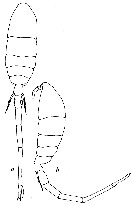 issued from : G.A. Heron & D.M. Damkaer in Smiths. Contr. Zool., 1969, 20. [p.16, Fig.20]. As Lubbockia glacialis. Male: a-b, habitus (dorsal and lateral, respectively). Scale single bar: 0.100 mm. Nota : Rostrum a small, rounded protrusion between bases of A1. Urosome 6-segmented, each segment with smooyh posterior margins ; 5th and anal segments with lateral margins slightly concave. Caudal rami about same length as genital segment. P5 simiilar to female, free segment elongate, extending two-figths the length of genital segment ; armature consists of 2 lanceolate setae, terminal seta about three times the length of lateral seta.
|
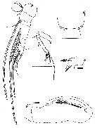 issued from : G.A. Heron & D.M. Damkaer in Smiths. Contr. Zool., 1969, 20. [p.17, Fig.21]. As Lubbockia glacialis. Male: a, A1; b, A2; c, Mx2; d, Mxp; e, posterior margin of genital segment (ventral). Scale single bar: 0.100 mm; double bars: 0.010 mm. Nota : A1 4 distinct segments.Faint line suggesting coalescence on proximal third of segment 3 corresponds to segmentation between segments III and IV of female. 4th segment of male corresponds to segments V, VI and VII of female. A2 subprehensile, 3-segmented. Basal segment unarmed ; 1 seta on short 2nd segment.Distal segment with terminal grouping of 7 setae, 1 three times the length of the remaining 6, 1 of which is plumose. Inner surface of 3rd segment with patches of hairs and 1 small spine near base of long seta ; 2 setae on outer margin. Md and Mx1 reduced to vestiges, represented only by the bases ; each with undefined margins. Mx2 with 2 terminal spines, 1 nude and 1 wiyh unilateral row of hairs ; 1 seta on lateral margin. Slightly sinuate spine, with unilateral patches of hairs, on small lobe protruding subapically. Mxp 3-segmented ; 2nd segment with spine and graduated row of coarse spinules on medial margin, followed by row of rugose striations. P6 probably represented by small triangular sclerotized ridge on ventrolateral posterior margin of genital segment, bearing 1 setule.
| | | | | NZ: | 8 | | |
|
Distribution map of Atrophia minuta by geographical zones
|
| | | | | | | | | | | | | Loc: | | | sub-Antarct. (SW Pacif.) , Strait of Magellan, off Cape Verde Is., Florida, Sargasso Sea, W Ireland, E Medit. (Lebanon Sea), Taiwan (E: Kuroshio Current), G. of Alaska, off British Columbia, Oregon | | | | N: | 10 | | | | Lg.: | | | (36) F: 1,3; M: 1,98-1,9; (139) M: 2,08; 1,94; (674) F: 1,5-1,24; M: 2,03-2; {F: 1,24-1,50; M: 1,90-2,08} | | | | Rem.: | epi-mesopelagic. | | | Last update : 21/01/2016 | |
|
|
 Any use of this site for a publication will be mentioned with the following reference : Any use of this site for a publication will be mentioned with the following reference :
Razouls C., Desreumaux N., Kouwenberg J. and de Bovée F., 2005-2025. - Biodiversity of Marine Planktonic Copepods (morphology, geographical distribution and biological data). Sorbonne University, CNRS. Available at http://copepodes.obs-banyuls.fr/en [Accessed August 27, 2025] © copyright 2005-2025 Sorbonne University, CNRS
|
|
|
|


;)
;)
;)
;)
;)
;)
;)
;)

