|
|
 |
|
Siphonostomatoida ( Order ) |
|
|
|
Pontoeciellidae ( Family ) |
|
|
|
Pontoeciella ( Genus ) |
|
|
| |
Pontoeciella abyssicola (T. Scott, 1894) (F,M) | |
| | | | | | | Syn.: | Artotrogus abyssicolus T. Scott, 1894 b (p.128, figs.F,M);
Ratania sp. : Rose, 1939 (p.24, Descr.F, figs.F, no M);
Carnegiella gracilis Wilson, 1942 a (p.176, figs.F = M);
Danodes plumata Wilson, 1942 a (p.182, figs.F); Minoda, 1971 (p.47, Rem.F);
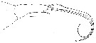
Danodes panikkari Ummerkutty, 1968 a (p.298, figs.F) | | | | Ref.: | | | Giesbrecht, 1899 (p.20, 83, 105, figs.F); Farran, 1926 (p.296); 1936 a (p.125, figs.F,M); Sewell, 1948 (p.398, Rem.); Saraswathy, 1966 (1967) (p.89, Rem.); Heron & Damkaer, 1969 (p.22, figs.F); Bourcier & al., 1969 (p.272); Boxshall, 1979 (p.242, figs.F,M, Rem.); Saraswathy, 1982 (p.635, Rem.F,M, chart); Huys & Boxshall, 1991 (p.444, 466, figs.F); Boxshall & Huys, 1998 (p.773, figs.F,M); Lapernat, 1999 (p.39, figs.F); Boxshall & Halsey, 2004 (p.811, p.812: figs.F,M); Vives & Shmeleva, 2010 (p.406, figs.F,M, Rem.) | 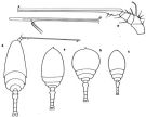 issued from : G.P. Farran in Great Barrier Reef Expedition 1928-29. Scientific Reports, V, No.3. Copepoda. 1936 [p.125, Fig.24]. Female: a, habitus (dorsal); b, idem (specimen from another station); c, idem. Nota: These specimens support the view held by Giesbrecht that this species shows great variation in the relative width of the cephalothorax. The variation also extends in small details of the form of the appendages. Male: d, habitus (dorsal); e, A1. Nota: Siphon absent. A1 apparently 6-segmented. A2 reduced as compared with those of the female. No traces of Md or Mx1. Mx2 and Mxp are of the same form as in female, but weaker and more slender. P5 consists of a single seta.. Urosome 5-segmented.
|
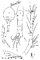
 issued from : M. Rose in Bull. Soc. zool. Fr., 1939, LXIV. [p.25]. Female (Bay of Alger): habitus (dorsal); A1, antennule; A2, antenna; Abd, urosome (dorsal). Nota: A1 8-segmented
|
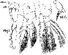 issued from : M. Rose in Bull. Soc. zool. Fr., 1939, LXIV. [p.26,]. Female: Mxp 1 ( = maxilla = Mx2); Mxp 2 (= maxilliped = Mxp); Md Mx, mandible and maxillule; swimming legs P2 and P3. Nota: Md and Mx1 appendices are heavily modified. It a kind of pointed tube from the joining of mandibles and in which a musculature powerful moves maxilla that appear to transformed into stylets.
|
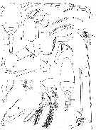
 issued from : A.N.P. Ummerkutty in Crustaceana, 1968, 15 (3). [p.299, Figs.1-7]. As Danodes panikkari. Female (from off Karwar, W coast of India): 1, habitus (dorsal); 2, urosome (enlarged); 3, A1; 4, A2; 5, siphon; 6, Md; 7, Mx1.
|
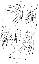 issued from : A.N.P. Ummerkutty in Crustaceana, 1968, 15 (3). [p.301, Figs.8-13]. As Danodes panikkari. Female: 8, Mx2; 9, Mxp; 10, P1; 11, P2; 12, P3, 13, P4.
|
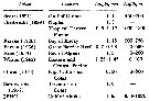
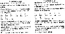 issued from : A.N.P. Ummerkutty in Crustaceana, 1968, 15 (3). [p.302, Table I]. As Danodes. Differences between Danodes (= Pontoeciella) plumata and Danodes ( = Pontoeciella) panikkari.
|
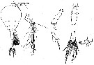
 issued from : G.A. Boxshall in Bull. Br. Mus. nat. Hist. (Zool.), 1979, 35 (3). [p.241, Fig.18]. Female (from Gulf of Guinea): A, habitus (dorsal); B, cephalosome (lateral); C, A1; D, A2; E, Md; F, Mx1; G, Mx2; H, Mxp. Male: I, habitus (dorsal); J, anterior portion of urosome (lateral); K, A1; L, A1 (from other specimen); M, A2; N, 3rd and 4th thoracic segments (lateral); O, Mx2; P, Mxp; Q, P1; R, P5 and P6. Scales 0.1 mm unless otherwise indicated.
|
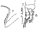 issued from : M. Saraswathy in Proc. Symp. Crustacea. - Symp. Ser. mar. biol. Ass. India, 1966 2 (1). [p.88, Fig.2 (7-8)]. Female (from Trivandrum coast, SW India): 7, Mx2; 8, P1. Nota: The description given by T. Scott (1894) applies to the present specimens except: the terminal segment of Mx2 has 1 spine and the proximal segment of the endopod of P1 carries 1 inner seta.
|
 issued from : G.A. Heron & D.M. Damkaer in Smiths. Contr. Zool., 1969, 20. [p.22, Fig.27]. Female: Mx1. Scale double bars: 0.010 mm.
|
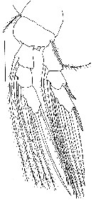 issued from : G.A. Heron & D.M. Damkaer in Smiths. Contr. Zool., 1969, 20. [p.23, Fig.28]. Female: P4. Scale bar: 0.100 mm.
|
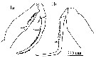 issued from : P.E. Lapernat & C. Razouls in Vie Milieu, 2002, 52 (1). [p.27, Pl. V, fig.1 a, b]. Female (from off Malta, Mediterranean Sea): 1a, Mxp; 1b, Mx2.
| | | | | Compl. Ref.: | | | V.N. Greze, 1963 a (tabl.2); De Decker & Mombeck, 1964 (p.13); Shmeleva, 1964 a (p.1068); Delalo, 1968 (p.139); Soenen, 1969 (p.122,180); Gamulin, 1971 (p.382, tab.3); Apostolopoulou, 1972 (p.329, 377); Bainbridge, 1972 (p.61, Appendix Table I: vertical distribution vs day/night); Deevey & Brooks, 1977 (p.156, tab.2, Station "S"); Kovalev & Shmeleva, 1982 (p.86); Scotto di Carlo & al., 1984 (p.1043); Guangshan & Honglin, 1984 (p.118, tab.); Greze & al., 1985 (p.8); Shih & Young, 1995 (p.75); Böttger-Schnack, 1995 (p.93); 1996 (p.1088); 1997 (p.410); Hure & Krsinic, 1998 (p.80, 103); Suarez-Morales & Gasca, 1998 a (p.114); Lapernat & Razouls, 2001 (tab.1); Lan & al., 2004 (p.332, tab.1); Isari & al., 2006 (p.241, tab.II); Hidalgo & al., 2010 (p.2089, Table 2); Mazzocchi & Di Capua, 2010 (p.430); Hsiao S.H. & al., 2011 (p.475, Appendix I); Tutasi & al., 2011 (p.791, Table 2, abundance distribution vs La Niña event); Uysal & Shmeleva, 2012 (p.909, Table I); Belmonte, 2018 (p.273, Table I: Italian zones) | | | | NZ: | 13 | | |
|
Distribution map of Pontoeciella abyssicola by geographical zones
|
| | | | | | | | | | | | 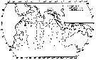 issued from : M. Saraswathy in J. Plankton Res., 1982, 4 (3). [p.634, Fig.1]. issued from : M. Saraswathy in J. Plankton Res., 1982, 4 (3). [p.634, Fig.1].
Distribution of P. abyssicola in the Indian Ocean.
Nota: Samples obtained from the upper 200m water colum at different season. |
 issued from : G.A. Heron & D.M. Damkaer in Smiths. Contr. Zool., 1969, 20. [p.22]. issued from : G.A. Heron & D.M. Damkaer in Smiths. Contr. Zool., 1969, 20. [p.22].
Records indicate a wide distribution and a variable length in female. |
| | | | Loc: | | | South Africa (E), G. of Guinea, G. of Mexico, off Bermuda: Station S (32°10N, 64°30W), Sargasso Sea, Bay of Biscay, Medit. (Alboran Sea, Algiers, Marseille, Ligurian Sea, Tyrrhenian Sea, off Malta, S Adriatic Sea, Ionian Sea, Aegean Sea, Lebanon Basin), Red Sea, Arabian Sea, W India, Indian Ocean, China Seas (East China Sea, South China Sea), Taiwan (E & NW), Pacif. (W equatorial), Australia (Great Barrier), Samoa Is., Pacif. (E tropical), Baja California, G. of Alaska, Galapagos-Ecuador, Chile (N-S) | | | | N: | 39 | | | | Lg.: | | | (34) F: 0,93- 0,7; (38) F: 1,15; (57) F: 1,1; (138) F: 1,65-0,7; (208) F: 1,49; (340) F: 1; (446) F: 1,45-1,25; M: 1,25; (530) F: 1; (676) F: 1,65; (716) F: 1,17-0,9; (717) F: 1,3; (793) F: 1,1-0,9; M: 0,9; {F: 0,70-1,65; M: 0,90-1,25} | | | | Rem.: | Meso-bathypelagic. (off Malta: 2000 m). Overall Depth Range in Sargasso Sea: 0-2000 m (Deevey & Brooks, 1977, Station "S"); after Boxshall (1979) the species is distributed in warm seas between 100 and 600 m
According to Heron & Damkaer (1969, p.22), Giesbrecht (1899) and Farran (1936) have discussed the variable shape of the prosome and irregularities in the armature of the swimming legs. Alaska's specimens and Scott's (1894) description of female appeared to be similar in most respects; both differ in some details from descriptions of Giesbrecht (1899) and Rose (1939): differences in proportion of urosomal segments; terminal spines of A2 vary in length and width; Md straight rather than curved; claw of Mx2 incined 65° (also shown by Saraswathy (1967) rather than 90°; P3 exopod spines are slightly longer than on Giesbrecht's and Rose's illustrations; P4 shows differences in spine of exopod 2 and seta of protopod 2; Mx1 differs from illustrations of Scott (1894) and Giesbrecht (1899), which resembled each other; P4 showed some minor differences in armature from descriptions given by Scott and Giesbrecht.
For Saraswathy (1982, p.637) the presence of a well developed siphon indicates its ability for a parasitic existence, but so far nothing is known about the probable host. The structure of the oral cone of the female, and the associated skeletomusculature was described by Boxshall (1990 a). Adult males are non-feeding and have an atrophied oral cone and mouthparts.
This species could be a relict form of the Tethys Sea.
First occurrence in Chilean waters by Hidalgo & al. (2010). | | | Last update : 09/12/2020 | |
|
|
 Any use of this site for a publication will be mentioned with the following reference : Any use of this site for a publication will be mentioned with the following reference :
Razouls C., Desreumaux N., Kouwenberg J. and de Bovée F., 2005-2026. - Biodiversity of Marine Planktonic Copepods (morphology, geographical distribution and biological data). Sorbonne University, CNRS. Available at http://copepodes.obs-banyuls.fr/en [Accessed April 01, 2026] © copyright 2005-2026 Sorbonne University, CNRS
|
|
|
|


;)
;)
;)
;)
;)
;)
;)
;)
;)
;)

;)
;)
