|
|
 |
|
Cyclopoida ( Order ) |
|
|
|
Oithonidae ( Family ) |
|
|
|
Dioithona ( Genus ) |
|
|
| |
Dioithona oculata Farran, 1913 (F,M) | |
| | | | | | | Syn.: | Oithona oculata Farran, 1913 a (p.188, Descr.F, figs.F); Rosendorn, 1917 (p.303); 1917 a (p.37, figs.F,M, Descr.M); Kiefer, 1929 g (p.10, Rem.F,M); Dakin & Colefax, 1933 (p.208); 1940 (p.116, fig.M); Sewell, 1947 (p.254, Rem.M); Tanaka, 1960 (p.60, figs.F); Björnberg, 1963 (p.76, Rem.: Juv.); Vervoort, 1964 a (p.25, Rem.); Gonzalez & Bowman, 1965 (p.273, figs.F,M, Rem.); Emery, 1968 (p.293, swarm); Wellershaus, 1970 (p.473); Björnberg, 1972 (p.82; fig., Rem. Nauplius); Kos, 1972 (Vol. I, figs.F, Rem.); Yeatman, 1976 (p.213, figs.F); Nishida & al., 1977 (p.139, figs.F, Rem.); Hamner & Carleton, 1979 (p.1, swarms); Dawson & Knatz, 1980 (p.9, figs.F,M); Ferrari & Bowman, 1980 (p.4, figs.F,M); Ueda & al., 1983 (p.165, Table 1, 2, 4, swarms); Ueda, 1985 (p.125); Nishida, 1985 a (p.63, Descr.F,M, figs.F,M, p.143, 144); Ferrari & Ambler, 1992 (p.275, Rem.: N); Yoo & Lim, 1993 (p.91, Rem.: p.95, 100, Table 1, fig.2); Kim & al., 1993 (p.271); Chihara & Murano, 1997 (p.938, Pl. 197, 202: F,M); Bradford-Grieve & al., 1999 (p.886, 966, figs.F,M); McKinnon, 2000 (p.108, Rem.); Ref. compl.: Sewell, 1948 (p.417) ; Sander & Moore, 1979 (p.215, zoogeographic chart); Hammer & Carleton, 1979 (p.1); Ueda & al., 1983 (p.165, Table 1, 2, 4, 5, swarms); Lindo, 1991 (tab.3); Yoo, 1991 (tab.1); McKinnon, 1991 (p.471); Madhupratap & al., 1991 (p.947, Rem.: p.951, 954, 956); Roff & al., 1995 (p.165,Table 6: bacteriovory); Webber & al., 1996 (tab.1); Hopcroft & al., 1998 (tab.2); Suarez-Morales & Gasca, 1998 a (p.112); Ueda & al., 2000 (tab.1); Ueda, 2001 (p.96, occurrence); Dunbar & Webber, 2003 (tab.1); Hernandez Molejon & Alvarez-Lajonchere, 2003 (p.471, predation); Rezai & al., 2004 (p.490, tab.2, 3, abundance, Rem., p.495, tab.8); Lo & al., 2004 (p.468, tab.2); Choi & al., 2005 (p.710: Tab.III); Sterza & Fernades, 2006 (p.95, Table 1, occurrence); Elliott & Kaufmann, 2007 (p.418); Ohtsuka & al., 2008 (p.115, Table 5); Hidalgo & al., 2010 (p.2089, Table 2); Medellin-Mora & Navas S., 2010 (p.265, Tab. 2); Fazeli & al., 2010 (p.153, Table 1); Maiphae & Sa-ardrit, 2011 (p.641, Table 2); Magris & al., 2011 (p.260, abundance, interannual variability); DiBacco & al., 2012 (p.483, Table S1, ballast water transport); in CalCOFI regional list (MDO, Nov. 2013; M. Ohman, comm. pers.); Araujo & al., 2016 (p.1, Table 3, abundance, %); Rosa J.C.L. & al., 2016 (p.71, Rem.); Ohtsuka & Nishida, 2017 (p.565, 578, Table 22.1, Rem.);
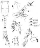
O. (Dioithona) oculata : Shuvalov, 1980 (p.172, figs.F,M); Conway & al., 2003 (p.199, figs.F,M, Rem.. | | | | Ref.: | | | Ferrari, 1993 (p.764); Ambler & al., 1991 (p.1257); 1996 (p.287); Buskey & al., 1996 (p.301, photoreception, swarming); Boxshall & Halsey, 2004 (p.611); Rasch & Wyngaard, 2006 (p.627: tab.1, genome size); Ferrari & Dahms, 2007 (p.23, 26, 29, 30, Rem. N, p.42, 57, 58, 59, 63, 87, figs. copepodites I-VI, fig.28: P3); Terbiyik Kurt, 2018 (p.569, table 2, fig.3, 4, 5, 6, 7, 8, morphological characters, , abundance, interannual variations) | 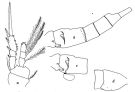 issued from : F.D. Ferrari & T.E. Bowman in Smiths. Contr. Zool., 1980, 312. [p.5, Fig.1]. As Oithona oculata. Female (from Caribbean area): a, P4; b, urosome (lateral left side); c, urosomal segment 1 and2 with spermatophore (lateral left side). Nota: Exopodal segment 2 of P4 with both setae modified, slightly curved toward their tips (proximal seta with serrate flange on distal 1/3 of medial edge, distal seta with serrate flange on distal 1/4); endopodal segment 3 proximal seta similarly modified with serrate flange on distal 1/6. Genital segment with knob near genital opening armed with anterodorsally curved spine, bearing tiny distinct teeth on posterior margin, below this spine a small point. Spermatophores attached ventrally on the genital segment, a small tubule passes dorsally from spermatophore (genital opening not identified, it may be located near the termination of the spermatophore tubule but it appears that the tubule empties into a sulcus or groove formed by two ridges of the integument; the ridges, depicted by heavy lines, extend dorsally on each side of the armed knob). Male: cephalothorax in lateral view produced into a small triangular extension near posterior ventral edge. Integumental organs scattered over the lateral surface (organs: see figure below).
|
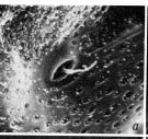 issued from : F.D. Ferrari & T.E. Bowman in Smiths. Contr. Zool., 1980, 312. [p.24, Fig.15 a]. Male (SEM): a, integumental organ of cephalosome (x 10,000). Nota: Organs with form of shallow pore without thickened peripheral ridge, small hair arises from the center of pore.
|
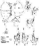
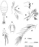 Issued from : S. Nishida in Bull. Ocean Res. Inst., Univ. Tokyo, 1985, No 20. [p.64, Fig.33]. As Oithona oculata. Female: a, habitus (dorsal); b, thoracic segment 5 and genital segment (ventral); c, anal segment and caudal rami (dorsal); d, A1; e, Md (mandibular palp); f, Md (biting edge); g, Mx1.
|
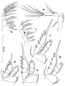
 Issued from : S. Nishida in Bull. Ocean Res. Inst., Univ. Tokyo, 1985, No 20. [p.65, Fig.34]. As Oithona oculata. Female: a, A2; b, Mx2; c, Mxp; d, P1; e, P2; f, P3; g, P4.
|
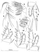
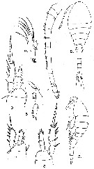
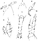
 Issued from : S. Nishida in Bull. Ocean Res. Inst., Univ. Tokyo, 1985, No 20. [p.66, Fig.35]. As Oithona oculata. Male: a, habitus (dorsal); b, forehead (lateral); c, thoracic segment 5 and genital segment (lateral right side); d, anal segment and caudal rami (dorsal); e, A1; f, Md; g, Mx1.
|
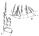
 Issued from : S. Nishida in Bull. Ocean Res. Inst., Univ. Tokyo, 1985, No 20. [p.67, Fig.36]. As Oithona oculata. Male: a, A2; b, Mx2; c, Mxp; d, P1; e, P2; f, P3; g, P4.
|
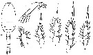
 issued from : G.P. Farran in Proc. zool. Soc. Lond., 1913. [Plate XXXI, Figs.2-9]. As Oithona oculata. Female (from Christmas Island, Indian): 2, habitus (dorsal); 3,idem (lateral); 4, A1; 5, Md (mandibular palp); 6, Mx1; 7-9, P1 to P3. Nota: Proportional lengths of urosomal segments and caudal rami 7:18:10:9:8:8. Caudal rami about two and half times as long as wide, with an outer edge seta located in the middle of the outer margin. Rostrum absent; cephalon slightly produced into a rostral prominence; the cephalic ganglia form a large bilobed mass, in front of which are situated, in the rostral prominence, a pair of small clear refractive areas. A1 reaches to the 2nd thoracic segment; proportional lengths of segments 11:6:16:11:12:12:16:7:13:4:9:6; 3rd segment with an imperfect separation near its proximal end, 4th segment with a small spine distally on its upper margin. P5 with 2setae For Farran, this species is closely allied to O (= Dioithona) rigida, differing mainly from it in the greater length of the terminal spines of the swimming legs: in rigida, as figured by Giesbrecht1897) and Cleve (1901), these spines are shorter than the terminal segments of the exopodites; another difference is the comparatively large size of the inner edge setae on the 1st segments of the exopodites in rigida, in oculata they are very small.
|
 issued from : G.P. Farran in Proc. zool. Soc. Lond., 1913. [Plate XXX, Figs.8-9]. As Oithona oculata. Female: 8, Mxp; 9, P4. Nota: Proportion of total lengths (p.cent) Prosome : 58.75, Urosome : 41.25 . Relative lengths of urosomal segments and caudal rami: 7 : 17 : 8 : 8 : 7 : 8. Setal formula of the exopod swimming legs P1 to P4 (Se = outer setae ; Si = inner setae), P1 : 1, 1, 3 Se ; 1, 1, 4 Si ; P2 : 1, 1, 3 Se ; 1, 1, 5 Si ; P3 : 1, 1, 3 Se ; 1, 1, 5 Si ; P4 : 1, 1, 2 Se ; 1, 1, 5 Si .
|
 issued from : I. Rosendorn in Wiss. Ergebn. dt. Tiefsee-Exped. \"Valdiviella\", 1917, 23. [p.38, Fig.23]. As Oithona oculata. Male: a, habitus (dorsal); b, Md mandibular palp); c, Mx1; d, exopod of P1; e, exopod of P2; f, exopod of P3; g, exopod of P4. Nota: Proportion of lengths (p.cent) Prosome : 61.54, Urosome : 38.46 . Relative lengths of urosomal segments and caudal rami: 12 : 14 : 11 : 11: 10 : 10 : 9. Setal formula of the exopod swimming legs P1 to P4 (Se = outer setae ; Si = inner setae), P1 : 1, 1, 3 Se ; 1, 1, 4 Si ; P2 : 1, 1, 3 Se ; 1, 1, 5 Si ; P3 : 1, 1, 3 Se ; 1, 1, 5 Si ; P4 : 1, 1, 2 Se ; 1, 1, 5 Si . Female: P5 with 2 setae.
|
 Issued from : S. Nishida, O. Tanaka & M. Omori in Bull. Plankton Soc. Japan, 1977, 24 (2). [p.140, Fig.13]. As Oithona oculata. Female (from Aburatsubo cove, Sagami Bay): a, habitus (dorsal), b, forehead (lateral); c, 4th thoracic segment, 5th thoracic segment with P5 and genital segment; d, Md; e, Mx1; f, P1; g, P2; h, P3; i, P4. Prosome length/urosome length = 1.45. Nota: Setae formula of exopod P1 to P4 are the same as those for Oithona nana.
|
 Issued from : O. Tanaka in Spec. Publs. Seto mar. biol. Lab., 10, 1960 [Pl. XXVI, 5-10]. As Oithona oculata. Female (from 35°09'S, 20°13'E): 5, head (lateral); 6, forehead (dorsal); 7, urosome (dorsal); 8, Md; 9, P1; 10, P4. Nota: Prosome and urosome in proportional lengths 58 to 32. Rostrum not visible from the dorsal, represented by a blunt process. The last thoracic segment has on each side a small spine on the postero-lateral margin. Urosomal segments and caudal rami in the proportional lengths 13 : 29 : 15 : 16 : 12 : 15 = 100.
|
 Issued from : O. Tanaka in Spec. Publs. Seto mar. biol. Lab., 10, 1960 [p.61]. As Oithona oculata. Female: Number of outer marginal spines and inner marginal setae on the exopodal segments of swimming legs P1 to P4.
|
 Issued from : O. Tanaka in Spec. Publs. Seto mar. biol. Lab., 10, 1960 [p.61]. As Oithona oculata. Female: Inner and outer marginal setae on the endopodal segments of swimming legs P1 to P4.
|
 Issued from K.-I. & D.-H. Lim in The Korean J. Syst. Zool., 1993, 9 (2). [Key of female]: ]Morphological characters of Oithona oculata (= Dioithona oculata) female in Korean waters :
1 - Anterior part of prosome rounded in dorsal view.
2 - Rostrum blunt. 3 - P5 with 2 setae. 4 - Length of exopod 3rd segment in legs P1-P4 shorter than that of terminal spine of exopod 3rd segment.
|
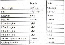 Issued from T. Terbiyik Kurt in Turk. J. Zool., 2018, 42 [p.570, Table 2]. Some morphological characteristics of female and male of Dioithona oculata from Iskenderun Bay (Turkey coast, Levantine Basin).
|
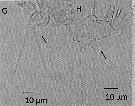 Issued from T. Terbiyik Kurt in Turk. J. Zool., 2018, 42 [p.572, Fig.4, G, H]. Male (from Iskenderun Bay): G, P5; H, Mx1.
|
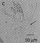 Issued from T. Terbiyik Kurt in Turk. J. Zool., 2018, 42 [p.571, Fig.3, C]. Female (from Iskenderun Bay): c, Mx1
|
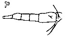 Issued from : K.-I. Yoo & D.-H. Lim in The Korean J. Syst. Zool., 1993, 9 (2) [p.99, Fig.2, P]. As Oithona oculata (= Dioithona oculata. Female (from East and West Korean Waters): P, abdomen (lateral view).
| | | | | Compl. Ref.: | | | Buskey, 1998 (p.13, Rem.: p.18, swarm & mating); 1998 a (p.425, swarming behavior vs energetic cost); Humphrey, 2008 (p.84: Appendix A); Marques-Rojas & Zoppi de Roa, 2017 (p.495, Table 1); Jerez-Guerrero & al., 2017 (p.1046, Table 1: temporal occurrence); Dias & al., 2018 a (p.189, Rem.: p.196); | | | | NZ: | 12 | | |
|
Distribution map of Dioithona oculata by geographical zones
|
| | | | | | | | | | | | 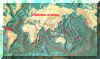 Chart of 1996 Chart of 1996 | |
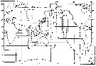 issued from in Bull. Ocean Res. Inst., Univ. Tokyo, 1985, No.20. [p.144, Fig.95]. As Oithona oculata. issued from in Bull. Ocean Res. Inst., Univ. Tokyo, 1985, No.20. [p.144, Fig.95]. As Oithona oculata.
Indo-Pacific georaphical distributio of Dioithona oculata. |
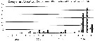
 Issued from: T. Terbiyik Kurt in Turk. J. Zool., 2018, 42 [p.573, Fig.6]. Issued from: T. Terbiyik Kurt in Turk. J. Zool., 2018, 42 [p.573, Fig.6].
Abundance distribution of D. oculata in the study area in Autumn. at Iskenderun Bay. (Stations 1 to 4 between about 36.8190°N-36.8143°N, 35.9327°E-35.9125°E) |
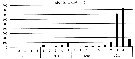
 Issued from: T. Terbiyik Kurt in Turk. J. Zool., 2018, 42 [p.574, Fig.7]. Issued from: T. Terbiyik Kurt in Turk. J. Zool., 2018, 42 [p.574, Fig.7].
The proportion of D. oculata in copepoda and zooplankton in autumn in the Iskendurun Bay.
Samples collected vertically (sampling depth ranged from 5 to 15 m) using a WP2 net (200 µm mesh aperture) at 5 stations in April, July, October and December of each year. Temperature and salinity measured by CTD probe. |
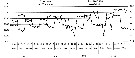 Issued from: T. Terbiyik Kurt in Turk. J. Zool., 2018, 42 [p.569, Fig.2]. Issued from: T. Terbiyik Kurt in Turk. J. Zool., 2018, 42 [p.569, Fig.2].
Seasonal changes of the sea water temperature and salinity values in the Iskenderun Bay (Levantine Basin, Turkish coast.
1 to 5: Stations in the Bay. Maximum depth of approximately 100 m at the opening of the bay.. Ceyhan River is a major freshwater source for the bay (180 m3 per sec.) |
 Issued from : J.C. Roff, J.T. Turner, M.K. Webber & R.R. Hopcroft in Aquat. microb. Ecol., 1995, 9. [p.172, Table 6]. Issued from : J.C. Roff, J.T. Turner, M.K. Webber & R.R. Hopcroft in Aquat. microb. Ecol., 1995, 9. [p.172, Table 6].
Summary observations on adult female of Oithona oculata (= Dioithona oculata) bacterovory on FLB (fluorescently labelled bacteria) a partir Escherichia coli (±0.7 µm3 cell volume) in Kingston Harbour (Jamaica).
Fluorescence levels: 0 = no materal for observation; - = no fluorescence observed; + = low fluorescence due to few FLB observe; * = highly fluorescence due to many FLB. |
| | | | Loc: | | | South Africa (off Cape of Good Hope), Brazil (.., Guanabara Bay, Vitoria Bay, Bracui estuary, Mucuri estuary); Barbada Is., Caribbean Colombia (Tayrona), Bahia de Mochima (Venezuela), Belize, Caribbean, Jamaica (Kingston Harbour), G. of Mexico, E Medit. (Turkey coast), Gulf of Oman, Arabian Gulf, Laccadives Is. (lagoons), Madagascar (Nosy Bé), Rodrigues Is., Indian, Nicobar Is. (Nankauri Harbour), Christmas Is., Straits of Malacca, China Seas (Yellow Sea, East China Sea), Taiwan (Tapong Bay), Korea (E, S & W), Okinawa (Kabira Bay, lagoons), Japan (Kyushu: Shijiki Bay), Palau Is., Australia (New South Wales, Great Barrier, Coral reef), New Caledonia, Caroline Is., Samoa Is., California (San Pedro Bay), Mission Bay, Bahia Cupica (Colombia), Chile (N) | | | | N: | 60 | | | | Lg.: | | | (66) F: 0,89; (104) M: 0,79; (155) F: 0,76; (179) F: 0,7-0,65; M: 0,6; (349) F: 0,9; (627) F: 0,68-0,62; M: 0,63-0,62; (634) F: 0,9; (649) F: 0,8; M: 0,65; (991) F: 0,62-0,8; M: 0,62-0,65; (1238) F: 0,59-0,68; M: 0,59-0,66; {F: 0,59-0,90; M: 0,59-0,79} | | | | Rem.: | epipelagic.
This species of Indo-pacific origin would have passed into the Atlantic by the Agulhas Current.
After Björnberg (1963, p.76) this species is easily recognized by the eye lenses. It was registered in the warmest waters off Brazil in the Soyh Equatorial Current.
Observed in ballasts of ships in San Francisco.
Sewell (1947, p.254: as Oithona oculata male) points to: The proportional lengths of the segments of the abdomen (Abd1 to caudal rami) as 23 : 18 : 17 : 16 : 14 : 12 = 100. Caudal rami twice as long as broad. In A1 it is extremely difficult to make out the total number of segments, but there appear to be 13 in all, of which the 2nd and 3rd and the 4th to 6 th respectively form overlapping groups; the 9th and 10th segments each bear a serrated spine on the anterior margin, segment 10 bears a single sensory filament. The kee-joint lies between segments 11 and 12, segment 12 bears a sensory filament about the middle of its length, and segment 13 bears 2 unequal sensory filaments, one slender and the other very stout at its distal end. In Mxp the 2nd and 3rd segments each bear a single stout S-shaped spine, that is fringed with fine needle-like spinules. In the swimming legs the terminal spines of the exopods are very long, exceeding in length the whole ramus. P5 bears 2 setae distally.
See in Oithona robertsoni Table 2 from McKinnon (2000, p.108).
First occurrence in Chilean waters by Hidalgo & al. (2010).
After Ueda & al., 1983 (p.167, Table 2 & p.169, Table 4, as Oithona oculata) at Tanabe Bay (A), Shijiki Bay (B) and Nagasaki Peninsula (C), respectively: (1)-Shape and diameter of swarm, (2)-depth (m), (3)- swarming position, (4)-number of samples examined, (5)-Mean % of adults (range), (6)- Mean % of females among adults (range).
A: (1)- continuous flat swarm; (2)- 6-14 m; (3)- over sandy flat bottom; (4)- 3; (5)- 37.6 (35.6-41.7); (6)- 61.5 (58.2-67.0).
B: (1)- irregular lalls (10 cm-3 m); (2)- 0.5-3 m; (3)- above algal patches in reef area; (4)-2; (5)- 78.0 (68.0-88.0); (6)- 71.5 (60.0-82/9).
C: (1)- irregular balls (10 cm-1.5 m); (2)- 1-10 m; (3)- above rocky shore with algal beds; (4)- 2; (5)- 36.8 (32/0-41.6); (6)- 57.3 (52.8-61.7) .
The species semm recorded mainly from the tropical waters, originating from the Pacific Ocean.
After McKInnon (2000, p.112) Oithona oculata is referred to the genus Dioithona Kiefer, 1935 on the basis of having 2 setae on the free segment of P5. Despite by Vervoort (1964), the generic rank of Dioithona has become widely used in recent litterature, species present in North Queensland coastal waters.
After Ohtsuka & Nishida (2017, p.578), this species forms dense swarms or schools during the daytime and disperses at night, as some other Acartia, Labidocera, Pontella, Tortanus (Atortus) (Ueda & al., 1983; Kimoto & al., 1988; Ohesuka & al., 2000 b).
Terbiyik Kurt (2018, p.568, figs.5, 6, 7) observed the first occurrence of this species in a coastal ecosystem of the Mediterranean Sea (Iskerendrum Bay, Levantine Basin) from 2013 and abundant in 2016.
See in DVP Conway & al., 2003 (version 1) | | | Last update : 22/10/2020 | |
|
|
 Any use of this site for a publication will be mentioned with the following reference : Any use of this site for a publication will be mentioned with the following reference :
Razouls C., Desreumaux N., Kouwenberg J. and de Bovée F., 2005-2026. - Biodiversity of Marine Planktonic Copepods (morphology, geographical distribution and biological data). Sorbonne University, CNRS. Available at http://copepodes.obs-banyuls.fr/en [Accessed February 27, 2026] © copyright 2005-2026 Sorbonne University, CNRS
|
|
|
|


;)
;)
;)
;)
;)
;)
;)
;)
;)
;)
;)
;)
;)
;)
;)

;)
;)
