|
|
 |
|
Cyclopoida ( Order ) |
|
|
|
Thaumatopsyllidae ( Family ) |
|
|
|
Caribeopsyllus ( Genus ) |
|
|
| |
Caribeopsyllus amphiodiae Ho, Dojiri, Hendler & Deets, 2003 (F,M) | |
| | | | | | | Ref.: | | | Ho, Dojiri, Hendler & Deets, 2003 (p.583, figs.F,M); Boxshall & Halsey, 2004 (p.691); Ferrari & Dahms, 2007 (p.25, 27, Rem. N, p.45, 57, 73, figs. copepodites I-VI); Ferrari & von Vaupel Klein, 2019 (p.179, figs.F, M, Rem.) | 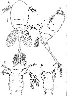 issued from : J.-s. Ho, M. Dojiri, G. Hendler & G.B. Deets in J. Crust. Biol., 2003, 23 (3). [p.584, Fig.1]. Female (from 33°59.33'N, 118°36.11'W): A, habitus with egg-sacs (dorsal); B, habitus (lateral); C, pedigers 4 and 5, and anterior part of genital double-somite (ventral); D, genital double-somite (dorsal). Scale bars: 0.1 mm (A, B); 0.05 mm (C, D). Nota : Cephalon bearing 2 dorsolateral conspicillae (large refractile lenses) and 1 median ventral conspicilla. Large red pigmented area in antetriormost region. Vestigial mouth and transverse cuticular structure, possibly labrum, present. A1 6-segmented (armature formula : 6, 2, 4, 2, 4, and 7 plus 1 aesthetasc ; setae sparsely pinnae or naked). A2 and oral appendages absent. Urosome 4-segmented. 4th pediger segment much smaller than 3rd pediger. Intercoxal plate well developed in P1 to P3, missing in P4. P1-P4 biramous, 3-segmented rami, except for P4. P5 bearing 1 small naked and 1 large pinnate terminalseta at each member. Genital double-somite broader in anterior region and carrying 2 small setae representing P6 in area of egg sac attachment. Multiseriate egg sacs with eggs measuring 0.05 mm in diameter. Anal somite long and slender, at least 4.8 times longer than broad, with concave lateral borders. Digestive tract and anus appear to be absent. Caudal ramus 2.85 times longer than wide, bearing 6 pinnate setae and inner distal row of setules.
|
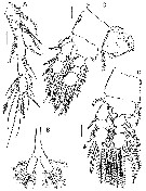 issued from : J.-s. Ho, M. Dojiri, G. Hendler & G.B. Deets in J. Crust. Biol., 2003, 23 (3). [p.586, Fig.2]. Female: A, A1; B, posterior part of urosome; C, P1 and intercoxal plate; D, P2. Scale bars: 0.05 mm (A, C, D; 0.1 mm (B).
|
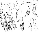 issued from : J.-s. Ho, M. Dojiri, G. Hendler & G.B. Deets in J. Crust. Biol., 2003, 23 (3). [p.587, Fig.3]. Female: A, P3 and intercoxal plate; B, P4; C, P5. Male: D, P4; E, P5. Scale bars: 0.05 mm in all drawings.
|
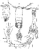 issued from : J.-s. Ho, M. Dojiri, G. Hendler & G.B. Deets in J. Crust. Biol., 2003, 23 (3). [p.588, Fig.4]. Male: A, habitus (dorsal); B, cephalothorax (ventral); C, 4th pediger and urosome (ventral); D, A1 (Roman numerals indicate ancestral segments). Scale bars: 0.1 mm (A, B, C); 0.05 mm (D). Nota : Body differs from female. A1 geniculate, 13-segmented (armature formula : 3, 1, 1, 1, 0, 0, 1, 4, 2, 2, 3 , 1 and 11) urosome 4-segmented. 5th pediger quadrangular, about 1.5 to 2.0 times broader than long. P1-P3 as in female. P4 as in female, but of smaller size. P5 situated on ventromedial surface of somite at posterior margin of pediger, with 2 members of pair fused medially at bases, intercoxal sclerite lacking ; each member elongate, about 4.3 times longer than broad, bearing 1 small and 2 large terminal setae. P6 lobiform, bearing 1 short and 1 long setae. Digestive tract and anus appear absent . vestigial mouth and transverse cuticular structure (labrum possibly) present.
|
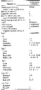 issued from : J.-s. Ho, M. Dojiri, G. Hendler & G.B. Deets in J. Crust. Biol., 2003, 23 (3). [p.589, Table 1]. Characters of female; Armature formulae for legs are presented as outer margin first, with Roman numerals for the number of spines, and Arabic numerals for setae.
| | | | | NZ: | 1 | | |
|
Distribution map of Caribeopsyllus amphiodiae by geographical zones
|
| | | | Loc: | | | California (Santa Monica Bay) | | | | N: | 1 | | | | Lg.: | | | (887) F: 1,51-1,89; M: 1,23-1,33; {F: 1,51-1,89; M: 1,23-1,33} | | | | Rem.: | Obtained after culture of nauplii collected in the stomac of the ophiuride Amphiodia urtica. | | | Last update : 08/02/2019 | |
|
|
 Any use of this site for a publication will be mentioned with the following reference : Any use of this site for a publication will be mentioned with the following reference :
Razouls C., Desreumaux N., Kouwenberg J. and de Bovée F., 2005-2026. - Biodiversity of Marine Planktonic Copepods (morphology, geographical distribution and biological data). Sorbonne University, CNRS. Available at http://copepodes.obs-banyuls.fr/en [Accessed January 08, 2026] © copyright 2005-2026 Sorbonne University, CNRS
|
|
|
|


;)
;)
;)
;)
;)
