|
|
 |
|
Calanoida ( Order ) |
|
|
|
Arietelloidea ( Superfamily ) |
|
|
|
Arietellidae ( Family ) |
|
|
|
Paraugaptiloides ( Genus ) |
|
|
| |
Paraugaptiloides magnus (Bradford, 1974) (M) | |
| | | | | | | Syn.: | Paraugaptilus magnus Bradford, 1974 a (p.523, figs.M); Heinrich, 1993 (p.7, figs.M) | | | | Ref.: | | | Ohtsuka & al., 1994 (p.123, Redescr.M, figs.M); Bradford-Grieve,1999 b (p.37, figs.M, figs.170, 189); Ohtsuka & al., 2005 (p.2503: Rem). | 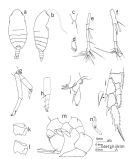 issued from : J.M. Bradford in N.Z. Jl mar. freshw. Res., 1974, 8 (3). [p.525, Fig.1]. As Paraugaptilus magnus. Male: a, habitus (dorsal); b, idem (lateral right side); c, rostrum; d, left caudal ramus; e, segments 19-21 of right A1; f, segments 17-19 of left A1; g, A2; h, Md (mandibular palp); i, Mx1; j, P2; k, basipod 2 of P3; l, basipod 2 of P4; m, P5; n, suctorian (large numbers of which attached mainly to segment 18 of left A1). Nota: This species has several distinctive characters, some of which may eventually necessitate its removal to a new genus.
|
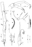 issued from : S. Ohtsuka, G.A. Boxshall & H.S.J. Roe in Bull. nat. Hist. Lond. (Zool.), 1994, 60 (2). [p.124, Fig.11]. Male (holotype, from S Indian Ocean): A, left A1 (segments XIX to XXVIII); B, idem (segments XXVI to WWVIII); C, A2; D, Md (endopod and exopod); E, 1st and 2nd praecoxal endites of Mx2; F, basal spine of Mx2; G, terminal seta on 4th endopod segment of Mx2. Scale bars in mm. Nota: Cephalosome and 1st pedigerous somite separate. Caudal ramus with seta II-VI well developed. Left A1 19-segmented. A2: 1st endopodal segment lacking inner seta, 2nd segment with 2 inner setae of unequal lengths subterminally and 5 setae and 1 setule terminally. Exopod indistinctly 8-segmented, setal formula 0, 1, 1, 1, 1, 1, 0, 3. Md: gnathobase with 3 cusped teeth, dorsalmost of which bifid at tip; the medial part is damaged and it is not known whether or not a tuft of setules is present; endopod rudimentary, 1-segmented, almost fused with basis, carrying 2 setae of unequal lengths; 1st exopodal segment with well developed seta, 5th segment with non-reduced outer seta. Mx2: 1st praecoxal endite with 2 setae and 1 vestigial element, 2nd with 2 spinulose setae; basal spine (fig.11F) with 2 rows of spinules; setae on endopod well developed, ornamented with row of long, simple spinules along inner margin (fig.11G)
|
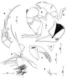 issued from : S. Ohtsuka, G.A. Boxshall & H.S.J. Roe in Bull. nat. Hist. Lond. (Zool.), 1994, 60 (2). [p.125, Fig.12]. Male: A, praecoxal arthrite, coxal endite, basal endite and endopod of Mx1 (basal seta indicated by arrowhead); B, 4th to 6th endopod segments of Mxp (innermost seta on 4th and 5th segments indicated by arrowhead); C, inner coxal seta of P4; D, P5 (posterior) (scar of element on 3rd exopod segment of left leg indicated by arrowhead); E, right endopod of P5; F, left endopod of P5; G, inner distal process on 2nd exopod segment of right P5. Scale bars in mm.
Nota: Mx1: praecoxal arthrite with 5 bare spines and 1 shorter process; coxal epipodite with 8 setae; coxal endite with long, spinulose seta; vestigial seta present (indicated by arrowhead); endopod bulbous, 1-segmented, bearing 2 relatively long, spinulose setae terminally.
Mxp: innermost seta on 4th and 5th endopodal segments (indicated by arrowhead) not reduced; seta a on 6th segment reduced; seta b relatively long; setae c and d simply ornamented with spinules along inner margin.
P1 with 2 outer spines on 3rd exopodal segment.
P4 with vestigial element on inner distal angle of coxa.
P5: coxae and intercoxal sclerite completely fused to form common base; coxa and basis incompletely fused in right leg and separate in left. Right leg endopod 1-segmented, spatulate, with minute sensillum on outer proximal margin and tubular prominences terminally; 1st exopdal segment produced on outer angle, with minute spine, 2nd segment almost completely separate from 3rd, with 2 tufts of fine setules at inner distal angle, minute sensillum at midlength of inner distal triangular process and outer terminal spiniform seta, 3rd segment triangular, tapering distally, with minute sensillum at outer middle margin and short vestigial element terminally; 3rd segment with well developed muscles proximally. Left leg endopod distinctly 2-segmented, 1st segment produced terminally, 2nd separate from first, spatulate, covered by numerous fine setules on outer surface, with attachment of muscles proximally; 1st exopodal segment similar to that of right leg, 2nd expanded inwards with outer seta subterminally, 3rd segment small, separate fron second, with 2 elongate, chitinized processes terminally and minute setule and scar of outer element proximally.
|
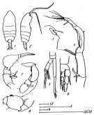 issued from : A.K. Heinrich in Zool. Zh., 1993, 72 (8). [p.8, Fig.2]. As Paraugaptilus magnus. Male (from SW Indian Ocean): 1, habitus (dorsal); 2, idem (lateral right side); 3, forehead (lateral); 4, left A1 (distal portion); 5, A2; 6, P1; 7-8, P5. Scale bars in mm: 1.0 (1, 3, 5, 8); 0.100 (4).
| | | | | Compl. Ref.: | | | Bradford-Grieve, 2004 (p.283) | | | | NZ: | 2 | | |
|
Distribution map of Paraugaptiloides magnus by geographical zones
|
| | | | | | | Loc: | | | New Zealand (North Island N), SW Indian (off S Madagascar) | | | | N: | 2 | | | | Lg.: | | | (74) M: 4,85; (306) M: 4,68; {M: 4,68-4,85} | | | | Rem.: | hyperbenthic ? (depth: 1060-1070 m; 1697 m). | | | Last update : 28/01/2015 | |
|
|
 Any use of this site for a publication will be mentioned with the following reference : Any use of this site for a publication will be mentioned with the following reference :
Razouls C., Desreumaux N., Kouwenberg J. and de Bovée F., 2005-2026. - Biodiversity of Marine Planktonic Copepods (morphology, geographical distribution and biological data). Sorbonne University, CNRS. Available at http://copepodes.obs-banyuls.fr/en [Accessed January 07, 2026] © copyright 2005-2026 Sorbonne University, CNRS
|
|
|
|


;)
;)
;)
;)

