|
|
 |
|
Calanoida ( Order ) |
|
|
|
Diaptomoidea ( Superfamily ) |
|
|
|
Acartiidae ( Family ) |
|
|
|
Acartia ( Genus ) |
|
|
|
Acanthacartia ( Sub-Genus ) |
|
|
| |
Acartia (Acanthacartia) sinjiensis Mori, 1940 (F,M) | |
| | | | | | | Syn.: | Acartia plumosa : Brodsky, 1948, 1950 (1967) (p.424, figs.F,M);
Acartia iseana : Ito, 1956 (p.468, fig.M);
Acartia bayli : Greenwood, 1972 (p.313, figs.F,M); 1978 (p.11);
Acartia sp. : Bayly, 1965 (p.327); Schipp & al., 1999 (p.81, culture method);
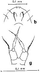
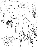
Acartia plumosa (sinjinensis) : Madhupratap & al., 1996 (p.77, Table 2: resting eggs); | | | | Ref.: | | | Mori, 1940 (p.329, figs.F,M); 1942 (p., figs.F,M); Ueda & Hiromi, 1987 (p.230, Descr.F,M, figs.F,M, Rem.); Yoo & al., 1991 (p.259, figs.F,M); Chihara & Murano, 1997 (p.671, Pl.20: F,M); Bradford-Grieve, 1999 b (p.217, figs.F,M, Rem., figs.186, 195); Mulyadi, 2004 (p.147, figs.F,M, Rem.); Ohtsuka & al., 2015 (p.123, Table 2). |  issued from : J.G. Greenwood in Crustaceana, 1972, 22 (3). [p.314, Fig.1]. As Acartia (Acanthacartia) baylyi. Female (from Brisbane River mouth, E Australia): a, habitus (dorsal); b, rostrum (ventral view); c, urosome (dorsal); d, basal region of P5 (lateral); e, idem (postrior view); f, P5 (posterior); g, posterior metasomal region and urosome (lateral right side). Scale line: 0.5 mm (a); 0.005 mm (all others). Nota: Metasome more than 3 times as long as wide; 4 times length of urosome. Head and 1st thoracic segment separate, 4th and 5th fused. Urosome 3-segmented. Caudal rami twice as long as wide. A1 when refleed reaching little beyond posterior border of metasome. Rounded posterolateral borders of metasome with variable number (3-7, usually 4) of small conical spines. Genital segment longer than wide with greatest width and depth about one-third of length from anterior end; posterodorsal rim with 6 to 8 spines on 6 loci, if 8, usually because spines in dorsolateral loci paired. 2nd urosomal segment similarly beset with 4 to 8 spines in 4 loci; these spines slightly larger than those on metasome or genital segment. Anal segment the shortest, without spines or setae. Caudal rami with lateral and medial borders fringed by hairs; 4 posterior and 1 lateral large plumose setae; smaller dorsal plumose seta.
|
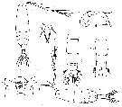
 issued from : J.G. Greenwood in Crustaceana, 1972, 22 (3). [p.316, Fig.2]. As Acartia (Acanthacartia) baylyi. Male: a, P5; b, habitus (dorsal); c, right A1 (hinge region); d, urosome (dorsal); e, posterior metasomal region and urosome (lateral left side). Scale line: 0.5 mm (b); 0.05 mm all others. Nota: Metasome 3.5 times length of urosome. A1 when reflexed reaching posterolateral border of 3rd thoracic segment. Posteriolateral borders of metasome similar in shape and spinulation to those of female, sometimes with 1 or 2 additional hairs. Urosome 5-segmented. 1st urosomal segment narrower than 2nd, without spines or setae ; 2nd segment the largest, posterodorsal rim with 6 widely separated conical spines, ventrolateral border on posterior half of segment with further 2 to 5 hair-like spines on each side ; 3rd segment with 4 to 6 spines at 4 loci on posterodorsal rim, two most dorsal positions frequently with a pair of spines ; 4th segment very short laterally, extending above and beyond anterior part of anal segment dorsally, posterodorsal rim with 2 large dorsal spines and a pair of smaller spinules on each side ; 5th segment (Anal ) unarmed. Caudal rami 1.3 times as long as wide, longer than anal segment. Inner border of basis of right P5 with shallow depression housing spinule and fine hairs, outer border with plumose seta ; exopodal segment 1 with finely plumose seta on posterior surface ; exopodal segment 2 with larger inner projection which is furrowed transversely at inner end so presenting distinct proximal and distal lobes, the latter slightly the smaller, distal border of flange with central conical spine ; exopodal 3 very long, curved, of irregular width but only slightly narrowed proximal neck region, outer margin without spines but with lobed lamellate extension proximal midpoint, inner margin smoothly curved except for single spine on slight projection near midpoint, segment terminating in similar small conical spine. Basis of left P5 with lamellate process developed from anteromedial surface in proximal half of segment, this process directed medially and terminating in a delicate acuminate spine with approximated fine hairs at its tip, plumose seta from distolateral border of this segment ; exopodal segment 1 without spines or setae ; exopodal segment 2 broad at base narrowing distally and curved medially to terminate in conical spine, inner surface with slight central concavity marked proximally by sparse group of fine hairs, long spine arises from centre of concavity, this spine slightly curved distally, with plumose margins, and lateral barb proximally.
|
 Issued from : K.A. Brodskii in Calanoida of the Far Eastern Seas and Polar Basin of the USSR. Opred. Fauna SSSR, 1950, 35 (Israel Program for Scientific Translations, Jerusalem, 1967) [p.424, Fig.299]. As Acartia plumosa. Female (from Sea of Japan): habitus (dorsal); urosome with spermatophore (dorsal); S5, P5. Male: habitus (dorsal); urosome (dorsal); S5, P5.
|
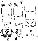
 issued from : H. Ueda & J. Hiromi in Crustaceana, 1987, 53 (3). [p.227, Fig.2, d-f]. Female: d, e, posterior metasome and urosome (dorsal and lateral, respectively); f, posterior metasome and 1st and 2nd urosomal segments (specimen with duplicate or triplicate spines on the posterior metasome). Nota: Posterior corner of metasome with 1 row of spines on each side; posteromedial region witout spinules. Genital segment with 1-14 posterodorsal spines, ventral surface without spinules. 2nd urosomal segment with 2-8 (usually 4) posterodorsal spines. 3rd ursomal segment without hairs. Caudal rami Length/Width 1.6-1.9. A1 spinule rows absent on any segment.
|
 issued from H. Ueda & J. Hiromi in Crustaceana, 1987, 53 (3). [p.228, Fig.b, g]. Female: b, forehead (ventral); g, P5 (anterior view).
|
 issued from : H. Ueda & J. Hiromi in Crustaceana, 1987, 53 (3). [p.229, Fig.4, c-e]. Male: c-d, urosome (dorsal and lateral, respectively); e, posterior urosome (specimen from Brisbane River mouth). Nota: 2nd urosomal segment with 3-8 posterodorsal spines; ventral surface with hair-like spines at 3-4 (usually 3) loci on each side (each locus usually with 1 spine but rarely with 2 or witout spines ( Two specimens from Brisbane River moth with medial 2 spines on urosomal segment 4 directed diagonally outward). Anal segment without hairs Caudal rami Length/width 1.0-1.1.
|
 issued from : H. Ueda & J. Hiromi in Crustaceana, 1987, 53 (3). [p.231, Fig.5, b, c]. Male: b, P5 (anterior view); c, left terminal segment under cover glass).
|
 issued from : H. Ueda & J. Hiromi in Crustaceana, 1987, 53 (3). [p.235, Table I]. Comparative list of characters in the Acartia plumosa - sinjiensis - tropica species group.
|
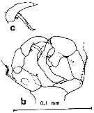
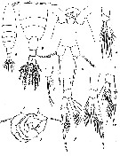
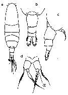
 issued from : T. Ito in Pacif. Sc., 1956, 10 (4). [p.Fig.1]. As Acartia iseana. Female (from middle Japan): a, habitus (dorsal); b, last thoracic segments and urosome (dorsal); C, forehead (ventral); d-g, P1 to P4; h-i, P5 (dorsal and lateral, respectively). Nota: Head and 1st thoracic segment separated, 4th and 5th fused. Lateral angle of last thoracic segment rounded, furnished with 4 spinules on either side. Genital segment not laterally dilated, about as long as other two abdominal segments combined, 2nd segment about twice as long as anal segment; first two abdominal segments each with 6 spinules along distal margin of dorsal surface. Caudal rami about 2 times as long as wide, inner margin with few hairs. A1 17-segmented, reaching end of cephalothorax. segment Male: j, P5. Nota: Abdomen 5-segmented, segment 4 well defined, but small. Caudal ramus 1.3 times as long as wide.
|
 issued from : T. Mori in Zool. Mag. Tokyo, 52 (8). [Figs.6-12]. Female (from Palao Islands): 6, habitus (dorsal); 7, P5; 10, urosome (dorsal); 11, forehead (ventral). Male: 8, P5; 9, urosome (dorsal); 12, habitus (dorsal).
|
 issued from : J.G. Greenwood in Crustaceana, 1972, 22 (3). [p.317, Table I. ]. As A. bayly. Comparison of A. bayli (= A. sinjiensis) with three closely related species. Acartia iseana Ito; 1956 was cnsidrerd as synomym of Acartia sinjiensis.
|
 issued from : T. Ito in Pacific Sc., 1956, 10 (4). [p.469, Fig.1]. As Acartia iseana. Female (from brackish ponds Ise Bay and Hegura Island, Japan): a, habitus (dorsal); b, posterior segments of metasome and urosome (dorsal); c, forehead (ventral); d,-g, P1 to P4, respectively); h-i, P5 (dorsal and lateral, respectively). Nota : A1 17-segmented, reaching end of prosome. Lateral angle of last thoracic segment rounded, with 4 spinules on either side. Genital segment not laterally dilated, about as long as other two abdominal segments combined ; 2nd segment about twice as long as anal segment ; first two abdominal segments each with 6 spinules along distal margin on dorsal surface. Caudal rami about 2 times as long as wide ; inner margin with a few hairs. P5 with basal segment longer than wide ; outer seta relatively short, shorter than terminal spine ; exopodite unsegmented, in form of a stout spine with rounded protuberance on ventral side of base ; terminal spine nearly straight, about 3 times as long as basal segment, armed distally with stout spinules on either side. Male: j, P5. Nota : Caudal rami shorter than in female, 1.3 times as long as wide. P5 : Right leg with basal segment longer than wide (about 2 :1) ; lengths (in micrometers) of basal segment and exopodites 1 and 2 : 25 :35 :30 ; inner margin of exopodite 1 smooth, but with row of fine hairs ; inner lobe of exopodite 2 very prominent , longer than wide, bifurcate at top ; exopodite 3 very narrow at base, curved, without spine on outer margin, but with a spine on inner edge. Left leg : lengths of segments (in micrometers) 35 :30 :41 ; exopodite 2 elongate, more than 4 times as long as wide, bearing a small spine on top, with a few hairs on inner margin distally.
|
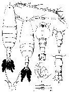 issued from : Mulyadi in Published by Res. Center Biol., Indonesia Inst. Sci. Bogor, 2004. [p.148, Fig.84]. As Acartia (Acartiura) sinjiensis.
Female (from Java): a, habitus (dorsal); b, forehead (ventral); c-d, posterior part of last thoracic segment and urosome (dorsal and lateral, respectively); e, P5.
Male: f, habitus (dorsal); h, P5.
|
 Issued from : M. Chihara & M. Murano in An Illustrated Guide to Marine Plankton in Japan, 1997. [p.678, Pl. 20, fig.8 a-d]. Female: a, habitus (dorsal); b, last thoracic segment and urosome (dorsal); c, same (lateral, right side); d, P5. Nota: numbers show characteristics of this species to compare with A. tsuensis.
| | | | | Compl. Ref.: | | | Ohtsuka & al., 1995 (p.157, 159); Ohno & al., 1996 (p.231, fig.2); McKinnon & Klumpp, 1998 (tab.1); Uye & al., 2000 (p.193, fig.7 : abundance vs t°, Sal.); Ueda & al., 2000 (tab.1); Ueda, 2001 (p.96, occurrence); Shimode & Shirayama, 2004 (tab.2); Lo & al., 2004 (p.468, tab.2); Knuckey & al., 2005 (p.339, diet, culture); Milione & Zeng, 2007 (p.656, effects of algal diets); 2008 (p.116, figs. 1, 2, 4, temperature and salinity effects); Ohtsuka & al., 2008 (p.115, Table 4, 5); Camus & Zeng, 2008 (p.220, effects of photoperiod); 2009 (p.145, egg production); Maiphae & Sa-ardrit, 2011 (p.641, Table 2); Itoh & al., 2011 (p.129, vertical distribution); Kang J.-H., 2011 (p.219, occurrences, inter-annual variability vs t° & Sal, Chl.a, Rem.: p.227.); Sakaguchi & al., 2011 (p.18, Table 1, 2, occurrences); Drillet & al., 2012 (p.155, Table 1, culture); Moon & al., 2012 (p.1, Table 1, seasonal & spatial variations); Gusmao & al., 2013 (p.279, Table 4, sex ratio); Gissi & al., 2013 (p.86, Table 3, copper toxicity); Tachibana & al., 2013 (p.545, Table 1, seasonal change 2006-2008); Suzuki, K.W. & al., 2013 (p.15, Table 2, 3, 4, estuaries, annual occurrence); Seo & al., 2013 (p.448, Table 1, occurrence); Trottet & al., 2017 (p. 7, Table 3: resting stage); Ohtsuka & Nishida, 2017 (p.565, 578, Table 22.1, Fig. 22.3: innermost station in Japanese waters); | | | | NZ: | 4 | | |
|
Distribution map of Acartia (Acanthacartia) sinjiensis by geographical zones
|
| | | | | | 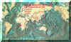 Chart of 1996 Chart of 1996 | |
 issued from : H. Ueda & J. Hiromi in Crustaceana, 1987, 53 (3). [p.232, Fig.6]. issued from : H. Ueda & J. Hiromi in Crustaceana, 1987, 53 (3). [p.232, Fig.6].
Distribution of the three species A. plumos, A. sinjiensis, A. topica. |
 Issued from : J.-H. Kang in Ocean Sci. J., 2011, 46 (4). [p.229, Fig.6] Issued from : J.-H. Kang in Ocean Sci. J., 2011, 46 (4). [p.229, Fig.6]
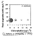
Abundance-total suspended solid-chlorophyll-a diagram of A. sinjiensis observed in the seaports from Korea during 3 years (2007-2009). |
 Issued from : J.-H. Kang in Ocean Sci. J., 2011, 46 (4). [p.230, Fig.7] Issued from : J.-H. Kang in Ocean Sci. J., 2011, 46 (4). [p.230, Fig.7]
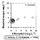
Abundance-dissolved oxygen-chlorophyll-a diagram of A. sinjiensis observed in the seaports from Korea during 3 years (2007-2009). |
 Issued from : J.-H. Kang in Ocean Sci. J., 2011, 46 (4). [p.228, Fig.5] Issued from : J.-H. Kang in Ocean Sci. J., 2011, 46 (4). [p.228, Fig.5]
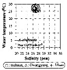
Abundance-temperature-salinity diagram of A. sinjiensis observed in the seaports from Korea during 3 years (2007-2009). |
| | | | Loc: | | | Manchouria (Poseta Bay), China Seas (East China Sea), S & W Korea, Incheon Harbour, Muan Bay, Japan (Naka-Umi: near Makata harbour, Sinji-Ko: brackish lake, Tokyo Bay, Tanabe Bay, Ise Bay, Lake in Hegura island, south estuaries, Honshu: Maizuru Bay, Lake Nakaumi), Okinawa (Naha Harbor), Taiwan (Tapong Bay), E Thailand, Cilacap Bay (S Java), off Tegal ( Java Sea), Singapore, Australia (E & NE : Brisbane estuary, Moreton Bay, Townsville, Haughton Riv., mouth of Ross River). | | | | N: | 38 | | | | Lg.: | | | (93) F: 1,1-0,9; M: 0,9-0,85; 1-0,99; (166) F: 1,09-1,05; M: 1,03-1; (189) F: 1,054-0,856; M: 0,905-0,868; (764) F: 1-0,9; M: 1-0,8; (866) F: 0,9-1,1; M: 0,8-1; (1122) F: 0,90; M: 0,80; {F: 0,856-1,10; M: 0,80-1,03} | | | | Rem.: | Littoral, Brackish.
For Ito (1956, p.470) A. iseana (= A. sinjiensis) is allied to A. bifilosa Giesbrecht, but it is distinguishable from bifilosa by the absence of the fine hairs on the dorsal surface of the abdomen and by the structures of P5 of both sexes.
According to Ueda & Hiromi (1987, p.232), contrary to the opinion of Tanaka (1965, p.387) Acartia plumosa and Acartia sinjiensis are not identical.
Mulyadi (2004, p.151) points to this species was originally described from the Lake of Nakaumi , a brackish lake connected with the Japan Sea, now occurs in the Gulf of Thailand, Cilacap estuary, Indonesia, and eastern coast Australia (as A. bayly Greenwood, 1978; Acartia sp. Bayly, 1965).
The species resembles Acartia tsuensis, but is distinguishable by the caudal ratio (2); exopodite of P5 in the female bearing a rounded lobe at base of terminal spine; inner prominence of exopodite 2 of right P5 in male bifurcate.
For Ohtsuka & al. (1995, p.157) A. sinjiensis is apparently a warm-water species, and distributed in the tropical waters of Singapore and Java as well as in the temperate and subtropical waters of Japan.
Ohtsuka & Nishida point to in the Inlet and brackish waters on the main islands of Japan, this species is dominant. | | | Last update : 02/10/2025 | |
|
|
 Any use of this site for a publication will be mentioned with the following reference : Any use of this site for a publication will be mentioned with the following reference :
Razouls C., Desreumaux N., Kouwenberg J. and de Bovée F., 2005-2026. - Biodiversity of Marine Planktonic Copepods (morphology, geographical distribution and biological data). Sorbonne University, CNRS. Available at http://copepodes.obs-banyuls.fr/en [Accessed February 27, 2026] © copyright 2005-2026 Sorbonne University, CNRS
|
|
|
|


 sinjiensis - Plate 1 of morphological figures',750,500);)
 sinjiensis - Plate 2 of morphological figures',750,500);)
 sinjiensis - Plate 3 of morphological figures',750,500);)
 sinjiensis - Plate 4 of morphological figures',750,500);)
 sinjiensis - Plate 5 of morphological figures',750,500);)
 sinjiensis - Plate 6 of morphological figures',750,500);)
 sinjiensis - Plate 7 of morphological figures',750,500);)
 sinjiensis - Plate 8 of morphological figures',750,500);)
 sinjiensis - Plate 9 of morphological figures',750,500);)
 sinjiensis - Plate 10 of morphological figures',750,500);)
 sinjiensis - Plate 11 of morphological figures',750,500);)
 sinjiensis - Plate 12 of morphological figures',750,500);)
 sinjiensis - Plate 13 of morphological figures',750,500);)
 sinjiensis - Plate 14 of morphological figures',750,500);)

 sinjiensis - Distribution map 2',650,500);)
 sinjiensis - Distribution map 2',750,500);)
 sinjiensis - Distribution map 3',750,500);)
 sinjiensis - Distribution map 4',750,500);)
 sinjiensis - Distribution map 5',750,500);)
 sinjiensis - Plate 1 of morphological figures)
 sinjiensis - Plate 2 of morphological figures)
 sinjiensis - Plate 3 of morphological figures)
 sinjiensis - Plate 4 of morphological figures)
 sinjiensis - Plate 5 of morphological figures)
 sinjiensis - Plate 6 of morphological figures)
 sinjiensis - Plate 7 of morphological figures)
 sinjiensis - Plate 8 of morphological figures)
 sinjiensis - Plate 9 of morphological figures)
 sinjiensis - Plate 10 of morphological figures)
 sinjiensis - Plate 11 of morphological figures)
 sinjiensis - Plate 12 of morphological figures)
 sinjiensis - Plate 13 of morphological figures)
 sinjiensis - Plate 14 of morphological figures)
 sinjiensis - Distribution map 2)
 sinjiensis - Distribution map 2)
 sinjiensis - Distribution map 3)
 sinjiensis - Distribution map 4)
 sinjiensis - Distribution map 5)
