|
|
 |
|
Calanoida ( Order ) |
|
|
|
Clausocalanoidea ( Superfamily ) |
|
|
|
Clausocalanidae ( Family ) |
|
|
|
Clausocalanus ( Genus ) |
|
|
| |
Clausocalanus jobei Frost & Fleminger, 1968 (F,M) | |
| | | | | | | Syn.: | Clausocalanus arcuicornis : T. Scott, 1894 b (p.73); Tanaka, 1960 (part., p.30, 31); Legaré, 1964 (Pl.3, fig.7); Vilela, 1965 (Pl.1, fig.2a); Calef & Grice, 1967 (p.89);
Clausocalanus farrani sp.1 Fleminger, 1964 a; 1967 a (tab. I) | | | | Ref.: | | | Frost & Fleminger, 1968 (p.55, figs.F,M, Rem.); Carli & Crisafi, 1969 (p.277, figs.F,M); Williams & Wallace, 1975 (p.176, fig.F); Dawson & Knatz, 1980 (p.7, figs.F); Alvarez-Marques, 1981 (p.157, figs.F, Rem.); Bradford-Grieve, 1994 (p.115, figs.F,M, fig.101); Bradford-Grieve & al., 1999 (p.878, 915, figs.F,M); Bucklin & al., 2003 (p.335, tab.2, fig.1, Biomol); Blanco-Bercial & Alvarez-marqués, 2007 (p.73, fig.2, Biomol.); Avancini & al., 2006 (p.77, Pl. 46, figs.F,M, Rem.); Vives & Shmeleva, 2007 (p.617, figs.F,M, Rem.) | 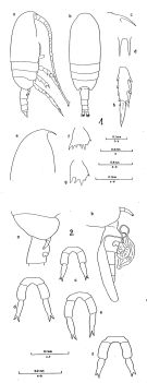 issued from : B. Frost & A. Fleminger in Bull. Scripps Inst. Oceanogr. Univ. California, San Diego, 1968, 12. [Pl.39, p.178-179; Pl.40, p.180-181]. Female: 1 a, habitus (right lateral view); 1 b, idem (dorsal view); 1 c, rostrum (right lateral); 1 d, rostrum (anteroventral); 1 e, frontal region (right lateral); 1 f, B2 of P2; 1 g, B2 of P3; 1 a,b,e from different specimens; 1 c,d,f,g,h from a fourth specimen; 2 a, Th.3 (posterior part), Th.4-5 and genital segment (right lateral); 2 b, Th.4-5 (posterior part) and urosome with spermatophore attached (right lateral); 2 c-f, P5; 2 a-f taken from different specimens.
|
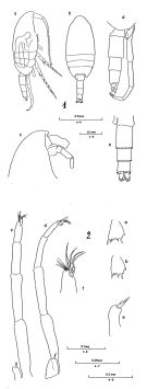 issued from : B. Frost & A. Fleminger in Bull. Scripps Inst. Oceanogr. Univ. California, San Diego, 1968, 12. [Pl.41, p.182-183; Pl.42, p.184-185]. Male: 1 a, habitus (right lateral view); 1 b, idem (dorsal view); 1 c, frontal region (right lateral); 1 d, Th.4-5 (posterior part) and urosome (right lateral); 1 e, urosome (dorsal view); 1 a-e taken from different specimens; 2 a, B2 of P2; 2 b, B2 of P3; 2 c, P5 (posterior); 2 d, P5 (right lateral); 2 e, right P5 (posterior); 2 f, 4P5 (distal part) and 5P5 (posterior); 2 a,b,f taken from the same specimen; 2 c,e from another. Nota: Armature of 5th segment of left P5 includes2 thick, spiniform setae; 2nd segment of P5 length more than 1.45 times as great as urosomal segment 2 width.
|
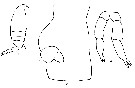
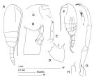 issued from : J.M. Bradford-Grieve in The Marine Fauna of New Zealand: Pelagic Calanoid Copepoda. National Institute of Water and Atmospheric Research (NIWA). New Zealand Oceanographic Institute Memoir, 102, 1994. [p.116, Fig.65]. Female: A, habitus (lateral right side); B, genital somite (lateral left side); C, forehead (lateral right side); D, basipod 2 of P3; E, P5. Male: F, habitus (lateral left side); G, P5; H, terminal part of right P5; I, terminal part of left P5.
|
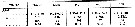 issued from : B. Frost & A. Fleminger in Bull. Scripps Inst. Oceanogr. Univ. California, 1968, 12. [p.46, Table 3a]. Clausocalanus jobei Females: Measurements and ratios . TL = total body length ; SL = spermatophore length ; P :U = ratio of prosome length to urosome length ; U :U1 = ratio of total urosome length to length of 1st urosomal segment (genital segment) ; S :U1 = ratio of spermatophore sac length to U1 length of female on which spermatophore is attached ; r = sample range; m = sample mean; n = number of specimens measured; s = sample standard deviation.
|
 issued from : B. Frost & A. Fleminger in Bull. Scripps Inst. Oceanogr. Univ. California, 1968, 12. [p.47, Table 3b]. Clausocalanus jobei Males: Measurements and ratios . TL = total body length; P :U = ratio of prosome length to urosome length ; P:U2 = ratio of prosome length to length od 2nd urosomal segment; U2:2P5 = ratio of U2 length to length of 2nd segment of longer ramus of P5; U2:3P5 = ratio of U2 length of 3rd segment of longer ramus of P5; 3P5:2P5 = ratio of length of 3P5 of longer ramus to length of 2P5 of longer ramus; r = sample range; m = sample mean; n = number of specimens measured; s = sample standard deviation.
|
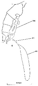 issued from : B. Frost & A. Fleminger in Bull. Scripps Inst. Oceanogr. Univ. California, 1968, 12. [p.102, P1.1, a]. Female: a, urosome with spermatophore attached (right lateral). cm = cementing material; am = accessory material associated with spermatophore neck; sn = spermatophore neck; ss = spermatophore sac.
|
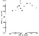 issued from : B. Frost & A. Fleminger in Bull. Scripps Inst. Oceanogr. Univ. California, 1968, 12. [p.24, Fig.7]. Ratio of 2nd segment of left P5 length to urosomal segment 2 width (ordinate) plotted against 2nd segment of left P5 length (abscissa) for males of Clausocalanus jobei.
|
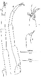
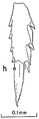 issued from : B. Frost & A. Fleminger in Bull. Scripps Inst. Oceanogr. Univ. California, 1968, 12. [p.179, Pl.39, h]. Female: h, exopodal segment 3 and terminal seta of P3.
|
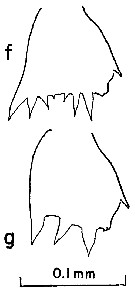 issued from : B. Frost & A. Fleminger in Bull. Scripps Inst. Oceanogr. Univ. California, 1968, 12. [p.179, Pl.39, f, g]. Female: f, basipodite 2 of P2; g, basipodite 2 of P3.
|
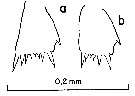
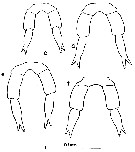 issued from : B. Frost & A. Fleminger in Bull. Scripps Inst. Oceanogr. Univ. California, 1968, 12. [p.181, Pl.40, c-f]. Female: c-f, P5 (from different specimens).
|
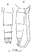 issued from : B. Frost & A. Fleminger in Bull. Scripps Inst. Oceanogr. Univ. California, 1968, 12. [p.183, Pl.41, d, e]. Male: d, posterior part of last thoracic segment and urosome (right lateral); e, urosome (dorsal).
|
 issued from : B. Frost & A. Fleminger in Bull. Scripps Inst. Oceanogr. Univ. California, 1968, 12. [p.185, Pl.42, c-d, e, f]. Male: c, P5 (posterior); d, P5 (right lateral; from another specimen); e, right P5 (posterior); f, 4th segment of P5 (distal part) and 5th segment of P5 (posterior).
|
 issued from : B. Frost & A. Fleminger in Bull. Scripps Inst. Oceanogr. Univ. California, 1968, 12. [p.185, Pl.42, a, b]. Male: a, basipodite 2 of P2; b, basipodite 2 of P3.
|
 Issued from F. Alvarez-Marques in Rev. Fac. Cienc. Univ. Oviedo (Ser. Biologia), (1979-80), 20-21, 1981. [p.165, Pl. II, Figs.1-3]. Female (from Gijon, NW Spain): 1, habitus (lateral); 2, genital segment and spermatheca (lateral); 3, P5.
| | | | | Compl. Ref.: | | | Binet & al., 1972 (p.71); Björnberg, 1973 (p.310, 385); Fleminger & Hulsemann, 1973 (p.340, chart); Peterson & Miller, 1976 (p.14, Table 1, 3, abundance vs interannual variations); 1977 (p.717, Table 1, seasonal occurrence); Carter, 1977 (1978) (p.35); Andronov & Maigret, 1980 (p.71, Table 3); Kovalev & Schmeleva, 1982 (p.83); Van der Spoel & Heyman, 1983 (p.44, fig.55); Greze & al., 1983 (p.17, Rem.: p.24); Turner & Dagg, 1983 (p.16); Scotto di Carlo & al., 1984 (p.1042); Regner, 1985 (p.11, Rem.: p.28); Brenning, 1985 a (p.28, Table 2); Greze & al., 1985 (p.7); Brinton & al., 1986 (p.228, Table 1); Diouf & Diallo, 1987 (p.260); James & Wilkinson, 1988 (p.249, Table 2, biomass, chemical composition); Pancucci-Papadopoulou & al., 1990 (p.199); Jimenez-Perez & Lara-Lara, 1988; Scotto di Carlo & al., 1991 (p.271); Palomares Garcia Vera, 1995 (tab.1); Hajderi, 1995 (p.542); Hure & Krsinic, 1998 (p.100); Suarez-Morales & Gasca, 1998 a (p109); Siokou-Frangou & al., 1999 (p.205, Table 5); Moraitou-Apostolopoulou & al., 2000 (tab.I); Fragopoulu & al., 2001 (p.49, tab.1); Uysal & al., 2002 (p.17, tab.1); Vukanic, 2003 (139, tab.1); Hsiao & al., 2004 (p.325, tab.1); Rezai & al., 2004 (p.489, tab.2, Rem.); Peralba & Mazzocchi, 2004 (p.645, figs.3,6); Lo & al., 2004 (p.89, tab.1); Isari & al., 2006 (p.241, tab.II); Zervoudaki & al., 2006 (p.149, Table I); Khelifi-Touhami & al., 2007 (p.327, Table 1); Valdés & al., 2007 (p.103: tab.1); Cabal & al., 2008 (289, Table 1); Ayon & al., 2008 (p.238, Table 4: Peruvian samples); C.E. Morales & al., 2010 (p.158, Table 1); Brugnano & al., 2010 (p.312, Table 2, 3, fig.8); Lidvanov & al., 2010 (p.356, Table 3); Hernandez-Trujillo & al., 2010 (p.913, Table 2); Schnack-Schiel & al., 2010 (p.2064, Table 2: E Atlantic subtropical/tropical, Fig.4, 6, 7, Tabe 4); Hidalgo & al., 2010 (p.2089, Table 2); Mazzocchi & Di Capua, 2010 (p.425); Mazzocchi & al., 2011 (p.1163, fig.6, long-term time-series 1984-2006); Isari & al., 2011 (p.51, Table 2, abundance vs distribution); Mazzocchi & al., 2012 (p.135, annual abundance 1984-2006); Uysal & Shmeleva, 2012 (p.909, Table I); Salah S. & al., 2012 (p.155, Tableau 1); Miloslavic & al., 2012 (p.165, Table 2, transect distribution); Brugnano & al., 2012 (p.207, Table 3); Hidalgo & al., 2012 (p.134, Table 2, 3, figs.6, 8, occurrence vs hydrology); in CalCOFI regional list (MDO, Nov. 2013; M. Ohman, comm. pers.); Lidvanov & al., 2013 (p.290, Table 2, % composition); Siokou & al., 2013 (p.1313, fig.4, 8, biomass, vertical distribution); Pansera & al., 2014 (p.221, Table 2, abundance); Zaafa & al., 2014 (p.67, Table I, occurrence); Mazzocchi & al., 2014 (p.64, Table 3, 4, 5, spatial & seasonal composition %) ; Benedetti & al., 2016 (p.159, Table I, fig.1, functional characters); El Arraj & al., 2017 (p.272, table 2, spatial distribution); Benedetti & al., 2018 (p.1, Fig.2: ecological functional group); Belmonte, 2018 (p.273, Table I: Italian zones) | | | | NZ: | 13 | | |
|
Distribution map of Clausocalanus jobei by geographical zones
|
| | | | | | | | | | | | 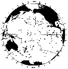 issued from : B. Frost & A. Fleminger in Bull. Scripps Inst. Oceanogr. Univ. California, San Diego, 1968, 12. [p.56, Chart 7, a]. issued from : B. Frost & A. Fleminger in Bull. Scripps Inst. Oceanogr. Univ. California, San Diego, 1968, 12. [p.56, Chart 7, a].
Occurrence of C. jobei in samples examined; closed circles represent samples examined; open circles represent samples in which adults were found; bars through open circles represent samples from which specimens were removed for measurements. |
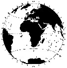 issued from : B. Frost & A. Fleminger in Bull. Scripps Inst. Oceanogr. Univ. California, San Diego, 1968, 12. [p.57, Chart 7, b]. issued from : B. Frost & A. Fleminger in Bull. Scripps Inst. Oceanogr. Univ. California, San Diego, 1968, 12. [p.57, Chart 7, b].
Occurrence of C. jobei in samples examined; closed circles represent samples examined; open circles represent samples in which adults were found; bars through open circles represent samples from which specimens were removed for measurements. |
| | | | Loc: | | | South Africa (E), S Atlant. (equatorial & subtropical), Congo, Ivorian shelf, Casamance, Morocco-Mauritania, Cap Ghir (Morocco), off S Cape Verde Is., Venezuela G. of Mexico, Florida, off New-York, SE Iceland, off W Ireland, Bay of Biscay, Gijon (NW Spain), Ibero-moroccan Bay, off W Tangier, Medit. (Alboran Sea, W Basin., Gulf of Annaba, Ligurian Sea, Tyrrhenian Sea, Lake Faro (Sicily), G. of Naples, Strait of Messina, Adriatic Sea, Aegean Sea, Thracian Sea, Lebanon Basin, Marmara Sea), Natal, SW Indian, Straits of Malacca, China Seas (East China Sea, South China Sea), Taiwan (E, N: Mienhua Canyon), Pacif. (E equatorial), Oregon (off Newport), S California, Baja California (Bahia Magdalena), G. of California, W Mexico, New Zealand, Cape Farewell-Taranaki Bight, Peru, Chile (N, Concepcion, off Santiago) | | | | N: | 80 | | | | Lg.: | | | (30) F: 1,56-1,01; M: 1,07-0,87; (1138) F: 1,53-1,17; {F: 1,01-1,56; M: 0,87-1,07}
The mean female size is 1.318 mm (n = 4; SD = 0.2710) and the mean male size is 0.97 mm. The size ratio establish on one sample is 0.76. | | | | Rem.: | epipelagic. Principally neritic.
For Frost & Fleminger (1968, p.58) C. jobei is distinct from all other Clausocalanus species except farrani in having a ventral protuberance on the female genital segment anterior to the genital pores. C. jobei and farrani females differ in the form of the rostrum in lateral view. C. jobei differs from minor in the form of the P5, and from paululus in body length, in the form and spacing of basipodite 2 spiniform processes 2 and 3 of P3, in the nature of the attachment of the spermatophore. Males of C. jobei are distinguished from males of farrani and minor by the ratio of the left 2nd segment of P5 length to the urosomal segment 2 and the armature of the distal segment of P5. C. jobei and paululus differ in body length, in the urosomal segment 2/2nd segment of P5, and 3rd segment/2nd segment of P5 length ratios, and in the form of basipodite 2 spiniform processes 2 and 3 of P3.
After Benedetti & al. (2018, p.1, Fig.2) this species belonging to the functional group 7 corresponding to small filter feeding herbivorous. | | | Last update : 28/10/2022 | |
|
|
 Any use of this site for a publication will be mentioned with the following reference : Any use of this site for a publication will be mentioned with the following reference :
Razouls C., Desreumaux N., Kouwenberg J. and de Bovée F., 2005-2026. - Biodiversity of Marine Planktonic Copepods (morphology, geographical distribution and biological data). Sorbonne University, CNRS. Available at http://copepodes.obs-banyuls.fr/en [Accessed March 22, 2026] © copyright 2005-2026 Sorbonne University, CNRS
|
|
|
|


;)
;)
;)
;)
;)
;)
;)
;)
;)
;)
;)
;)

;)
;)
