|
|
 |
|
Calanoida ( Order ) |
|
|
|
Clausocalanoidea ( Superfamily ) |
|
|
|
Aetideidae ( Family ) |
|
|
|
Aetideus ( Genus ) |
|
|
| |
Aetideus acutus Farran, 1929 (F,M) | |
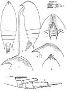
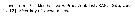
| | | | | | | Syn.: | Euaetideus acutus : Farran, 1936 a (p.87); Sewell, 1948 (p.553); Vervoort, 1957 (p.6, 51, figs.F, Rem.); Grice, 1962 (p.190, figs.F, Rem.); Grice & Hart, 1962 (p.287, 293: Rem.); Brodsky, 1962 c (p.119, figs.F); De Decker & Mombeck, 1964 (p.12); Chen & Zhang, 1965 (p.51, figs.F); Grice & Hulsemann, 1965 (p.223, as actus); 1967 (p.15); Fleminger, 1967 a (tabl.1, as E. acuta); Vinogradov, 1968 (1970) (p.79, 277); Park, 1968 (p.545, figs.F, Rem.); 1970 (p.475); Tanaka & Omori, 1970 (p.110); Corral Estrada, 1970 (p.151, figs.F); Kos, 1972 §Vol. I, figs.F, Rem.); Roe, 1972 (p.277, tabl.1, tabl.2); 1972 a (p.332); Corral Estrada & Pereiro Muñoz, 1974 (tab.I); Carter, 1977 (1978) (p.35); Dawson & Knatz, 1980 (p.6, figs.F); Björnberg & al., 1981 (p.630, figs.F); Zheng & al., 1982 (p.31, figs.F); Guangshan & Honglin, 1984 (p.118, tab.); De Decker, 1984 (p.316); Brinton & al., 1986 (p.228, Table 1); Chen Y.-Q., 1986 (p.205, Table 1: abundance %); Madhupratap & Haridas, 1986 (p.105, tab.1); Yoo, 1991 (tab.1); Kim & al., 1993 (p.269); Shih & Young, 1995 (p.66); Park & Choi, 1997 (Appendix); Noda & al., 1998 (p.55, Table 3, occurrence); Mulyadi, 2004 (p.59, figs.F, Rem.); | | | | Ref.: | | | Farran, 1929 (p.208, 228, Descr.F, figs.F); Tanaka, 1957 a (p.36, figs.F); Bradford, 1971 (p.30, Descr.M, figs.F,M); Park, 1974 (p.217, figs.F,M, Rem.); Bradford & Jillett, 1980 (p.14, figs.F,M, fig. 73, distribution chart); Markhaseva, 1996 (p.12, figs.F,M); Chihara & Murano, 1997 (p.682, Pl.32: F); Lapernat, 1999 (p.7, 40, 55, fig.F); Bradford-Grieve & al., 1999 (p.879, 920, figs.F,M); Vives & Shmeleva, 2007 (p.543, figs.F,M, Rem.); Lim & al., 2011 (p.36, figs.F, molecular sequence) | 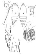 Issued from : T.S. Park in Fishery Bull. Fish Wild. Serv. U.S., 1968, 66 (3). [p.543, Pl.5, Figs.8-14]. As Euaetideus acutus. Female: 8, habitus (dorsal); 9, idem (left lateral side); 10, rostrum (frontal view); 11, last thoracic segment and urosome (left lateral side); 12, Mx2; 13, P4; 14, basipod of P4. Nota: The coxa of P4 has about 5 acute spinules at the insertion of the internal seta.
|
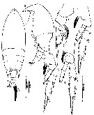
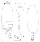 issued from : J.M. Bradford & J.B. Jillett in Mem. N.Z. Oceanogr. Inst., 86, 1980. [p.15, Fig.5]. Female: A, habitus (dorsal); B, rostrum. Male: C, habitus (ventral); D, P5.
|
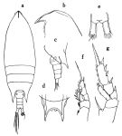 issued from : O. Tanaka in Publ. Seto Mar. Biol. Lab., 1957, VI (1). [p.36, Fig.25]. Female: a, habitus (dorsal); b, forehead (lateral); c, last thoracic segment and urosome (lateral left side); d, rostrum; e, anal segment and furca; f, P1; g, P2. Nota: forehead with a well-marked chitinous crest which slightly at the apex, but not so strongly arched as described and figured by Farran (1929). Rostrum with two knobs at its base. Proportional lengths of urosomites and furca 34:18:13:13:22 = 100. A1 exceeds slightly distal end of furca.
|
 issued from: Q.-c Chen & S.-z. Zhang in Studia Marina Sinica, 1965, 7. [Pl.13, 6-7]. As Euaetideus acutus. Female (from E China Sea): 6, forehead (dorsal); 7, urosome (lateral right side).
|
 issued from : J. Corral Estrada in Tesis Doct., Univ. Madrid, A-129, Sec. Biologicas, 1970. [Lam.42]. As Euaetideus acutus. Female (from Canarias Is.): 1, habitus (dorsal); 2, idem (left lateral side); 3, forehead (frontal view); 4, idem (lateral); 5, idem (dorsal); 6, P4; 7, forehead (lateral, another individual).
|
 issued from : J. Corral Estrada in Tesis Doct., Univ. Madrid, A-129, Sec. Biologicas, 1970. [Lam.41, figs.8-11]. As Euaetideus acutus. Female: 8, posterior part cephalothorax and urosome (lateral); 9, P1; 10, P2; 11, P3.
|
 issued from : W. Vervoort in B.A.N.Z. Antarctic Reseach Expedition, Report Ser. B, Vol. III., 1957 [Fig.28]. As Euaetideus acutus. Female (from Malay Archipelago): a-b, habitus (lateral and dorsal, respectively); c, forehead (lateral); d-e, posterior part cephalothorax and urosome (dorsal and lateral, respectively).
|
 issued from : W. Vervoort in B.A.N.Z. Antarctic Reseach Expedition, Report Ser. B, Vol. III., 1957 [Fig.29]. As Euaetideus acutus. Female: a, right P1 (anterior); b-d, P2 to P4 (left legs); e, left Mx2; f, left Md (mandibular palp).
|
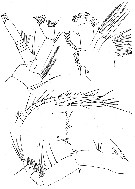 issued from : W. Vervoort in B.A.N.Z. Antarctic Reseach Expedition, Report Ser. B, Vol. III., 1957 [Fig.30]. As Euaetideus acutus. Female: a, left A2; b, left Mx1; c-d, right A1; e, Md (cutting edge); f, right Mxp.
|
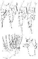
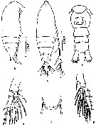 issued from : Z. Zheng, S. Li, S.J. Li & B. Chen in Marine planktonic copepods in Chinese waters. Shanghai Sc. Techn. Press, 1982 [p.32, Fig.18]. As Euaetideus acutus. Female: a-b, habitus (dorsal and lateral, respectively); c, rostrum; d, urosome (ventral); e, P1; f, P4. Scale bars in mm.
|
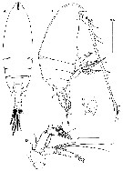 issued from : B.-J. Lim, Song S.J. & Min G.-S. in Korean J. Syst. Zool., 2011, 27 (1). [p.37, Fig.1]. Female (from 36°30'N, 131°20'E): A-B, habitus ( dorsal and lateral, respectively); C, rostrum (frontal view); D, A2. Scale bars: 0.5 mm (A-C); 0.1 mm (D). Nota : Prosome about 3.8 times as long as urosome. Cephalosome and 1st pedigerous somite fused, 4th and 5th fused. Head with dorsal crest. Rostrum strong and bifurcate ; concave area near bases of furcations with a pair of small, rounded projections. A1 24-segmented, reaching slightly beyond end of caudal rami Posterior corners of last pedigerous somite extending to 2/3 region of 2nd abdominal somite.
|
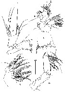 issued from : B.-J. Lim, Song S.J. & Min G.-S. in Korean J. Syst. Zool., 2011, 27 (1). [p.38, Fig.2]. Female: A, Md; B, Mx1; C, Mx2; D, Mxp; E, Md (cutting edge of the gnathobase). Scale bar: 0.1 mm (A-D). Nota : Endopod of A2 2-segmented. Gnathobase of Md with 14 teeth and 1 long and 1 short setae. Endopod 2-segmented ; exopod 5-segmented. Mx1 : arthrite with 5 posterior setae and 9 spines ; coxal epipodite with 7 large and 2 minute setae ; coxal endite with 3 setae ; basal endites with 4+4 setae ; endopod with 12 setae ; exopod with 11 setae. Mx2 : 1st and 2nd praecoxal endites and 1st coxal endite each with 2 long and 1 short spinulose setae and row of spinules on one side. 2nd coxal endite with 1 long and 1 short spinulose setae and 1 thickened seta, and row of spinules on one side. 1st basal endite with 3 setae, one of them thickened and claw-like. Endopod with 6 setae. Mxp : Coxa with 8 setae. Basis with 3 medial setae. Endopod 6-segmented, 1st segment with 2 setae, 2nd to 6th segments with 4, 4, 3, 3+1, and 4 setae respectively.
|
 issued from : B.-J. Lim, Song S.J. & Min G.-S. in Korean J. Syst. Zool., 2011, 27 (1). [p.39, Fig.3]. Female: A-D, P1 to P4; E, posterior coxal margin of P4. Scale bar: 0.1 mm (A-D).
|
 issued from : B.-J. Lim, Song S.J. & Min G.-S. in Korean J. Syst. Zool., 2011, 27 (1). [p.36]. Female: Armature formula of swimming legs P1 to P4. Roman numerals = spines; Arabic numerals = setae.
|
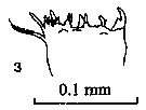 issued from : P.E. Lapernat & C. Razouls in Vie Milieu, 2002, 52 (1). [p.28, Pl. VI, fig.3]. Masticatory edge of Md gnathobase female (from off Malta, Mediterranean Sea). Nota: Itoh's index: 349 (number of teeth : 10).
|
 issued from : P.E. Lapernat & C. Razouls in Vie Milieu, 2002, 52 (1). [p.20, Pl. I, fig.4]. Masticatory edge of Md gnathobase female (from off Malta, Mediterranean Sea).
|
 issued from : Mulyadi in Published by Res. Center Biol., Indonesia Inst. Sci. Bogor, 2004. [p.60, Fig.33]. As Euaetideus acutus. Female: a, habitus (dorsal); b, forehead (lateral); c, posterior end of last thoracic segment and urosome (lateral left side); d-g, P1 to P4, respectively.
|
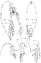
 issued from : E.L. Markhaseva in Proc. Zool. Inst. RAN, St. Petersburg, 1996, 268. [p.13, Fig.2]. Female (from 29°17'N, 142°47'E).
|
 issued from : E.L. Markhaseva in Proc. Zool. Inst. RAN, St. Petersburg, 1996, 268. [p.13, Fig.2]. From Pars, 1974. Male (fron G. of Mexico).
|
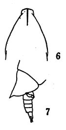
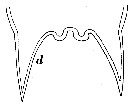 issued from : W. Vervoort in B.A.N.Z. Antarctic Reseach Expedition, Report Ser. B, Vol. III., 1957 [Fig.20 d]. As Euaetideus acutus. Female: distal portion of rostral plate.
|
 Aetideus acutus Aetideus acutus female: 1 - Posterior corners of last thoracic segment prolonged into wing-like lobes pointed at topos, reaching at least the middle of urosomal segment 2. Anterior part of cephalon with crest 2 - Excavation between rostral rami with 2 thickenings. 3 - Duct between ventral and dorsal parts of spermatheca not narrowed. Rostal base visible in dorsal view.
|
 Aetideus acutus Aetideus acutus male: 1 - Last thoracic segment with points. 2 - Posterior corners of last thoracic segment with points exceeding posterior border of urosomal segment 1 (in dosrsal view). 3 - Caudal rami 1.64-1.83 times as long as wide.
|
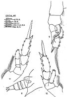
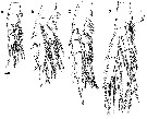
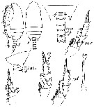
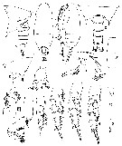 Issued from : T. Park in Fish. Bull., 1974, 72 (1). [p.219, Fig.3]. Female (from G. of Mexico): A, posterior part of body (lateral). Nota: The appendages are similar to those of A. pacificus Park, 1968, except that Mx1 carries 3 + 3 +6 setae on the endopod. Male: B-C, habitus (dorsal and lateral, respectively); D, posterior part of body (dorsal); E, last metasomal and genital segments (lateral); F, 129th segment of A1; G, A2; H, Md; I, Mx1; J, Mxp; K-N, P1 to P4 (anterior views); O, P5 (anterior).
|
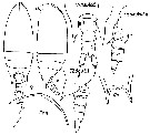
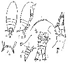 Issued from : M.S. Kos in Field guide for plankton. Zool Institute USSR Acad., Vol. I, 1972. After Brodsky, 1962. As Euaetideus acutus. Female: 1-2, habitus (dorsal and lateral, respectively); 3, forehead (lateral); 4, rostrum; 5, corner of the last thoracic segment and abdomen (lateral), 6, urosome (ventral); 7, P1.
| | | | | Compl. Ref.: | | | Furuhashi, 1966 a (p.295, vertical distribution in Kuroshio region, Table 9, 10); Vives, 1982 (p.290); Brenning, 1983 (p.2, Rem.); Cummings, 1984 (p.163, Table 2); Brenning, 1985 a (p.28, Table 2); Ambler & Miller, 1987 (tab.2, 3, 4, 5); Lozano Soldevilla & al., 1988 (p.58); Suarez-Morales & Gasca, 1998 a (p.107); Lapernat & Razouls, 2001 (p.123, tab.1); Shimode & Shirayama, 2004 (tab.2); Hsiao & al., 2004 (p.325, tab.1); Pusch & al., 2004 (251, tab.3); Lo & al., 2004 (p.89, tab.1); Lavaniegos & Jiménez-Pérez, 2006 (tab.2, 4, Rem.); Dur & al., 2007 (p.197, Table IV); Fernandes, 2008 (p.465, Tabl.2); Lan Y.C. & al., 2008 (p.61, Table 1, % vs stations, Table 2: indicator species); C.-Y. Lee & al., 2009 (p.151, Tab.2); Williamson & McGowan, 2010 (p.273, Table 3, Pacific central gyres: N and S); Cornils & al., 2010 (p.2076, Table 3); Schnack-Schiel & al., 2010 (p.2064, Table 2: E Atlantic subtropical/tropical); Medellin-Mora & Navas S., 2010 (p.265, Tab. 2); W.-B. Chang & al., 2010 (p.735, Table 2, 4, abundance); Hsiao S.H. & al., 2011 (p.475, Appendix I) ; in CalCOFI regional list (MDO, Nov. 2013; M. Ohman, pers. comm.); Hirai & al., 2013 (p.1, Table I, molecular marker); Lidvanov & al., 2013 (p.290, Table 2, % composition); Marques-Rojas & Zoppi de Roa, 2017 (p.495, Table 1). | | | | NZ: | 12 | | |
|
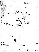
Distribution map of Aetideus acutus by geographical zones
|
| | | | | | | | | | | |  issued from : J.M. Bradford in N.Z. Jl Mar. Freshw. Res., 1971, 5 (1). [p.34, Fig.13]. issued from : J.M. Bradford in N.Z. Jl Mar. Freshw. Res., 1971, 5 (1). [p.34, Fig.13].
Southwest Pacific Ocean showing distribution of four Aetideus species. |
| | | | Loc: | | | South Africa (E), Atlant. (tropical), off Senegal, Morocco-Mauritania, Great Meteor Seamount, Canary Is., off Madeira, Portugal, Cuba, S Brazil, off Amazon, Venezuela, Bahia de Mochima, Caribbean Colombia, Caribbean Sea, G. of Mexico, E Medit. (off Malta), Natal, Indian, Bay of Bengal, Indonesia-Malaysia, Flores Sea, S Celebes Sea, China Seas (Yellow Sea, East China Sea, South China Sea), Taiwan (S, SW, E, N: Mienhua Canyon), Korea (S, E), Japan (Kuchinoerabu Is., Izu, Tanabe Bay), off SE Japan, Izu-Bonin Trench, N Pacif. (central), off Hawai (NW, NE), California, W Baja California, G. of California, Pacif. (W equatorial), Pacific (central gyres: N and S), Australia (Great Barrier), New Zealand | | | | N: | 67 | | | | Lg.: | | | (25) F: 1,7; (34) F: 1,62-1,56; M: 1,3-1,23; (35) F: 1,8-1,68; (37) F: 1,8-1,48; M: 1,58-1,22; (39) F: 1,66; (72) F: 1,78-1,65; (101) F: 1,7-1,55; (113) F: 1,78-1,56; (150) F: 1,68-1,57; M: 1,98-1,68; (199) F: 1,67-1,52; (201) F: 1,8-1,5; M: 1,58-1,22; (202) F: 1,7-1,8; M: 1,23-1,3; (204) F: 1,65-1,5; M: 1,58; 1,42; (290) F: 1,6-1,65; (340) F: 1,75-1,55; (866) F: 1,5-1,8; M: 1,22-1,58; (1023) F: 1,7; (1090) F: 1,75; (1122) F: 1,6; {F: 1,48-1,80; M: 1,22-1,98}
The mean female size is 1.655 mm (n =: 33; SD = 0.1030), and the mean male size is 1.437 mm (n = 14; SD = 0.2321). The size ratio (male : female) is 0.878 (n = 7; SD = 0.1291). | | | | Rem.: | epi-mesopelagic; (off Malta: 2000-3000 m). 0-217 m at Station T-1 (E Tori Is., E Japan) from Furuhashi (1966 a).
According to Lim & al. (2011, p.36) this species is quite rare in Asian waters.
R. Stephen: Data sheets of NIO, Kochi, India (on line). | | | Last update : 03/12/2020 | |
|
|
 Any use of this site for a publication will be mentioned with the following reference : Any use of this site for a publication will be mentioned with the following reference :
Razouls C., Desreumaux N., Kouwenberg J. and de Bovée F., 2005-2026. - Biodiversity of Marine Planktonic Copepods (morphology, geographical distribution and biological data). Sorbonne University, CNRS. Available at http://copepodes.obs-banyuls.fr/en [Accessed April 01, 2026] © copyright 2005-2026 Sorbonne University, CNRS
|
|
|
|


;)
;)
;)
;)
;)
;)
;)
;)
;)
;)
;)
;)
;)
;)
;)
;)
;)
;)
;)
;)
;)

;)
